
1. Introduction
The tibial tubercle osteotomy (TTO), first described by Dolin (Dolin 1983) and later modified by Whiteside (Whiteside and Ohl 1990), is a well-established technique used in the setting of revision total knee arthroplasty (RTKA), as it provides improved exposure while protecting the extensor mechanism from avulsion (Mendes et al. 2004). Although TTO is a well-documented procedure, complications after TTOs include tibia fracture, loss of fixation/migration of the osteotomy, non-union, delayed union, deep vein thrombosis, and infection (Lehane et al. 2025; Lundeen et al. 2023). Previously reported methods for osteotomy fragment fixation have included screws, sutures, wires, or combinations of these, with no method currently established as the optimal technique (Kitridis et al. 2020). In the setting of RTKA, the rate of proximal migration of the osteotomy fragment ranges from 2.3 to 29% (Young et al. 2008; Kitridis et al. 2020; Choi, Burke, et al. 2012; Choi, Kwon, et al. 2012; Ries and Richman 1996; Wolff et al. 1989; Chalidis and Ries 2009; Mendes et al. 2004; van den Broek et al. 2006; Cance et al. 2023). Proximal migration can contribute to poor extension through shortening of the patellar tendon, and the potential need for revision fixation (Young et al. 2008).
This study describes a novel fixation method for TTOs in RTKAs and details the human factor evaluation of the fixation method in cadavers as it informs further development and modification of the device. This study focused on the first prototype of the tibial tubercle osteotomy plate (TTOP 1.0) and its current fit relative to the proximal tibia in order to inform development of a modified plate (TTOP 2.0) prior to further implant production for biomechanical testing.
We aimed to determine (a) whether the anatomic shape of the medial and lateral aspects of the proximal tibia would accommodate a symmetric plate or if right and left-sided plates are needed, (b) if there was enough variability in the location of the distal aspect of the tibial tubercle to suggest the need for multiple lengths of the plate and/or (c) more than one crosspiece or crosspiece positions on the plate, and (d) if the proximal and distal widths of the patellar tendon footprint supported the current proximal width of the plate.
2. Material and Methods
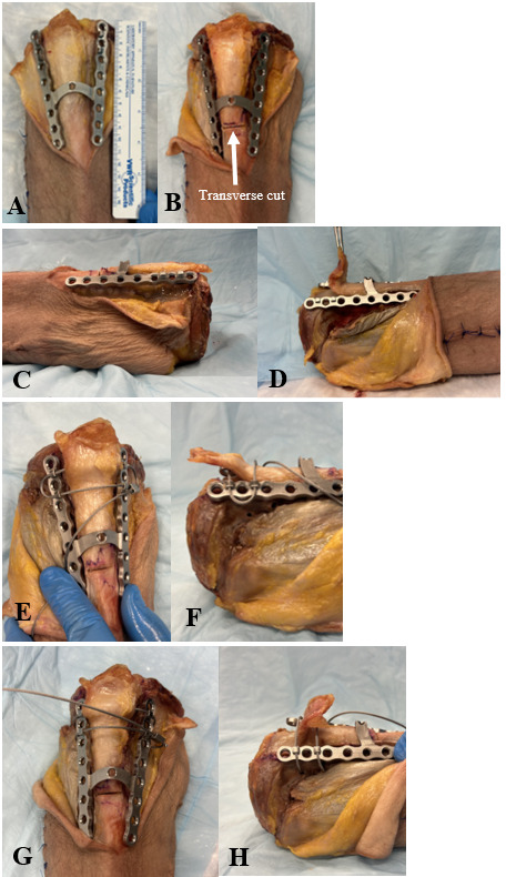
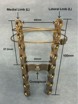
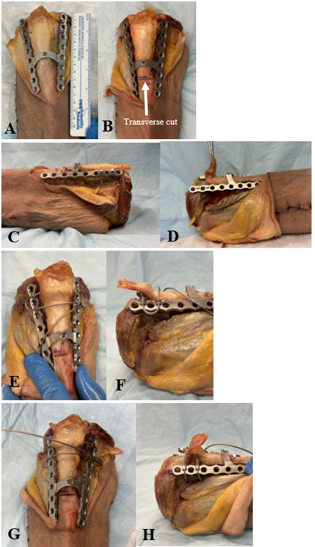
A single machined prototype of the device (TTOP 1.0) was used for evaluation on cadaver tibia. Design features of this first version incorporated the key elements outlined in the patent application (Fig 1). The size of the plate was informed by our examination of anatomic relationships of the proximal tibia based on x-rays obtained after total knee arthroplasty (DeCicco et al. 2025). Key elements incorporated into the TTOP 1.0 include two distally converging limbs with screw holes, a crosspiece connecting the limbs, also with screw holes, and slightly raised extensions with tunnels on the anterior surface of the proximal portion of the two limbs to provide transverse passage of cerclage type devices such as FiberWire or cables. The rationale for the side-by-side vertically oriented limbs was to provide fixation away from the central intramedullary stem of the tibial revision component, whether through screws around the stem or through locking unicortical screws, onto the intact non-osteotomized bone medial and lateral to the vertical osteotomy bone cuts. The crosspiece is designed to prevent anterior displacement of the distal portion of the osteotomy fragment with pull of the extensor mechanism and to provide the opportunity for, but not requiring, screw fixation through the crosspiece. The vertically oriented holes that accommodate the cerclage devices either through or posterior to the patellar tendon are designed to prevent proximal migration of the tubercle fragment.
2.1. Study Population
An anatomic cadaver study was conducted on 44 knees from 22 fresh human cadavers (28 female, 16 male). The mean age of cadaver donors at death was 81.9 years old (range, 41-101 years). The mean height of cadavers was 161.9cm (range, 145-183cm), the mean weight was 50.1kg (range, 35-64kg), and the mean BMI was 18.9 kg/m2 (range, 13.2-27.6 kg/m2). Most of the cadavers were Caucasian.
The specimens were previously dissected for instructional purposes. Additional sharp dissection was required to properly expose the insertion point of the patellar tendon as well as the patellar tendon footprint to collect measurements.
2.2. Data Collection
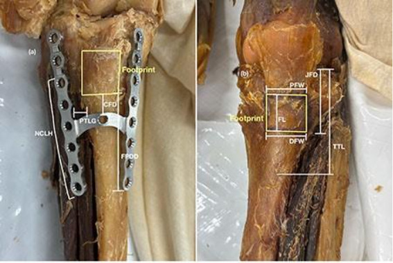
Eight standardized measurements were collected on each knee. Three of the measurements were collected with the TTOP 1.0 plate in place on the tibia to evaluate the relationship between the patellar tendon footprint and the position of the plate as well as how the plate contoured to the lateral aspect of the proximal tibia (Figure 2a). The other five measurements were taken without the plate in place; they recorded the length and width of the patellar tendon footprint as well as the distances from the joint line to the distal aspect of the patellar tendon footprint and to the distal aspect of the tibial tubercle (Figure 2b). The measurements were then used to judge the appropriateness of the current TTOP 1.0 dimensions (Fig. 3). The number of screw holes that were not in contact with the lateral aspect of the tibia was also recorded.
_measurements_taken_with_the_ttop_1.0_on_the_tibia_and_(b)_with.png)
_with_dimensions_and_labeled_components.png)
2.3. Data Analysis
Once measurements were collected, data was analyzed using IBM SPSS Statistics (Version 30, Chicago, IL). The data was first tested for normality using the Shapiro-Wilk test. Based on normality, an independent sample t-test or Mann-Whitney U test was then used to determine statistical significance between measurements.
2.4. Plate Modification
Data analysis was then used to propose modifications to TTOP 1.0. Modifications focused on achieving optimal apposition of the plate to the proximal tibia around a TTO to improve ease of application and secure fixation. Although the goal of these modifications is ultimately to minimize rates of malunion and nonunion and reduce proximal migration of the TTO, in this step of the plate development, no biomechanical testing was done.
3. Results
Measurements were taken on 44 knees from 22 cadavers (28 female, 16 male) for this study. Descriptive statistics were calculated for each of the eight measurements to evaluate the current dimensions of TTOP 1.0. Measurements were also compared between sex for statistical significance. All measurements showed statistical significance (p<0.05) between males and females except for Distal Footprint Width (p=0.070). An important data point to note was the gap between the lateral limb of the plate and the lateral aspect of the proximal tibia, called the Plate-Tibia Lateral Gap (PTLG). The mean PTLG was 6.7mm (range, 1.2-11.6mm), demonstrating a need to narrow the distal configuration of the plate by translating the distal aspect of the lateral limb medially to achieve better contact between the plate and the anterolateral aspect of the tibia.
The data was then used to answer the study questions and inform the design of the next version of the tibial tubercle osteotomy plate (TTOP 2.0). To determine if (a) a right-sided and left-sided plate was required and to demonstrate the difference in the shape of the medial and lateral aspects of the proximal tibia, data from the Non-Contact Lateral Holes (NCLH) variable was used. On average, 6 of the 8 holes on the lateral limb of the plate were not in contact with the tibia, suggesting a need to modify the length, contour, and angulation of the lateral limb of the TTOP 1.0.
To determine if (b) there was enough variability in the distance to the distal aspect of the patellar tendon footprint to suggest multiple lengths of the plate, the Tibial Tubercle Length (TTL) was examined. The mean TTL was 76.2mm (range, 56.6-99.1mm). The current plate has a length of 100mm that was selected based on our initial radiographic imaging study examining x-rays after primary total knees, demonstrating an appropriate distance to adequately bypass the distal aspect of the tibial tubercle in all cases (DeCicco et al. 2025). Therefore, with the current anatomic and human factor analysis described, a single length plate is sufficient, but in clinical practice, access to more than one plate length would likely be optimal. Further, the current length allows the plate to secure the tubercle distally, helping to prevent proximal migration of the osteotomy.
When evaluating (c) if there was substantial variability in the location of the distal aspect of the patellar tendon footprint to consider adding a second crosspiece, Joint Line to Footprint Distance (JFD) was evaluated. When evaluating this measurement, it was important to note the crosspiece should be positioned distal to the patellar tendon footprint but remain securely on the tibial tubercle. Therefore, with the current distance from the proximal aspect of the plate to the crosspiece measuring 57.2mm and the average JFD measuring 41.3mm (range, 26.4-61.8mm), the results show that the current crosspiece position bypasses the footprint in 40 of the 44 knees (91%) and that one crosspiece should be sufficient. Also, to ensure the screw placed in the crosspiece still falls on the tibial tubercle, we reviewed the TTL data, showing that the current crosspiece distance is sufficient and falls within the tibial tubercle in 43 of 44 tibias (98%).
Lastly, to determine if (d) the current proximal width of the TTOP 1.0 provides sufficient breadth to span the osteotomy limbs if they are made at the widest portion of the patellar tendon footprint, we analyzed the Proximal Footprint Width (PFW) and the Distal Footprint Width (DFW). The mean PFW was 23.3mm (range, 17.7-30.3mm), and the mean DFW was 21.1mm (range, 13.8-27.0mm). With the current proximal width of the plate being 44mm and the inner distance at the crosspiece being 35mm, the current width of the plate is sufficient to support all tibias in this study.
4. Discussion
The annual number of revision total knees in the United States is projected to reach 268,200 by 2030, a 600% increase from 2005 (Iorio et al. 2008). TTOs are a valuable technique in revision total knees to achieve better exposure in difficult cases, but complications are well documented. At current rates, TTOs are used in 3-5% of revision TKA (Sheng et al. 2006). As the need and familiarity with the technique increases, those numbers are likely to increase. In the absence of a single widely accepted, safe, reliable, effective, and efficient fixation technique, the need exists for an optimal fixation device that minimizes migration of the tibial tubercle osteotomy fragment, provides immediate secure fixation with the prospect for earlier active motion, and streamlines fixation of a technically challenging surgical technique specifically designed for use in revision of total knees. The initial steps in development of the TTOP have been focused on mitigating associated complications through intelligent plate design.
The popularized technique for performing a TTO includes a 6-8cm long, 2cm wide, and 1cm deep (proximally) to 0.5cm deep (distally) osteotomy. A TTO can be indicated for better exposure in several scenarios, including both non-arthroplasty cases and primary and revision TKA procedures (Mendes et al. 2004; Garvin et al. 1995). As an alternative or supplement to TTO, a quadriceps snip (QS) procedure also affords exposure during similar procedures. The simplicity of a QS procedure, performed by making a 45-degree cut in the quadriceps tendon, is an advantage over a TTO. However, QS relies on soft tissue healing of the tendon, which can be less reliable than the bone healing that occurs in a TTO and does not provide direct exposure to the intramedullary proximal tibia to facilitate extraction of TKA tibial components (Garvin et al. 1995). Studies comparing TTO and QS in RTKA have shown statistically significant improvement in range of motion and PROMs for patients in both groups, but one study found a higher percentage of RTKA patients developed post-operative extensor lag when a QS was performed (45% vs. 13%) (Bruni et al. 2013; Sun et al. 2015).
One novel technique to address this problem was published recently. A custom metaphyseal cone that had adaptable screw holes built in to secure the tubercle has been reported in the setting of revision and re-revision TKAs with substantial tibia bone loss. Use of the custom cone device yielded no instances of proximal migration of the TTO fragment (Piovan et al. 2022). However, thus far, this device is available only on a custom design basis and is restricted to when a metaphyseal cone is needed.
Studies comparing current TTO fixation methods both underscore the need for an answer to the question of best fixation in this setting and identify important design elements that should be incorporated in such a TTO fixation device. Stevens et al. described a standard plate and screw construct, but it was designed for use in standard realignment TTO rather than in the setting of a RTKA and would not accommodate the presence of an intramedullary stem in the latter setting (Stevens et al. 2020). In the setting of patellar instability TTO fixation techniques, biomechanical testing favored the combination of screws and suture tape in a figure-of-eight configuration over screw fixation alone. The significantly lower TTO fragment displacement in the augmented suture tape fixation supports the use of the combination screw and cable or suture fixation accommodated by the TTOP (Frame et al. 2021). In a biomechanical study testing six different TTO fixation methods that were variations of screws only or a screw and plate construct, the least amount of displacement in the setting of realignment TTOs was seen with a combination of plate and screws rather than screws alone (Guneri et al. 2022). These findings also support the plate fixation concept incorporated in the TTOP.
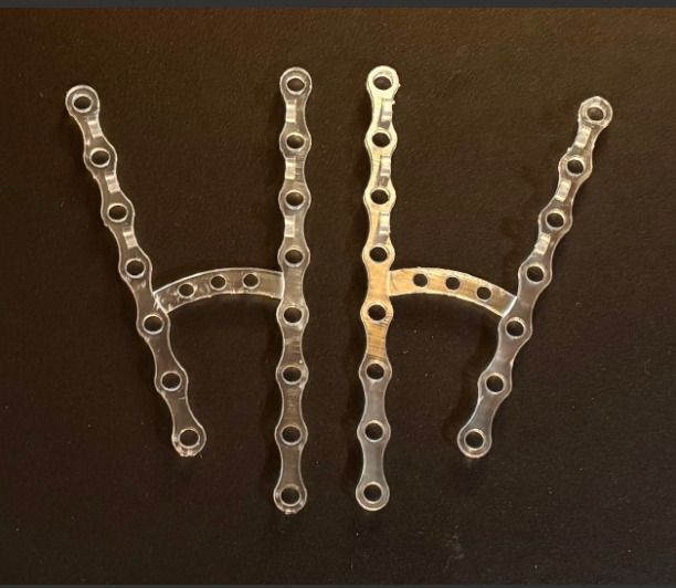
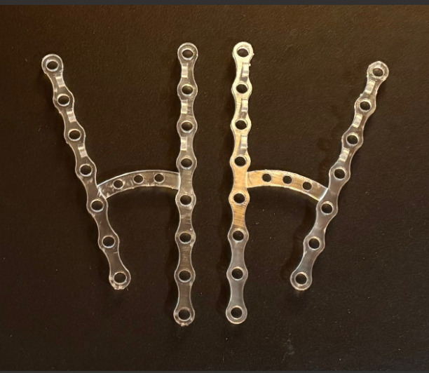
When analyzing the data collected in this study and considering the original design of the TTOP 1.0, modifications were identified that were felt to improve the effectiveness of the plate. In the next iteration of the TTOP 2.0, it would be beneficial to converge the plate more distally on the lateral aspect of the plate to lessen the gap between the plate and the lateral proximal tibia (PTLG).
Furthermore, when modifying the TTOP 2.0, it would also be beneficial to shorten the plate by one hole on the lateral limb and contour the lateral aspect of the plate to allow for the concavity of the lateral aspect of the proximal tibia, therefore necessitating side-specific plates. Although the lateral side will be shortened, the medial limb will remain at 10cm as it adequately extends past the osteotomy site currently recommended to exit the anterior cortex between 8 and 10cm distal to the joint to achieve optimal fixation (Whiteside and Ohl 1990; DeHaan et al. 2016). Although statistically significant differences were documented between sexes, the TTOP 2.0 design will cover a range of variation that accommodates differences in sex, eliminating the need for sex-specific plates. Other modifications not specifically informed by this study include a lower profile, low-contact plate, and variable angle locking screws to facilitate angulation of screws around the stem or the use of unicortical locking screws and more screw holes in the crosspiece to broaden the fixation options in the distal aspect of the osteotomy fragment (Fig. 4).
Future research on this topic will include anatomic fit and human factor testing of TTOP 2.0 to evaluate what further modifications are needed to optimize the positioning of the plate in a cadaver setting. Most importantly, biomechanical studies will be required to examine effectiveness of fixation compared to other techniques.
Limitations of this study include the narrow cross-section of potential patients represented by our mostly elderly and Caucasian cadaver population, although the age range reflects the population typically operated on for RTKA. Also, the study lacked interobserver reliability testing of the measurements as they were conducted by one study team member. Further, in addition to evaluating the current design based on the above quantitative data, certain aspects of the design required consideration of qualitative factors such as the potential need for contouring the lateral limb informed the decision to shorten the lateral limb of the plate by one hole. Moreover, the challenge of dealing with the anterior compartment soft tissues abutting the concave lateral surface of the proximal tibia presents a challenge in the current design. However, when possible, the lateral arm of the plate would be best placed over the periosteum and soft tissue sleeve maintained laterally. Thus, the clinical decision on use of the plate would have to incorporate a decision balancing these two competing goals of maintaining the completely unadulterated lateral based soft-tissue vascularity as much as possible versus achieving fixation that prevents proximal migration, the occurrence of which would likely disrupt the blood supply in a more delayed fashion. Finally, this human factor testing and cadaver anatomic measurement study does not provide biomechanical data supporting the purported biomechanical advantages of the plate design, but that will require machining TTOP 2.0 plates with variable angle locking screw holes in order to test the actual spectrum of fixation constructs possible with the new design.
The development of the TTOP 1.0 as a novel device for use in the setting of revision TKA combines an H-shaped plate affixed to the bone with screws with optional cerclage (wire, suture, or cable) fixation in a configuration that allows secure fixation around the presence of an intramedullary stemmed tibial component with or without augments and cones. The lateral and medial limbs provide secure fixation and the crosspiece, combined with the extension of the limbs past the osteotomy both proximally and distally, provide distal and proximal fixation, noted to be important in resisting translation and elevation of the tubercle (Stevens et al. 2020). Moreover, the addition of raised transverse anterior surface holes in the proximal aspect of the medial and lateral limbs allows for cerclage devices to be passed through in various configurations to also aid in fixation of the fragment, further counteracting the proximal vector of the extensor mechanism. The current study informed the development of the TTOP 2.0, with dedicated right and left sided plates with a shorter, more acutely angled lateral limb to improve bone apposition and conform to nearly all tibias.
5. Conclusion
Overall, the data collected and analyzed in this study further informs novel fixation plate design. The described analysis of the TTOP 1.0 advances the development of the TTOP 2.0 by demonstrating the need for better contouring the unique anatomy of the proximal tibia as well as the value of creating sided implants to better adapt to the anatomy.