
Introduction
First described by Lichtenstein in 1938 (Lichtenstein 1938), fibrous dysplasia (FD) is a benign fibro-osseous lesion that is estimated to represent 5% to 7% of all benign bone tumours (Coley 1960; Campanacci 1999). FD may manifest as either monostotic (involving one bone) or polyostotic (involving several bones). The monostotic type is seen in up to 75% of FD cases (Riddle and Bui 2013). It commonly affects the femur, tibia and humerus but may also involve the ribs, clavicle and craniofacial skeleton (Ippolito et al. 2003). The polyostotic type is less common and may be associated with endocrine disorders such as McCune Albright Syndrome.
FD is thought to arise from a failure of complete bone remodelling, resulting in immature trabeculae stuck in dysplastic fibrous tissue. As such, the affected immature bone mineralises abnormally and cannot realign to mechanical stress. Due to the lack of mechanical strength, FD may present with pain, deformity or after a pathologic fracture (DiCaprio and Enneking 2005). FD is not inherited but instead results from a postzygotic somatic mutation of the GNAS (guanine nucleotide-binding protein/α-subunit) gene (Riminucci et al. 2010).
Monostotic fibrous dysplasia (MFD) is typically more amenable to conservative management by observation as it less likely to progress especially after skeletal maturity. However, surgery may be indicated if symptoms remain persistent or if there has been a pathologic fracture, to prevent refracture. While curettage with bone grafting is typically the treatment of choice for both weight and non-weight-bearing areas, graft resorption and recurrence are common (Guille et al. 1998; Harris et al. 1962).
Evidently, there is a need for a reproducible, fast and effective method that satisfactorily eliminates remnant tumour progenitor cells to prevent recurrence. While there was promising research by Segev et al (Segev et al. 2002). in 2002, in which cryoablation in addition to curettage was used successfully in the treatment of FD, there has been little progress in treatment options since then.
Additionally, benign bone tumours such as FD have been accessed using historical surgical approaches that disrupt the periosteum and sacrifice viable cortex (Campanacci 1999). Typically, the bone surface closest to the tumour or where the tumour bulges out from, is excised completely as part of the approach to get to the tumour. Newer approaches such as the “trapdoor” technique adopted in osteonecrosis of the femoral head (ONFH), conversely aim to accelerate healing by preserving the cortical window and periosteum. This has not yet been adapted in the surgical treatment of FD.
We herein describe a case series of 3 patients successfully treated with a novel surgical technique which involves adjuvants with a cortex-preserving approach, with promising results.
Methods
This study was approved by the institution ethics review board (CIRB Ref: 2023/2559). This was a retrospective chart review of patients with a histopathologic diagnosis of fibrous dysplasia who presented to our paediatric orthopaedic service from the period of 2018 to 2024. Chart review was performed to assess each patients’ presentation, surgical treatment and outcomes. The inclusion criteria were any patient with a diagnosis of monostotic fibrous dysplasia (not associated with other conditions), who were treated with this combination therapy. In total, 3 consecutive patients were included.
All 3 patients initially presented to children’s emergency with a pathological fracture and an incidental discovery of a focal expansile lucent lesion on radiographs. They were then immobilised using slabs or casts for fracture healing and followed-up in clinic with serial radiographs. Surgery versus observation was offered after the fracture had healed. All 3 patients eventually consented and underwent surgery using a similar novel surgical technique.
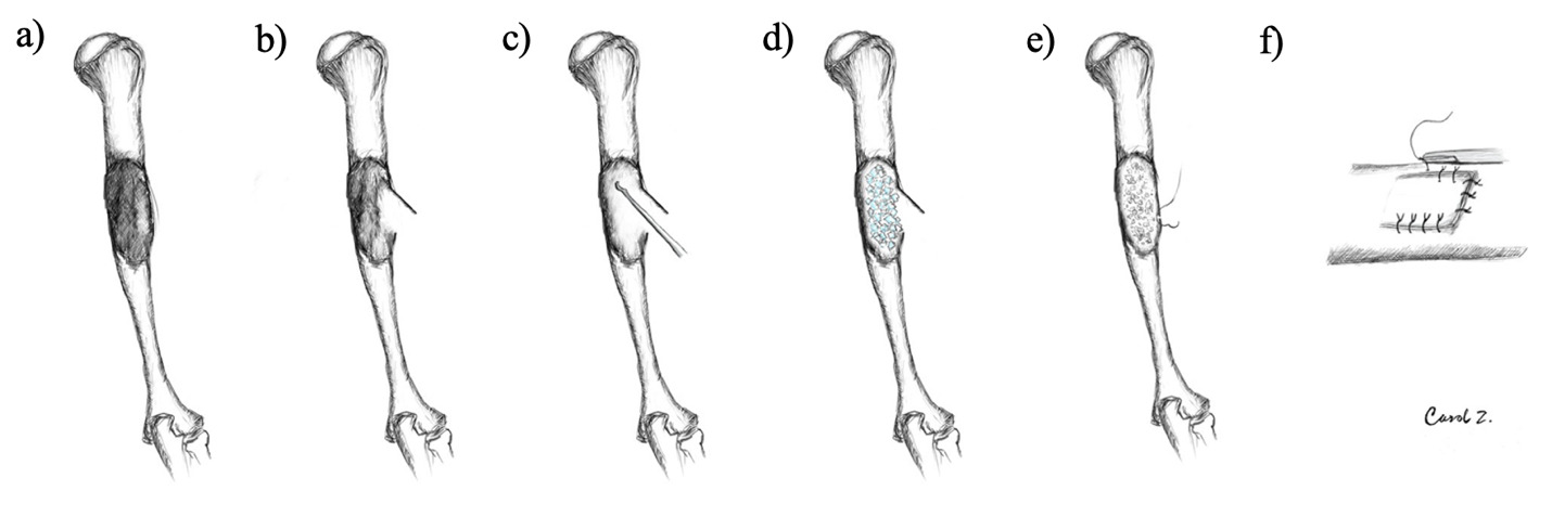
The surgical approach varied based on the anatomical location of the lesion. After dissecting through the soft tissue safely, a bony trapdoor was created either using a burr or scalpel to gain access to the lesion. Extensive curettage was then performed systematically. Two types of adjuvant therapy were then used sequentially. Firstly, dehydrated alcohol (95% ethanol, 5% water) was poured into the cavity and left in situ for 3 minutes. Cryoablation was then performed using a dry method. Liquid nitrogen was added to 20 mL of Methylated Spirit Industrial (Denatured Alcohol) BP 70% until ice crystals formed. These ice crystals were then packed into the cavity and left in situ for 1 minute. The ice crystals subsequently turn into a slurry then liquid and removed with suction. This same method of cryoablation was used for all cases. Following adjuvant treatment, allograft cancellous bone chips are packed generously into the cavity. Suture osteosynthesis of the bone flap is then performed to close the trapdoor and the operation concludes with closure by layers. The surgical technique is illustrated by Figure 1.
**_mfd_lesion_affecting_the_mid-humeral_shaft_**b)**_trapdoor_created_using_either_bur.png)
Results
The average follow-up duration of each patient was 3.4 years. The initial age of presentation, anatomical location of MFD and age of operation are shown in Table 1.
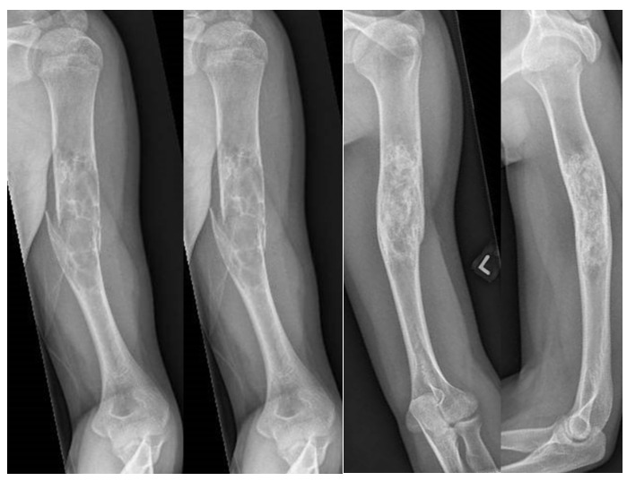
Patient 1: On postoperative day (POD) 8, the wound had healed well with good shoulder and elbow range of motion (ROM). At 5-week follow-up, radiographs revealed satisfactory healing of the trapdoor with adequate ossification. At 4-month review, the patient had mild left elbow pain, specifically stiffness when carrying loads. However, shoulder abduction power was full, and radiographs showed continued healing. The patient was counselled on doing light exercise to reduce stiffness from immobilisation but to avoid contact sports. At 10-month review, the patient was able to carry his own luggage and now played football. The patient’s scar had also healed well with radiographs showing further healing/remodelling at the left humeral shaft fracture at the site of fibrous dysplasia. At the 1 year 10-month review, the patient had good left arm ROM with no pain and radiographs also showed good thickness with no recurrence (Figure 2.). As of 2 year 11-month review, there was no recurrence of MFD.
.png)
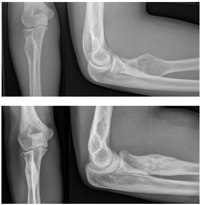
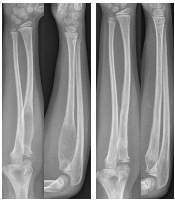
Patient 2: On POD 5, the wound had healed. However, there was pain over the proximal radius. The right extensor pollicis longus and other digit extension was weak, with right 1st dorsal webspace numbness. On 3-week review, the patient’s pain had reduced to occasional pain on moving his arm up and down with stiffness over the right upper limb. The numbness and weakness while still present had improved. Radiographs showed good healing. The patient was prescribed right upper limb stretching exercises and hand joint mobilisation. On 9-week review, the patient was well with full and equal forearm supination however pronation was still limited at 45 degrees. On the 22-week review in August 2023, radiographs showed good graft incorporation and the patient was doing well in school (Figure 3.). The patient was scheduled for follow up with repeat radiographs at 2-year review. As of 2-year 8-month review, there was no recurrence of MFD.
.png)
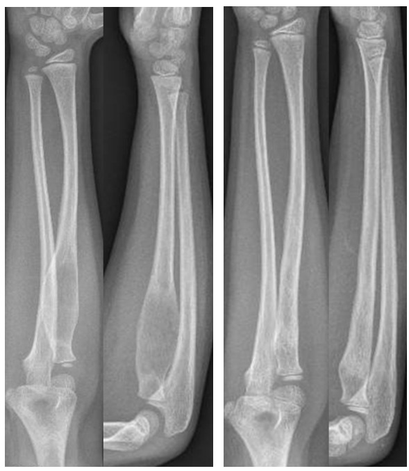
Patient 3: At 2-week follow-up, the wound had healed well. At 7-week follow-up, the patient was able to supinate to 90 degrees and pronate to 70 degrees. At 4-month follow-up, the wound was well healed, and radiographs showed that the trap door had closed with more trabeculae seen. At 11-month follow-up, there was no recurrence on radiographs and the bone had remodelled well, pronation and supination were full, and the patient was allowed to return to activities. At 1-year 9-month post-operation, the patient was readmitted to children emergency after falling from 4 steps of stairs with outstretched hand on the right, there was however no fracture. At 2-years 11-month follow up, the bone had remodelled almost to normal bone with no growth arrest (Figure 4.). The patient had full supination and pronation. The patient was scheduled for follow-up with repeat radiographs at 3-year 11-month review. As of 4 year 8-month review, there was no recurrence of MFD.
Discussion
In this treatment algorithm, all MFD lesions could be surgically managed without the need for hardware. While all patients initially presented with pathologic fractures, adequate time was given for fracture healing with conservative casting before surgery was even discussed. The involvement of non-weight bearing areas such as the radius and humerus also allowed for satisfactory resection and grafting with good stability, despite the lack of fixation.
Unfortunately, recurrence rates for FD have been traditionally high regardless of surgical treatment (Guille et al. 1998; Harris et al. 1962). Li et al (Li et al. 2014). hypothesised that this high recurrence rate associated with en bloc resection and grafting was due to the incomplete removal of dysplastic tissue containing mutated cells. Our multimodality approach with adjuvant alcohol therapy and cryoablation aims to address this concern directly.
In a study of 73 patients with giant cell tumour (GCT), 42 patients were treated with adjuvant anhydrous alcohol after resection while 31 patients did not receive adjuvant therapy (Oh et al. 2006). At an average follow-up of 4.1 years, only 9.5% in the adjuvant alcohol group experienced recurrence while 48.4% of the non-adjuvant group succumbed to recurrence. Despite its success, adjuvant alcohol has never been described for use in fibrous dysplasia until now.
Separately, the mechanism of cryoablation is thought to be via inducing cell death and the disruption of cellular integrity during freezing and thawing (Gage et al. 2009; Baust et al. 2014). A multimodality protocol involving cryoablation for FD was first described by Segev et al (Segev et al. 2002) in 2002, which involved either directly pouring liquid nitrogen (LN) into the cavity (Marcove and Miller 1969) or via cold conduction using metal probes (Bickels et al. 1999). In 6 years, only 1 of 16 patients had recurrence, showing the potential of such an approach. However, research in cryoablation for FD seems to have stalled since then.
We elected to use an alternative method of cryoablation using ice crystals to avoid the potential spillage of LN onto normal tissue and bone when pouring into a deep cavity. This method has been described for use in aneurysmal bone cysts (Wong and Mahadev 2021), a similar method described as “freezing nitrogen ethanol composite (FNEC)” has also been shown to be comparable to liquid nitrogen in vivo and in vitro for giant cell tumours (Wu et al. 2017). A safety study comparing FNEC to LN showed that LN caused more damage to surrounding normal tissue (Chen et al. 2018). Pertinently, none of the patients in our study experienced unintended damage to normal tissue or fractures from cryoablation.
We also opted to use a trapdoor technique, adapted from the treatment of osteonecrosis of the femoral head (ONFH) (Mont et al. 1998). In fact, in Mont et al.'s series utilising the trapdoor technique, 20 out of 24 stage III ONFH had achieved good or excellent Harris hip scores despite cortical collapse (Mont et al. 1998). There has been no study directly comparing outcomes and complications of curettage and grafting for FD using the trapdoor technique versus conventional cortex-sacrificing approaches. However, we believe that such an approach reduces time spent on osteosynthesis and could potentially allow for earlier bone healing of the cortex due to the minimisation of periosteal disruption.
Our method is highly repeatable and reproducible, due to the relative ease of access to dehydrated alcohol, liquid nitrogen, denatured alcohol and bone chips. The bony trapdoor technique also presents a shallow learning curve.
Additionally, our method proved to be effective regardless of skeletal maturity. Case 3 described a skeletally immature girl with FD involving the meta-diaphysis, who achieved complete bone remodelling with no growth arrest. This patient had recurrent reattendance to children’s emergency for falls and even bumping her elbow onto the dining table without wearing her removable cast. As such, early surgical intervention may be recommended for younger children to avoid undue worry for both patient and family.
Bisphosphonates were also considered in the management of these patients. While effective at pain relief, bisphosphonates have little effect on structural deformity. Hence these patients were all offered surgery as definite treatment (Regmi and Niraula 2025). A pertinent area of further investigation would be the combination of surgical curettage with biologics such as denosumab (de Castro et al. 2023) in the treatment of fibrous dysplasia.
Limitations
The main limitation of this study was its small sample size and relatively short follow-up.
Additionally, this series only included cases of isolated MFD involving the non-weight-bearing upper limb. As such, the authors cannot recommend this technique for non-isolated FD and MFD involving other locations such as the weight-bearing lower limb.
Finally, a combination of technique such as cryoablation and adjuvant alcohol were used. Hence it is difficult to differentiate whether cryoablation or adjuvant alcohol influenced the satisfactory results.
Conclusion
A multimodality approach should be taken to treat MFD. In our limited case series of 3 patients in which curettage supplemented with adjuvant alcohol and cryoablation was used, promising results were seen. Ultimately due to the relatively short follow-up period, the long-term efficacy of such a treatment will require further investigation.