
Introduction
The posterior cruciate ligament (PCL) is a major soft tissue restraint in the knee. The PCL enjoys an important functional role that is derived from its structure and orientation from the medial femoral condyle to the posterior central tibia. It prevents posterior tibial translation throughout flexion, restrains internal rotation of the tibia, and facilitates femoral rollback during knee flexion (Steinbrück et al. 2014). Successful total knee arthroplasties (TKA) have accurate and generally symmetric balancing through the range of motion and a large proportion of TKA surgeons resect this ligament during posterior-stabilized knee arthroplasty.
Without controversy, the PCL is understood to contribute to flexion stability, but prior investigations into the effect of PCL resection are inconsistent (Baldini et al. 2004; Foge et al. 2019; Kayani et al. 2019; Mihalko and Krackow 1999; Nowakowski et al. 2012; Park et al. 2009; Schnurr et al. 2012; Warth et al. 2021). Historical teaching predicts a symmetric increase of 4 mm in the flexion space from PCL resection, however, the literature describes a wide range of 1-6mm of increased flexion gap, with some studies showing symmetry of the gap, and others showing a greater or smaller effect on the medial side as compared to the lateral side (Baldini et al. 2004; Mihalko and Krackow 1999; Park et al. 2009; Warth et al. 2021). Some studies were performed on non-arthritic cadaveric specimens (Mihalko and Krackow 1999; Nowakowski et al. 2012) or had small sample sizes (Mihalko and Krackow 1999; Park et al. 2009). Some assess compartment gaps manually, while others use newer technologies (Mihalko and Krackow 1999; Nowakowski et al. 2012; Park et al. 2009). Some make assessments before bone cuts (Nowakowski et al. 2012), while others measure after cuts are made (Park et al. 2009; Schnurr et al. 2012; Warth et al. 2021), thereby introducing relevant variables like tibial slope, femoral sizing, joint line positioning, and whether a PCL bone block is preserved.
With the development of robotic-assisted surgery and other technologies, mediolateral flexion-extension gaps can be more precisely measured, allowing surgeons to achieve a more well-balanced TKA. A better understanding of the association between PCL resection and flexion and extension gap balance could allow the surgeon to adjust the final reconstructive plan to account for the changes in knee mechanics. Thus, this study aimed to describe the effects of PCL resection using intra-operative robotic gap assessment during primary robotic-assisted TKA in two consecutive, nonrandomized cohorts of cruciate-retaining (CR) and posterior-stabilized (PS) TKA. We hypothesized that those patients in the PS cohort would have a significant increase in flexion gap space with no significant difference in extension gap space. Furthermore, we sought to determine if this expected difference would present symmetrically between the medial and lateral compartments.
Material and methods
Patient Selection
Institutional review board (IRB) approval was obtained prior to initiation of this study (AAAT6504). Records were retrospectively reviewed for 98 consecutive patients who underwent primary robotic-assisted total knee arthroplasty (raTKA) utilizing the ROSA System (Zimmer-Biomet; IN, USA) by two fellowship-trained arthroplasty surgeons at a single academic medical center between July 2020 and March 2021. This study did not require any recruitment materials and a waiver of consent was provided given no more than minimal risk to study participants. The ROSA system was utilized for all patients in this time period, however, if the ROSA system could not achieve proper registration, those patients were excluded and conventional manual instrumentation was instead utilized. All records for whom robotic data was available were included in this study, without exclusions. Both varus and valgus knees were included in this study, though unfortunately full length standing radiographs were not available for all patients. Patient demographic and medical information was collected from the electronic medical record, which included age, gender, body mass index (BMI), and American Society of Anesthesiologists (ASA) classification. Integrity of the PCL was not formally assessed prior to surgery.
Surgical Techniques
All surgical procedures were performed using a standard incision and midvastus or medial parapatellar arthrotomy. A standard medial soft tissue exposure was performed, and all visible osteophytes were removed. One surgeon retained the PCL at this step utilizing a bone block technique, and one surgeon completely resected the ligament utilizing a single prong retractor and anterior tibial subluxation; this distinction generated the two groups studied. Percutaneous self-drilling, self-tapping pins were then inserted into the tibial and femoral diaphysis using the recommended technique and tracking arrays (ROSA, Zimmer-Biomet; IN, USA) were attached. Notably, standard cruciate retaining (CR) polyethylene bearings were used in the PCL retention group while posterior stabilized (PS) inserts were used in the PCL resection group.
ROSA Gap Measurement Registration
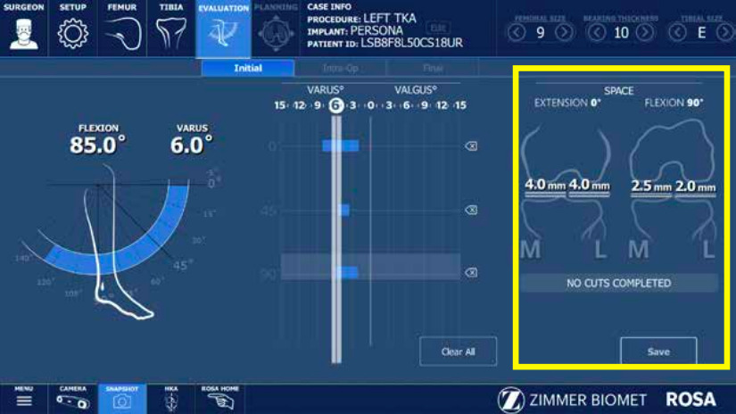
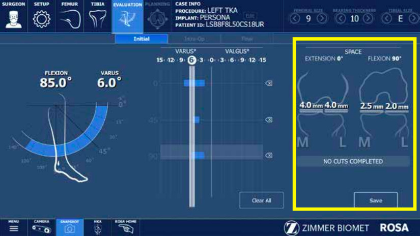
Registration was performed following the recommended technique, without image guidance. Femoral landmarks included the femoral head center, femoral canal entry, posterior condyles, trochlear groove, medial and lateral epicondyles, medial and lateral distal femur, and anterior cortex. Tibial landmarks included the medial and lateral malleoli, medial third tubercle, tibial canal entry, PCL insertion, and medial and lateral plateaus (Zimmer Biomet, n.d.). The patella was then reduced from its subluxated position. The knee was brought through a dynamic ROM with manual stressed varus and valgus forces applied by the attending surgeon at or near full extension and at 90 degrees of flexion. No standardized tensiometer or automated distraction device was utilized; the gaps represent the maximal manual stress applied by the surgeon based on their clinical judgment. This provided quantitative information, in millimeters, about the medial and lateral compartment gaps in flexion and extension (Figure 1). All measurements collected with ROSA were performed following surgical approach and soft tissue releases but prior to any bone cuts.
Statistical Analysis
Medial and lateral compartment gaps in both flexion and extension were compared using multiple adjusted linear regression analysis, adjusted for demographics. Demographic characteristics were compared using univariate analysis of variance, and between-subjects effects. Significance was considered as a P-value less than 0.05.
Results
98 primary consecutive ra-TKA had sufficient data for inclusion in this study. PCL was resected in 39 cases and retained in 59 knees. There were no significant differences in demographic characteristics between the two cohorts, including age, sex, BMI, or ASA classification (Table 1).
There were no significant differences in the pre-operative coronal alignment comparing the PCL resection group to PCL retention group as measured by the ROSA system prior to any bony resection.
We found a statistically significant difference in the medial flexion gap between the resection group and the retention group (7.16mm vs 3.7mm [Δ3.4mm], p<0.001, 95%CI=2.3 to 4.4mm). To a smaller effect, we found a statistically significant difference in the medial extension gap (resection, 5.8mm vs. retention, 4.01mm [Δ1.7mm], p=0.005). There were no significant differences between groups found on the lateral side, either in extension or flexion (Table 2).
Discussion
The most important finding of this study was that the PCL-resected/PS cohort demonstrated larger medial gaps compared to the PCL-retained/CR cohort, with a bigger effect in flexion (3.4mm) than extension (1.7mm). While we hypothesized an increase in the flexion space with no change in the extension space, our findings demonstrated a significant, albeit smaller, increase in the medial extension gap. This unexpected finding highlights the complex role the PCL plays throughout the knee’s entire range of motion.
The PCL is the primary restraint of posterior tibial translation. It is comprised of two bundles, the anterolateral bundle and the posteromedial bundle. In flexion, the anterolateral bundle is tensioned and provides most of the resistance to posterior translation, whereas in extension the posteromedial bundle does this (Kennedy et al. 2013). The PCL also resists internal rotation of the tibia, as well as external rotation in conjunction with the posterolateral corner. Eliminating the PCL necessarily eliminates its functional role, yet the literature does not definitively demonstrate a clinically significant difference in outcomes between PS and CR TKA. However, from a biomechanical and intra-operative perspective, a 3.4mm increase in the medial flexion gap may be highly relevant as flexion instability is a leading cause of early TKA failure. A gap difference of this magnitude during trial reduction may necessitate intra-operative adjustments, such as up-sizing the femoral component, altering the posterior tibial slope, or utilizing a thicker polyethylene insert to appropriately balance the flexion space and prevent instability.
A 2019 study by Kayani et al. also assessed gap balancing using a robotic platform (Kayani et al. 2019). Importantly, their study utilized an internally controlled, intra-patient design, measuring the same knee before and after PCL resection, whereas our study compared two distinct cohorts separated by surgeon technique. Despite this methodological difference, their findings in the extension space closely mirrored our own, demonstrating a relatively symmetric increase (1.5 mm medially and 1.2 mm laterally). In flexion, however, they noted an asymmetric effect in the opposite direction, with a greater increase laterally (3.3 mm) than medially (2.3 mm). While our inability to detect an effect on the lateral flexion space is unclear, their finding of a larger lateral gap is also difficult to explain given the anatomical orientation of the PCL. We suspect these discrepancies may stem from other technical surgical variables, particularly differences in baseline medial laxity following surgical exposure.
In other studies, the gaps were measured after bone cuts were made. This method introduces additional variables like tibial slope, femoral sizing, joint line positioning, and whether a PCL bone block is preserved. Warth et al. examined 129 TKA cases by 2 surgeons, using a calibrated tension device to measure gaps before and after the PCL was resected (Warth et al. 2021). These measurements were made after all bony cuts and no PCL block was preserved. They found an asymmetric effect in flexion, in the opposite direction from our study, similar to Kayani et al. (1.58mm medially and 3.3mm laterally). In extension, they found a 1.76 mm change medially and no significant change laterally, similar to our study.
Schnurr et al. examined 50 consecutive, internally-controlled patients using imageless computer navigation by one surgeon (Schnurr et al. 2012). After bone cuts were made, a tensiometer was placed through the range of motion and the gaps were monitored by the computer. They found no difference in extension gaps, but an asymmetric difference in flexion (1.3 mm medially and 0.7 mm laterally). Foge et al. examined 10 cadaver specimen using both navigation and calipers while sequentially releasing the PCL. At 90 degrees of flexion, after full release, they found an asymmetric increase in flexion gap (1.11 mm medially and 0.5 mm laterally) (Foge et al. 2019).
These studies are similar to ours in terms of the asymmetry of the flexion space increase. Considering our findings relative to these and other reports in the literature, the wide variability of the effect of releasing the PCL on gaps is evident. This may not be surprising considering the variability of PCL integrity (as some patients do not have an intact PCL pre-operatively), function, and contribution to knee mechanics, especially when considering variabilities in osteophyte formation, flexion contractures, coronal deformity, and scarring from injury or prior surgery. For example, if large osteophytes were tenting the PCL at the medial femoral notch and/or the posterior medial tibia, then the release of that PCL with associated osteophytectomy would be expected to result in a greater increase in gaps than a similar PCL without the osteophytes. Ultimately, the totality of literature examining the PCL may best be interpreted as reflecting the variability of knees in general. Given the variety of intra-operative methods used to measure flexion and extension gaps in these studies, a rational conclusion would be supportive of the use of higher level technologies, like robotics, to help surgeons tune flexion balancing specific to individual patients. The use of robotic assistance to quantify these gaps is supported by recent literature validating the accuracy of the ROSA system. Cadaveric studies have demonstrated high intraoperative precision, with bony resections and gap measurements reliably executed within 1 mm and 1 degree of the preoperative plan (Parratte et al. 2019). Furthermore, the system has demonstrated excellent inter- and intra-rater reliability specifically regarding the dynamic soft-tissue laxity assessments utilized in this workflow (Charette et al. 2022).
Several important limitations must be considered when interpreting the findings of this study. Most notably, the retrospective, non-randomized study design introduces a significant risk of surgeon technique bias. The cohorts were separated entirely by surgeon preference (Surgeon A exclusively performing CR, Surgeon B exclusively performing PS). Consequently, this study compares two different surgeons performing two different techniques, rather than evaluating the effect of PCL resection within the same knee. Therefore, the differences observed can only be described as an association, not a causal effect of PCL resection. The variations in gap laxity could be significantly confounded by inter-surgeon differences in soft-tissue handling, the extent of medial release, or the effect of posterior osteophytes on soft-tissue tensioning.
A second major limitation of this study is the lack of standardized force used to test the gaps. While the ROSA system provides highly precise measurements, the distraction forces applied to the knee were manual and subjective. As prior literature has demonstrated that gap magnitude is heavily force-dependent, relying on manual stress without a tensiometer introduces significant inter-surgeon variability and serves as a major confounder in our results (Ma et al. 2017).
Third, due to the retrospective nature of this data, which was collected during the early adoption phase of this robotic platform at our institution, the robotic outputs primarily captured the initial knee state. The surgeons did not routinely utilize post-resection validation tools or manual caliper checks after the final implants were placed. As a result, we are unable to report whether the gap differentials persisted after final implant placement, nor do we have sufficient long-term follow-up to correlate these initial gap measurements with TKA survival or subsidence rates. Therefore, surgeons should note the clinical relevance of these gap differences when setting resection planes and implant balancing during robotic-assisted TKA.
Fourth, the integrity of the PCL was not formally assessed with advanced imaging or specific intraoperative grading prior to surgery. Degenerative PCL attenuation or partial incompetence in some patients could have blunted the observed effects. Finally, our regression analysis did not strictly control for preoperative coronal deformity, as dedicated full length standing radiographs were not universally available. The PCL would be expected to function differently depending on the presenting deformity and associated osteophytes.
Future prospective studies should utilize internally controlled, intra-patient designs to assess gap changes before and after PCL resection within the same knee. Furthermore, the incorporation of standardized robotic tensiometers is necessary to eliminate the confounding variability of manual surgeon stress. Finally, utilizing this robotic gap-assessment technology within a controlled cadaveric model could isolate the kinematic effects of resecting the PCL or other critical stabilizing structures, such as the medial collateral ligament, without the confounding patient and surgeon variables present in in-vivo clinical studies.
Conclusion
Robotic gap assessment data demonstrates PCL resection may be associated with significant increases in medial flexion and extension laxity with negligible changes in lateral flexion-extension laxity. However, due to study design limitations, these differences cannot be causally attributed to the PCL alone and likely reflect a combination of ligamentous release and surgeon-specific technique. PCL resection should be performed prior to gap assessment during robotic or manual PS TKA, and surgeons should be aware of these potential asymmetric mechanical differences to allow for appropriate adjustments in the final reconstructive plan.