Loading [Contrib]/a11y/accessibility-menu.js

This website uses cookies

We use cookies to enhance your experience and support COUNTER Metrics for transparent reporting of readership statistics. Cookie data is not sold to third parties or used for marketing purposes.

Skip to main content
null
J Orthopaedic Experience & Innovation
  • Menu
  • Articles
    • Brief Report
    • Case Report
    • Case Series
    • Conference Proceedings
    • Data Paper
    • Editorial
    • Meeting Reports/Abstracts
    • Methods Article
    • Product Review
    • Research Article
    • Review Article
    • Review Articles
    • Systematic Review
    • All
  • For Authors
  • Editorial Board
  • About
  • Issues
  • Blog
  • "Open Mic" Topic Sessions
  • Advertisers
  • Recorded Content
  • CME
  • JOEI KOL Connect
  • search
  • RSS feed (opens a modal with a link to feed)

RSS Feed

Enter the URL below into your favorite RSS reader.

https://journaloei.scholasticahq.com/feed
ISSN 2691-6541
Review Article
Vol. 7, Issue 1, 2026May 07, 2026 EDT

Open Latarjet Versus Arthroscopic Free Bone Block for Anterior Shoulder Instability: A Comparative Review

Paul Sethi, M.D., Shariff K Bishai, D.O., M.S., Frank Alberta, M.D., Parth Kamdar, M.D., John Sharpe, M.D., Tim Saunders, B.S., Ellie Sheehan,
ShoulderInstabilityGlenoid Bone LossLatarjet
Copyright Logoccby-nc-nd-4.0 • https://doi.org/10.60118/001c.147402
J Orthopaedic Experience & Innovation
Sethi, Paul, Shariff K Bishai, Frank Alberta, et al. 2026. “Open Latarjet Versus Arthroscopic Free Bone Block for Anterior Shoulder Instability: A Comparative Review.” Journal of Orthopaedic Experience & Innovation 7 (1). https://doi.org/10.60118/001c.147402.
Save article as...▾
Download all (6)
  • Figure 1. Optimized screw fixation of open Latarjet procedure, the screws are parallel to the joint surface, with an alpha angle less than 10 degrees.
    Download
  • Figure 2a and 2b. Figure A demonstrates less ideal screw fixation, with higher alpha angle after Latarjet procedure. 2B demonstrates bone resorption with prominent hardware after allograft procedure.
    Download
  • Figure 3. Arthroscopic DTA procedure.
    Download
  • Download
  • Download
  • Download

Error

Sorry, something went wrong. Please try again.

If this problem reoccurs, please contact Scholastica Support

Error message:

undefined

View more stats

Abstract

Anterior inferior shoulder instability with glenoid bone loss presents a significant clinical challenge with respect to recurrent instability. Arthroscopic labrum repair alone when performed with glenoid bone loss may have high failure rates. Several surgical solutions have been proposed to treat glenoid bone loss. This review will focus on bone restoration solutions for this problem, with a comparison of Latarjet and free bone bock procedures. The Latarjet procedure remains the gold standard due to its triple blocking effect and demonstrated efficacy. Conversely, arthroscopic free bone block techniques have emerged as a promising alternative, offering comparably low recurrence rates and the potential for fewer complications.

Introduction

Anterior inferior shoulder instability when complicated by glenoid bone loss remains a challenging problem for patients and surgeons alike. Recurrent instability negatively affects patient quality of life, often necessitating complex surgical stabilization, and lost productivity in vocational and athletic endeavors. Many surgical techniques to restore glenoid bone loss have been employed historically, but currently coracoid transfer (Latarjet) and free bone block (FBB) procedures are popular choices for addressing these difficult problems due to the unacceptably high failure rates in arthroscopic soft tissue based procedures.

Arthroscopic and open enhancements of labral repair, including remplissage, dynamic anterior stabilization and labral scaffolds, have yielded promising results without utilizing bone augmentation (Keeling et al. 2023). In cases of subcritical glenoid bone loss among non-collision athletes undergoing primary stabilization, arthroscopic and open Bankart repair with remplissage remain effective treatment options; the addition of remplissage may decrease recurrent instability from 30% to 9.6% (Woodmass et al. 2024). Nonetheless, numerous studies have demonstrated inferior results of soft tissue stabilization when greater than 20-25% of glenoid bone loss is present (Shin et al. 2016). Some authors support bony procedures, such as Latarjet or FBB, with bone loss as low as 13.5% (Dickens et al. 2017). Other indications include revisions following failure of primary soft tissue repair, high Instability Severity Index Scores (ISIS), and involvement in high-contact or collision sports. The Latarjet has a long-standing history of successful stabilization and involves the transfer of the coracoid process with an attached conjoint tendon. Conversely, FBB procedures anatomically reconstruct the glenoid defect and emphasize preservation of native soft tissues and anatomic relationships. What is clear, however, is that appropriate surgical management with bony augmentation in the setting of significant bone loss remains paramount to reduce recurrent glenohumeral instability and posttraumatic osteoarthritis (Dickens et al. 2017; Leroux et al. 2014).

This review will compare the benefits of open Latarjet versus arthroscopic FBB procedures in the athlete with respect to surgical efficiency, complications and efficacy. This review will focus on the current state of knowledge, highlight areas of contention, and provide an overview for clinicians navigating the decision on how best to address glenoid bone loss.

The Role for Open Latarjet

The Latarjet technique has been lauded as the gold standard for conferring shoulder stability via Patte’s proposed triple blocking effect. First, the coracoid autograft recreates the anterior arc of the glenoid (osseous blocking). Second, the rerouted conjoint tendon tensions the subscapularis, creating a sling, which provides soft tissue stability in external rotation and abduction (musculotendinous blocking). Finally, repair of the anterior capsule and inferior glenohumeral ligaments aids in stability by mimicking a capsulolabral repair (capsulolabral blocking).

Yamamoto et al. demonstrated restoration of translational force to the intact-condition level after Latarjet in cadaveric biomechanical studies (Yamamoto et al. 2013). The triple blocking effect was validated with the sling effect and anterior capsular repair conferring 77% and 23% of stability, respectively, in end-range arm position. In mid-range arm position, the sling effect and glenoid cavity reconstruction contributed 51% to 62% and 38% to 49% of stability, respectively. These findings are further supported by Giles et al. who showed that the dynamic sling effect prevented dislocations in 100% of specimens compared to 75% of specimens without the sling effect (Giles et al. 2013). Despite these biomechanical advantages, the clinical benefit of the sling has been challenged in a recent meta-analysis. (Hao et al. 2025).

After open Latarjet procedures, recurrent instability and dislocation rates remain below 10%, underscoring the effective restoration of shoulder stability (Hurley, Jamal, et al. 2019; Mizuno et al. 2014). Furthermore, postoperative Rowe and SANE scores are reported to approach 90 (Hurley, Lim Fat, et al. 2019; Mizuno et al. 2014). However, long term follow up studies have identified a risk of development or progression of osteoarthritis in approximately 23% of patients (Mizuno et al. 2014). Despite reports suggesting a reduction in external rotation, Smith et al. observed postoperative restoration of range of motion in all directions in as early as six months, when compared to both the ipsilateral and contralateral shoulder (Smith et al. 2024).

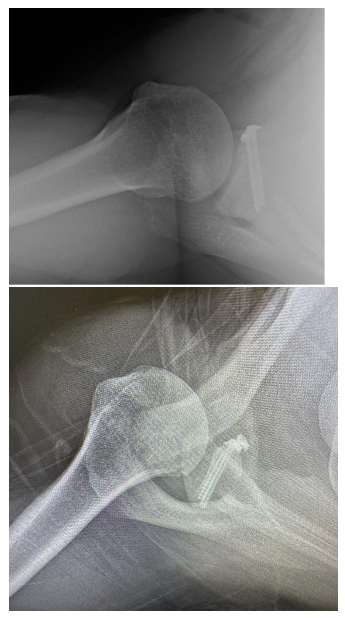
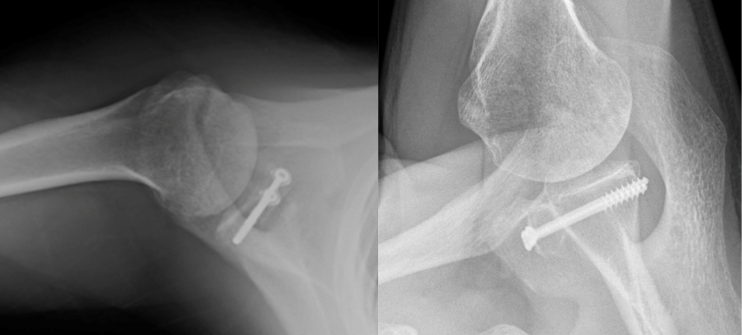
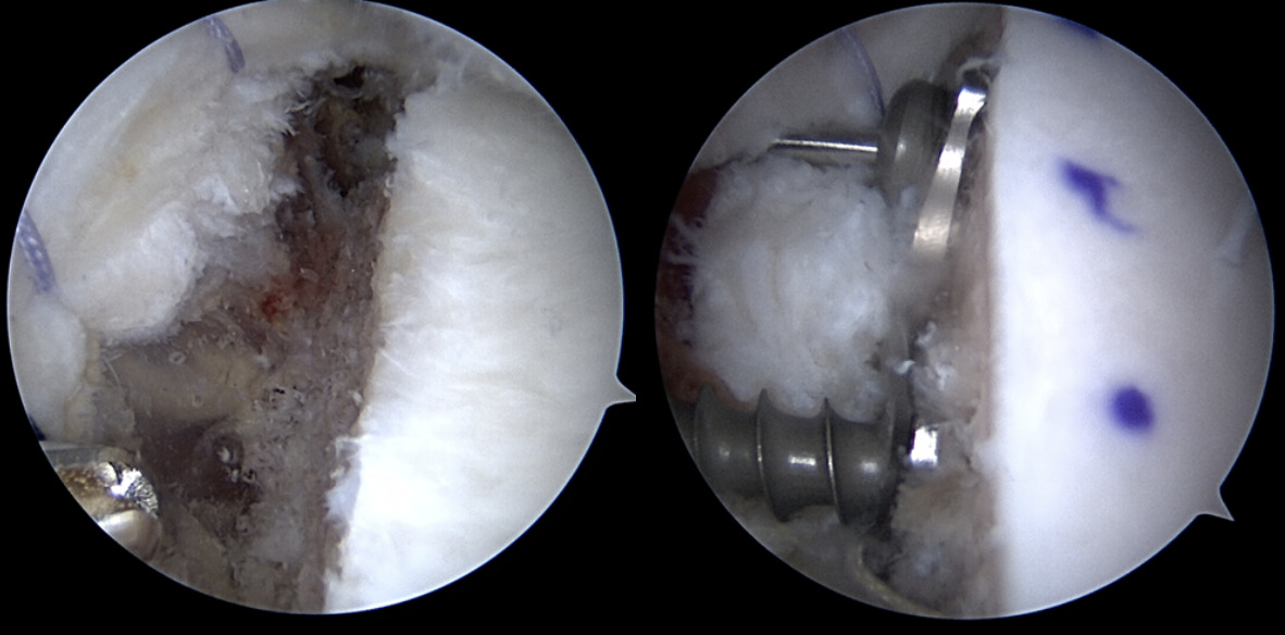
Success of the Latarjet may be predicated on surgical technique. Screw and graft placement are vital components of successful glenoid reconstruction. The ideal screw positioning is perpendicular to the anterior wall of the glenoid and parallel to the joint line (Figure 1). Screws should be placed at an alpha angle less than or equal to 10° to avoid suprascapular nerve injury and minimize graft displacement (Lädermann et al. 2012; Hsu et al. 2022). Improper screw orientation can negatively affect graft healing, cause neurovascular complications, and increase symptomatic hardware rates (Figure 2). Malpositioned screws can increase the rate of screw removal due to loosening caused by oblique compression between graft and glenoid. Medially oriented screws typically have poorer purchase due to the thinner posterior glenoid cortex. Additionally, superior and medial screws have an increased likelihood of causing suprascapular nerve palsy (Maquieira et al. 2007). Accurate graft positioning is also crucial, with the ideal placement being 1.5-3 mm lateral to the flush position. Excessive medialization increases the risk of recurrent instability. Excessive lateralization elevates contact pressure between the glenoid and humeral head, increasing the risk of osteoarthritis (Martins et al. 2022). Additionally, bone grafts placed at 4 o’clock on the anteroinferior glenoid rim reduce displacement in external rotation and anterior translation (Nourissat et al. 2014).

Figure 1
Figure 1.Optimized screw fixation of open Latarjet procedure, the screws are parallel to the joint surface, with an alpha angle less than 10 degrees.
Figure 2a and 2b
Figure 2a and 2b.Figure A demonstrates less ideal screw fixation, with higher alpha angle after Latarjet procedure. 2B demonstrates bone resorption with prominent hardware after allograft procedure.

Long term subscapularis function remains another theoretical concern after the Latarjet procedure. One study utilized magnetic resonance imaging (MRI) to evaluate subscapularis functional integrity with a mean follow up of approximately eight years and demonstrated no evidence of partial or full thickness tears. Additionally, negative belly-press and lift off tests demonstrated no decrease in internal rotation strength after eight years (Azizi et al. 2022). Both Caubere et al. and Azizi et al. demonstrated no fatty infiltration of the subscapularis at one year and eight years, respectively (Caubère et al. 2017; Azizi et al. 2022). These studies suggest that despite the subscapularis split performed during surgery, range of motion, function and muscular integrity of the tendon are preserved.

Prior to shoulder stabilization surgery, many patients identify the ability to return to sport as their greatest concern (Warth et al. 2013). A systematic review of open Latarjet with ten year follow-up reported a return to sport rate of 85%, with 76% returning to preinjury level (Hurley, Jamal, et al. 2019). When comparing outcomes between competitive and recreational athletes, Rowe scores significantly increased in competitive athletes after open Latarjet (Baverel et al. 2018). Overhead athletes have successful outcomes after Latarjet, with 73% of European handball players reporting no pain while throwing and returning to ball training and competitive play at 3.2 and 4.9 months, respectively (Bauer et al. 2024). A separate study of non-overhead athletes found no difference in throwing velocity or distance following open Latarjet when compared to a control group. Although external rotation at the side was reduced in this study, external rotation at 90° abduction remained unaffected, likely explaining the preservation of throwing ability (Yokoi et al. 2024).

Open Latarjet has however been associated with longer operative times, higher minor and major complications and an increased risk for return to the OR when compared to standard arthroscopic Bankart techniques. Other authors have attempted to define the learning curve and time to surgical proficiency for the procedure. Operative time decreased significantly after the first 15 cases and plateaued after 30 cases in this single surgeon series (Papalia et al. 2024). Complications, while high at nearly 11%, do not appear to be affected by hospital or surgeon volume in some large studies (Alkaduhimi et al. 2023).

The success of the Latarjet procedure relies on precise execution of the surgical technique, specifically accurate screw and graft placement, which ensures stable fixation, minimized complications, and reduced recurrent instability. In addition to low rates of persistent glenohumeral instability, this procedure yields excellent clinical outcomes, including a restored range of motion. When performed with meticulous attention to screw and graft positioning, the open Latarjet continues to serve as a reliable option for restoring shoulder stabilization and returning to sport in a timely manner.

While the Latarjet remains the gold standard for managing shoulder instability with glenoid bone deficiency, there remains a 10-15% complication rate (Athwal et al. 2016; Domos et al. 2018). The complication risk and relatively long learning curve highlights the need to assess other stabilization options. FBB procedures, although lacking the benefit of the sling effect, may confer joint stability without anatomical alteration associated with conjoint transfer and may help reduce the complication risks of Latarjet reconstruction. Arthroscopic Latarjet deserves mention as an effective alternative to open Latarjet, with theoretically lower complications. A full discussion of this goes beyond the scope of this article. Nonetheless, North American adoption of the arthroscopic technique has lagged due to the technical difficulties for surgeons without a high volume of this procedure (Hurley, Lim Fat, et al. 2019).

Arthroscopic Free Bone Block Procedures

The first described FBB procedure was reported in 1918 with an iliac crest autograft (Eden 1918). Several autograft options have since been described, including scapular spine, distal clavicle and hardware-free sculpted grafts (Xiang et al. 2021; Auffarth et al. 2008). In 2008, Provencher popularized use of fresh distal tibial allografts (DTA) as an alternative to autografts (Menendez et al. 2023; Provencher et al. 2009). In 2015, Wong described the use of an arthroscopic osteochondral FBB to manage glenoid bone deficiency (Wong and Urquhart 2015). Currently, FBB graft options include fresh or frozen DTA or distal radius allografts.

Arthroscopic FBBs serve as an alternative to traditional Latarjet with both theoretical and practical advantages. Proponents of arthroscopy contend that the articular surface visualization and graft placement are improved compared to its open counterpart. Furthermore, sling procedures are non anatomical and may complicate future surgery (Figure 3) (Lafosse et al. 2010). The relevance of graft position is critical, as 1 mm of graft lateralization can lead to poorer outcomes (Ernstbrunner et al. 2023). Avoiding harvest of the coracoid minimizes the risk of neurovascular injury as well as intraoperative coracoid fracture. Additionally, the use of osteochondral allografts removes prior size limitations given the fixed size of the coracoid autograft. Finally, the shape and contour of the DTA may provide a more anatomic match with the native glenoid (Provencher et al. 2009).

Figure 3
Figure 3.Arthroscopic DTA procedure.

Comparison

The results of open Latarjet and arthroscopic FBB both yield satisfactory outcomes, with a recurrent instability rate of less than 10% (Hurley, Lim Fat, et al. 2019). In comparison studies, both Frank et al. and Gilat et al. found no significant difference in recurrent instability rates between Latarjet and FBB procedures (Frank et al. 2018; Gilat and Haunschild et al. 2021).

Graft incorporation is slower in FBB, with allografts requiring approximately 6.9 months versus Latarjet autografts requiring 6-12 weeks (Dawe et al. 2025; Kordasiewicz et al. 2019). While achieving bony union of the glenoid bone block may not be absolutely required for clinical success, a malunited, nonunited, or overly resorbed bone block may contribute to recurrent instability, pain or hardware complications. While both graft types experience resorption, the degree of resorption is substantially higher in FBB (Wong et al. 2018; Delgado et al. 2024). Excessive resorption may contribute to screw prominence and consequent hardware-related complications. Despite resorption of ≥50%, one study reported patients can still achieve excellent clinical outcomes, suggesting that such resorption levels are not consistently associated with poor outcomes or increased rates of revision surgeries (Wong et al. 2020).

Fixation of the graft is typically achieved with screws, buttons or cerclage fixation. A systematic review of arthroscopic versus open Latarjet stabilization with attention to hardware complications demonstrated a higher rate of screw removal in the arthroscopic group (Lacouture-Suarez et al. 2023). When comparing open Latarjet screws and arthroscopic buttons, Nascimento et al. found comparable clinical and radiological outcomes; however, a letter to the editor highlighted that 16% of the button group had postoperative complications (primarily neurologic injury) compared to 2% in the screw group (Nascimento et al. 2025; Lädermann 2025). Similarly, Pancura et al. compared button and screw fixation in arthroscopic FBB and found that patients who underwent button fixation were significantly more likely to suffer from recurrent dislocations. Over 40 percent of the button group in the study had recurrent dislocations versus zero dislocations in the screw group with one of the conclusions cautioning against using buttons in allograft FBB (Pancura et al. 2025).

Graft and screw positioning after open and arthroscopic Latarjet have been analyzed using computed tomography (CT) imaging. Neyton et al. performed a retrospective multicenter study demonstrating a significantly lower mean angulation in both superior and inferior screws in the open group versus the arthroscopic group. Graft positioning also differed between each technique on both axial and sagittal views. In the axial plane, arthroscopic grafts were placed significantly more laterally. In the sagittal plane, the open group showed more reliable placement of grafts below the equator (Neyton et al. 2018). These findings are corroborated by Marion et al. who noted more lateral and inferior graft placement in arthroscopic Latarjet when compared to the open technique (Marion et al. 2017). While these previously mentioned studies are from arthroscopic Latarjet, and not FBB studies, certain similarities may be drawn. This highlights the technical challenges of graft position and fixation in the arthroscopic techniques.

Arthroscopic bone restoration of the glenoid is technically demanding and can be difficult to master. Multiple studies have demonstrated a substantial improvement in proficiency and corresponding decrease in operative time for arthroscopic Latarjet and DTA from an initial range of 103 to 183 minutes to a range of 76 to 95 minutes after 20-45 arthroscopic procedures (Leuzinger et al. 2019; Cunningham et al. 2016; Bishai et al. 2022; Moga et al. 2018; Castricini et al. 2013; Kany et al. 2016; Getz and Joyce 2020; Ekhtiari et al. 2018). A multisurgeon, large volume analysis showed achievement of an operative time steady state after 30-50 cases (Valsamis et al. 2020). However, Zhu et al. observed that, regardless of surgical experience, arthroscopic Latarjet remained 30 to 40 minutes longer than the open approach (Zhu et al. 2017). Arthroscopic FBB techniques are less frequently described in the literature, but may be associated with a faster learning curve, as the procedure may be comparatively easier to perform. Surgeons who endeavor to perform arthroscopic glenoid bone procedures may consider this when their operative case load (15-30 per year) supports the time commitment to the associated learning curve.

When choosing between open Latarjet and arthroscopic FBB other important considerations include cost and graft procurement. The open Latarjet provides a zero-cost bone block compared to the thousands of dollars associated with fresh or frozen DTA. Uffmann et al. performed a cost minimization analysis comparing DTA, open Latarjet, and arthroscopic Bankart repair. Open Latarjet was approximately 66% and 15% less costly than DTA and arthroscopic Bankart repair, respectively (Uffmann et al. 2019). In addition to cost, access to allograft bone in FBB procedures can pose logistical challenges, particularly in centers without an onsite bone bank. Autograft iliac crest, distal clavicle and scapular spine remain viable economical options. A more detailed analysis of total costs when taking recurrence, complications, and additional surgery are all considered, however, has not been performed to our knowledge.

Return to sport rates following arthroscopic FBB approach 80%, with most non-returning patients citing reasons such as reprioritization of work and life (Ojaghi et al. 2024). A systematic review showed a higher return to sport rate for FBB procedures for autografts than allografts in both open and arthroscopic procedures (Gilat et al. 2021). In a cohort of competitive collision or overhead athletes, open Latarjet demonstrated a faster return to sport (5.07 months), compared to the arthroscopic approach (5.87 months) (Abdul-Rassoul et al. 2019).

Conclusion

While the open Latarjet procedure remains a reliable surgical option for anteroinferior instability, FBB procedures present a compelling alternative. The Latarjet’s high historic success rate is appealing, especially for surgeons well-versed in the open approach, but is not without its challenges in terms of complications, learning curve and long-term alteration of anatomy. Consequently, arthroscopic FBB has become an attractive option, addressing many concerns with its subscapularis-sparing technique, lower neurovascular risk, and favorable learning curve for the arthroscopic surgeon. Long term follow up is not yet available. FBB procedures conserve patients’ anatomy, specifically the subscapularis and coracoid, which are relevant if and when additional reconstructive surgery may become necessary. Conversely, allograft bone is costly, has a longer return to sport, is subject to greater resorption and the arthroscopic technique is not without its challenges in graft positioning or fixation.

Ultimately, in the hands of a skilled surgeon, both techniques are safe and effective. As more long-term prospective data is collected, the specific indications for each will be refined.

Sethi Final Take

My instability patient population shapes my preference for the surgical procedures I offer. While many instability operations (remplissage, DAS) gain popularity, there remains to be a role for glenoid bone restoration in the young collision athletes and revision cases that make up my practice. I value the advantages of an all arthroscopic bone block, subscapularis sparing operation that does not alter the shoulder’s neurovascular anatomy. However, at this time, allograft bone procedures exhibit greater resorption and have higher failure rates when screw fixation is not used. Furthermore, a predictably faster return to sport, and the additional stability benefit conferred by the conjoint tendon make Latarjet my choice operation in the competitive collision athlete. As these gaps in recovery are inevitably narrowed, I imagine further adoption of the Arthroscopic Anatomic Glenoid Reconstruction (AAGR) techniques for the collision athlete.

Bishai Final Take

When treating patients with bone loss shoulder instability, the pathology often dictates the surgical treatment plan. Although the coracoid serves as a local graft with no cost, the amount of bone that can be made up for the glenoid may not be enough. In these cases the distal tibial allograft utilizing the AAGR technique is a great tool in the armamentarium. Secondly, the ability to maintain the remaining labrum and capsule to help augment the repair and attach it to the paleoglenoid helps with keeping the humeral head positioned more posterior as a secondary rein. The AAGR also maintains the integrity of the subscapularis which allows for decreased concerns of subscapularis failure for those doing open peels and tenotomies or even a split. These patients with shoulder instability with bone loss can be very difficult to treat therefore it is paramount to have the ability to stratify these patients into the correct treatment algorithm whether it be an autograft or allograft reconstruction with techniques like the Latarjet and AAGR respectively.

Alberta Final Take

Recurrent instability with bone loss remains one of the unsolved challenges in my practice. While it’s hard to argue with the long term results and success of Latarjet reconstruction (both published and personally experienced), there are limitations in terms of graft size and patient selection that remain. Reconstruction is limited to the width of the local coracoid graft in Latarjet and while that addition of the sling effect is beneficial, it is at the cost of severely altered anatomy which presents many future challenges. The return to sports in the highest level athletes also tends to dictate the procedure when all else is equal. Because the return to sport rates, recurrent dislocation rates and time to return to sports tend to favor open Latarjet in this group, that is typically my choice, especially in contact/collision athletes. In recreational athletes, laborers, seizure disorder patients, and those with defects too large to be reconstructed with the local coracoid, I favor AAGR with allograft. In either case, extreme attention to detail with accurate graft placement and solid fixation with screws is mandatory. In both procedures, what has become clear is that resorption is common, especially when the graft exceeds the native glenoid size and that does not necessarily affect clinical outcomes. Both procedures are highly effective at solving the recurrent instability problem which is the primary goal and it is up to us as surgeons to offer our patients the procedures we can reliably perform with their best interest in mind. And ultimately it is our responsibility to take advantage of all of the many learning tools available to us to become better for our patients.

Open Latarjet Arthroscopic Free Bone Block
Mechanism of Stability Triple blocking effect: anterior glenoid reconstruction, dynamic sling effect, capsular repair Anatomic glenoid reconstruction without dynamic sling effect
Return to Sport 85% return rate, faster return (4 months), excellent outcomes in contact and overhead athletes 80% return rate, slower return (6 months)
Graft Choice Coracoid autograft with conjoint tendon, faster union (6-12 weeks), fixed autograft size Allograft bone blocks (DTA, distal radius), autograft scapular spine , distal clavicle or Iliac Crest
Surgical Complexity Moderate learning curve, 30-40 minutes faster, more reliable graft placement High technical difficulty, longer operative times, improved articular surface visualization
Cost Zero-cost autograft, ~66% less expensive than DTA Significant cost for fresh or frozen DTA
Pearls Pitfalls
Arthroscopic FBB Halifax portal allows for ideal screw placement.
Trapezoidal shape of graft may reduce resorption.
Prepare and contour the anterior glenoid and bony bed of the graft to achieve an anatomical osteochondral match.
Oversized grafts can lead to increased resorption.
Graft and Screw Placement Screws positioned perpendicular to the anterior wall of the glenoid and parallel to the joint line prevent screw loosening and symptomatic hardware.
Grafts placed at 1.5 medial to the flush position and 4 o’clock on the anterior rim reduce anterior glenohumeral instability.
Screws with an alpha angle greater than 10° increase the risk of suprascapular nerve injury.
2 button fixations increase risk of neurologic injury, recurrent instability, and pullout.
Excessive medialization of the graft risks recurrent instability.
Excessive lateralization of the graft risks degenerative changes.
Open Latarjet Preserve the blood supply to the coracoid graft along the medial aspect of the tendon.
Perform a subscapularis split to minimize violation of the subscapularis as opposed to partial tenotomy to preserve internal rotation strength.
Confirm at least 2 cm of coracoid autograft is obtained to safely place 2 screws.
Small coracoids or abnormal morphology may lack adequate autograft or increase the risk of coracoid fracture.

Submitted: August 05, 2025 EDT

Accepted: November 15, 2025 EDT

References

Abdul-Rassoul, H., J. W. Galvin, E. J. Curry, J. Simon, and X. Li. 2019. “Return to Sport After Surgical Treatment for Anterior Shoulder Instability: A Systematic Review.” Am J Sports Med 47 (6): 1507–15. https:/​/​doi.org/​10.1177/​0363546518780934.
Google Scholar
Alkaduhimi, H., N. W. Willigenburg, R. N. Wessel, et al. 2023. “Ninety-Day Complication Rate Based on 532 Latarjet Procedures in Dutch Hospitals with Different Operation Volumes.” J Shoulder Elbow Surg 32 (6): 1207–13. https:/​/​doi.org/​10.1016/​j.jse.2022.11.015.
Google Scholar
Athwal, G. S., R. Meislin, C. Getz, D. Weinstein, and P. Favorito. 2016. “Short-Term Complications of the Arthroscopic Latarjet Procedure: A North American Experience.” Arthroscopy 32 (10): 1965–70. https:/​/​doi.org/​10.1016/​j.arthro.2016.02.022.
Google Scholar
Auffarth, A., J. Schauer, N. Matis, B. Kofler, W. Hitzl, and H. Resch. 2008. “The J-Bone Graft for Anatomical Glenoid Reconstruction in Recurrent Posttraumatic Anterior Shoulder Dislocation.” Am J Sports Med 36 (4): 638–47. https:/​/​doi.org/​10.1177/​0363546507309672.
Google Scholar
Azizi, S., L. Urbanschitz, S. Bensler, C. G. Lenz, P. Borbas, and K. Eid. 2022. “Structural and Functional Results of Subscapularis and Conjoint Tendon After Latarjet Procedure at 8-Year Average Follow-Up.” Am J Sports Med 50 (2): 321–26. https:/​/​doi.org/​10.1177/​03635465211061599.
Google Scholar
Bauer, S., L. Neyton, P. Collin, and M. Zumstein. 2024. “The Open Latarjet-Patte Procedure for the Treatment of Anterior Shoulder Instability in Professional Handball Players at a Mean Follow-up of 6.6 Years.” J Shoulder Elbow Surg 33 (4): 924–31. https:/​/​doi.org/​10.1016/​j.jse.2023.07.029.
Google Scholar
Baverel, L., P. E. Colle, M. Saffarini, G. Anthony Odri, and J. Barth. 2018. “Open Latarjet Procedures Produce Better Outcomes in Competitive Athletes Compared With Recreational Athletes: A Clinical Comparative Study of 106 Athletes Aged Under 30 Years.” Am J Sports Med 46 (6): 1408–15. https:/​/​doi.org/​10.1177/​0363546518759730.
Google Scholar
Bishai, S.K., G.R.S. Ball, C. King, et al. 2022. “Arthroscopic Latarjet Learning Curve: Operating Time Decreases After 25 Cases.” Arthrosc Sports Med Rehabil 5 (1): e179–84. https:/​/​doi.org/​10.1016/​j.asmr.2022.11.010.
Google Scholar
Castricini, R., M. De Benedetto, N. Orlando, M. Rocchi, R. Zini, and P. Pirani. 2013. “Arthroscopic Latarjet Procedure: Analysis of the Learning Curve.” Musculoskelet Surg 97 Suppl 1: 93–98. https:/​/​doi.org/​10.1007/​s12306-013-0262-3.
Google Scholar
Caubère, A., D. Lami, P. Boileau, S. Parratte, M. Ollivier, and J. N. Argenson. 2017. “Is the Subscapularis Normal after the Open Latarjet Procedure? An Isokinetic and Magnetic Resonance Imaging Evaluation.” J Shoulder Elbow Surg 26 (10): 1775–81. https:/​/​doi.org/​10.1016/​j.jse.2017.03.034.
Google Scholar
Cunningham, G., S. Benchouk, O. Kherad, and A. Lädermann. 2016. “Comparison of Arthroscopic and Open Latarjet with a Learning Curve Analysis.” Knee Surg Sports Traumatol Arthrosc 24 (2): 540–45. https:/​/​doi.org/​10.1007/​s00167-015-3910-3.
Google Scholar
Dawe, N., J. Ma, and I. Wong. 2025. “Allograft Resorption Following Arthroscopic Anatomic Glenoid Reconstruction Is Part of Remodeling to Restore the Native Glenoid Size and Shape 6.9 Months Postoperatively.” Arthroscopy, ahead of print. https:/​/​doi.org/​10.1016/​j.arthro.2025.03.048.
Google Scholar
Delgado, C., E. Calvo, J. Díaz Heredia, P. Cañete, M. García Navlet, and M. A. Ruiz Ibán. 2024. “Graft Position, Healing, and Resorption in Anterior Glenohumeral Instability: A Comparison of 4 Glenoid Augmentation Techniques.” Orthop J Sports Med 12 (6): 23259671241253163. https:/​/​doi.org/​10.1177/​23259671241253163.
Google Scholar
Dickens, J. F., B. D. Owens, K. L. Cameron, et al. 2017. “The Effect of Subcritical Bone Loss and Exposure on Recurrent Instability After Arthroscopic Bankart Repair in Intercollegiate American Football.” Am J Sports Med 45 (8): 1769–75. https:/​/​doi.org/​10.1177/​0363546517704184.
Google Scholar
Domos, P., E. Lunini, and G. Walch. 2018. “Contraindications and Complications of the Latarjet Procedure.” Shoulder Elbow 10 (1): 15–24. https:/​/​doi.org/​10.1177/​1758573217728716.
Google Scholar
Eden, R. 1918. “Zur Operation der habituellen Schulterluxation unter Mitteilung eines neuen verfahrens bei Abriß am inneren Pfannenrande.” Deutsche Zeitschrift f Chirurgie 144 (3–4): 269–80. https:/​/​doi.org/​10.1007/​BF02803861.
Google Scholar
Ekhtiari, S., N. S. Horner, A. Bedi, O. R. Ayeni, and M. Khan. 2018. “The Learning Curve for the Latarjet Procedure: A Systematic Review.” Orthop J Sports Med 6 (7): 2325967118786930. https:/​/​doi.org/​10.1177/​2325967118786930.
Google Scholar
Ernstbrunner, L., D. L. Robinson, Y. Huang, et al. 2023. “The Influence of Glenoid Bone Loss and Graft Positioning on Graft and Cartilage Contact Pressures After the Latarjet Procedure.” Am J Sports Med 51 (9): 2454–64. https:/​/​doi.org/​10.1177/​03635465231179711.
Google Scholar
Frank, R. M., A. A. Romeo, C. Richardson, et al. 2018. “Outcomes of Latarjet Versus Distal Tibia Allograft for Anterior Shoulder Instability Repair: A Matched Cohort Analysis.” Am J Sports Med 46 (5): 1030–38. https:/​/​doi.org/​10.1177/​0363546517744203.
Google Scholar
Getz, C. L., and C. D. Joyce. 2020. “Arthroscopic Latarjet for Shoulder Instability.” Orthop Clin North Am 51 (3): 373–81. https:/​/​doi.org/​10.1016/​j.ocl.2020.02.002.
Google Scholar
Gilat, R., S.E. Wong, O. Lavoie-Gagne, et al. 2021. “Outcomes Are Comparable Using Free Bone Block Autografts versus Allografts for the Management of Anterior Shoulder Instability with Glenoid Bone Loss: A Systematic Review and Meta-Analysis of ‘The Non-Latarjet.’” Knee Surg Sports Traumatol Arthrosc 29 (7): 2159–74. https:/​/​doi.org/​10.1007/​s00167-020-06194-z.
Google Scholar
Giles, J. W., H. W. Boons, I. Elkinson, et al. 2013. “Does the Dynamic Sling Effect of the Latarjet Procedure Improve Shoulder Stability? A Biomechanical Evaluation.” J Shoulder Elbow Surg 22 (6): 821–27. https:/​/​doi.org/​10.1016/​j.jse.2012.08.002.
Google Scholar
Hao, K. A., T. R. Buchanan, V. E. Bindi, et al. 2025. “Is the ‘Sling Effect’ of the Conjoint Tendon in Latarjet Procedures Real? A Systematic Review and Descriptive Synthesis of Controlled Laboratory and Comparative Clinical Studies.” J Shoulder Elbow Surg 34 (8): 2044–57. https:/​/​doi.org/​10.1016/​j.jse.2024.11.007.
Google Scholar
Hsu, K. L., M. L. Yeh, F. C. Kuan, et al. 2022. “Biomechanical Comparison between Various Screw Fixation Angles for Latarjet Procedure: A Cadaveric Biomechanical Study.” J Shoulder Elbow Surg 31 (9): 1947–56. https:/​/​doi.org/​10.1016/​j.jse.2022.02.031.
Google Scholar
Hurley, E. T., M. S. Jamal, Z. S. Ali, C. Montgomery, L. Pauzenberger, and H. Mullett. 2019. “Long-Term Outcomes of the Latarjet Procedure for Anterior Shoulder Instability: A Systematic Review of Studies at 10-Year Follow-Up.” J Shoulder Elbow Surg 28 (2): e33–39. https:/​/​doi.org/​10.1016/​j.jse.2018.08.028.
Google Scholar
Hurley, E. T., D. Lim Fat, S. K. Farrington, and H. Mullett. 2019. “Open Versus Arthroscopic Latarjet Procedure for Anterior Shoulder Instability: A Systematic Review and Meta-Analysis.” Am J Sports Med 47 (5): 1248–53. https:/​/​doi.org/​10.1177/​0363546518759540.
Google Scholar
Kany, J., O. Flamand, J. Grimberg, et al. 2016. “Arthroscopic Latarjet Procedure: Is Optimal Positioning of the Bone Block and Screws Possible? A Prospective Computed Tomography Scan Analysis.” J Shoulder Elbow Surg 25 (1): 69–77. https:/​/​doi.org/​10.1016/​j.jse.2015.06.010.
Google Scholar
Keeling, L. E., N. Wagala, P. M. Ryan, R. Gilbert, and J. D. Hughes. 2023. “Bone Loss in Shoulder Instability: Putting It All Together.” Ann Jt 8 (July): 27. https:/​/​doi.org/​10.21037/​aoj-23-6.
Google Scholar
Kordasiewicz, B., K. Małachowski, M. Kiciński, et al. 2019. “Intraoperative Graft-Related Complications Are a Risk Factor for Recurrence in Arthroscopic Latarjet Stabilisation.” Knee Surg Sports Traumatol Arthrosc 27 (10): 3230–39. https:/​/​doi.org/​10.1007/​s00167-019-05400-x.
Google Scholar
Lacouture-Suarez, J. D., M. Azar, C. M. Brusalis, R. Ranieri, M. Brotat-Rodriguez, and P. Boileau. 2023. “Screw-Related Complications May Occur at a Greater Rate After Arthroscopic Versus Open Latarjet Procedure: A Systematic Review.” Arthrosc Sports Med Rehabil 5 (4): 100726. https:/​/​doi.org/​10.1016/​j.asmr.2023.04.010.
Google Scholar
Lädermann, A. 2025. “Letter to the Editor Regarding ‘Latarjet Procedure: Open with Screws or Arthroscopic with Cortical-Buttons.’” Journal of Shoulder and Elbow Surgery 34 (6): e400–401. https:/​/​doi.org/​10.1016/​j.jse.2024.11.037.
Google Scholar
Lädermann, A., P. J. Denard, and S. S. Burkhart. 2012. “Injury of the Suprascapular Nerve during Latarjet Procedure: An Anatomic Study.” Arthroscopy 28 (3): 316–21. https:/​/​doi.org/​10.1016/​j.arthro.2011.08.307.
Google Scholar
Lafosse, L., S. Boyle, M. Gutierrez-Aramberri, A. Shah, and R. Meller. 2010. “Arthroscopic Latarjet Procedure.” Orthop Clin North Am 41 (3): 393–405. https:/​/​doi.org/​10.1016/​j.ocl.2010.02.004.
Google Scholar
Leroux, T., D. Wasserstein, C. Veillette, et al. 2014. “Epidemiology of Primary Anterior Shoulder Dislocation Requiring Closed Reduction in Ontario, Canada.” Am J Sports Med 42 (2): 442–50. https:/​/​doi.org/​10.1177/​0363546513510391.
Google Scholar
Leuzinger, J., R. Brzoska, P. Métais, et al. 2019. “Learning Curves in the Arthroscopic Latarjet Procedure: A Multicenter Analysis of the First 25 Cases of 5 International Surgeons.” Arthroscopy 35 (8): 2304–11. https:/​/​doi.org/​10.1016/​j.arthro.2019.03.035.
Google Scholar
Maquieira, G. J., C. Gerber, and A. G. Schneeberger. 2007. “Suprascapular Nerve Palsy after the Latarjet Procedure.” J Shoulder Elbow Surg 16 (2): e13–15. https:/​/​doi.org/​10.1016/​j.jse.2006.04.001.
Google Scholar
Marion, B., S. Klouche, J. Deranlot, T. Bauer, G. Nourissat, and P. Hardy. 2017. “A Prospective Comparative Study of Arthroscopic Versus Mini-Open Latarjet Procedure With a Minimum 2-Year Follow-Up.” Arthroscopy 33 (2): 269–77. https:/​/​doi.org/​10.1016/​j.arthro.2016.06.046.
Google Scholar
Martins, R., C. Quental, J. Folgado, A.C. Ângelo, and C. de Campos Azevedo. 2022. “Influence of Graft Positioning during the Latarjet Procedure on Shoulder Stability and Articular Contact Pressure: Computational Analysis of the Bone Block Effect.” Biology (Basel) 11 (12): 1783. https:/​/​doi.org/​10.3390/​biology11121783.
Google Scholar
Menendez, M. E., I. Wong, J. M. Tokish, and P. J. Denard. 2023. “Free Bone Block Procedures for Glenoid Reconstruction in Anterior Shoulder Instability.” J Am Acad Orthop Surg 31 (21): 1103–11. https:/​/​doi.org/​10.5435/​JAAOS-D-22-00837.
Google Scholar
Mizuno, N., P. J. Denard, P. Raiss, B. Melis, and G. Walch. 2014. “Long-Term Results of the Latarjet Procedure for Anterior Instability of the Shoulder.” J Shoulder Elbow Surg 23 (11): 1691–99. https:/​/​doi.org/​10.1016/​j.jse.2014.02.015.
Google Scholar
Moga, I., G. Konstantinidis, C. Coady, S. Ghosh, and I. H. Wong. 2018. “Arthroscopic Anatomic Glenoid Reconstruction: Analysis of the Learning Curve.” Orthop J Sports Med 6 (11): 2325967118807906. https:/​/​doi.org/​10.1177/​2325967118807906.
Google Scholar
Nascimento, A. T., C. S. Checchia, J. H. Assunção, et al. 2025. “Latarjet Procedure: Open with Screws or Arthroscopic with Cortical Buttons? A Retrospective Cohort Comparison of Outcomes and Complications.” J Shoulder Elbow Surg 34 (6): e390–99. https:/​/​doi.org/​10.1016/​j.jse.2024.08.049.
Google Scholar
Neyton, L., J. Barth, G. Nourissat, et al. 2018. “Arthroscopic Latarjet Techniques: Graft and Fixation Positioning Assessed With 2-Dimensional Computed Tomography Is Not Equivalent With Standard Open Technique.” Arthroscopy 34 (7): 2032–40. https:/​/​doi.org/​10.1016/​j.arthro.2018.01.054.
Google Scholar
Nourissat, G., C. Delaroche, B. Bouillet, L. Doursounian, and F. Aim. 2014. “Optimization of Bone-Block Positioning in the Bristow-Latarjet Procedure: A Biomechanical Study.” Orthop Traumatol Surg Res 100 (5): 509–13. https:/​/​doi.org/​10.1016/​j.otsr.2014.03.023.
Google Scholar
Ojaghi, R., S. Remedios, and I. Wong. 2024. “Poster 116: When Do Patients Return to Sports? Arthroscopic Anatomic Glenoid Reconstruction Versus Bankart Repair.” Orthop J Sports Med 12 (7 suppl2): 2325967124S00068. https:/​/​doi.org/​10.1177/​2325967124S00068.
Google Scholar
Pancura, D., F. Licht, and I. Wong. 2025. “Screw Fixation Has Better Outcomes, Lower Incidence of Redislocation, and Lower Bone Resorption Than Button Fixation for Arthroscopic Anatomic Glenoid Reconstruction With Distal Tibia Allograft: A Matched Cohort Analysis.” Arthroscopy, ahead of print. https:/​/​doi.org/​10.1016/​j.arthro.2025.02.034.
Google Scholar
Papalia, A.G., P.V. Romeo, M.G. Alben, A. Cecora, D. Ragland, and M.S. Virk. 2024. “Learning Curve for the Open Latarjet Procedure: A Single-Surgeon Study.” Clin Shoulder Elb 27 (4): 400–406. https:/​/​doi.org/​10.5397/​cise.2024.00199.
Google Scholar
Provencher, M. T., N. Ghodadra, L. LeClere, D. J. Solomon, and A. A. Romeo. 2009. “Anatomic Osteochondral Glenoid Reconstruction for Recurrent Glenohumeral Instability with Glenoid Deficiency Using a Distal Tibia Allograft.” Arthroscopy 25 (4): 446–52. https:/​/​doi.org/​10.1016/​j.arthro.2008.10.017.
Google Scholar
Shin, S. J., Y. W. Koh, C. Bui, et al. 2016. “What Is the Critical Value of Glenoid Bone Loss at Which Soft Tissue Bankart Repair Does Not Restore Glenohumeral Translation, Restricts Range of Motion, and Leads to Abnormal Humeral Head Position?” Am J Sports Med 44 (11): 2784–91. https:/​/​doi.org/​10.1177/​0363546516656367.
Google Scholar
Smith, A.F., P. Collin, A. Elsenbsy, et al. 2024. “Latarjet Procedure Restores Range of Motion at 6 Months Postoperatively: A Prospective Cohort Study Using Motion Capture Analysis.” J Shoulder Elbow Surg 33 (12): 2878–85. https:/​/​doi.org/​10.1016/​j.jse.2024.03.070.
Google Scholar
Uffmann, W. J., G. V. Christensen, M. Yoo, et al. 2019. “A Cost-Minimization Analysis of Intraoperative Costs in Arthroscopic Bankart Repair, Open Latarjet, and Distal Tibial Allograft.” Orthop J Sports Med 7 (11): 2325967119882001. https:/​/​doi.org/​10.1177/​2325967119882001.
Google Scholar
Valsamis, E. M., J. Kany, N. Bonnevialle, et al. 2020. “The Arthroscopic Latarjet: A Multisurgeon Learning Curve Analysis.” J Shoulder Elbow Surg 29 (4): 681–88. https:/​/​doi.org/​10.1016/​j.jse.2019.10.022.
Google Scholar
Warth, R. J., K. K. Briggs, G. J. Dornan, M. P. Horan, and P. J. Millett. 2013. “Patient Expectations before Arthroscopic Shoulder Surgery: Correlation with Patients’ Reasons for Seeking Treatment.” J Shoulder Elbow Surg 22 (12): 1676–81. https:/​/​doi.org/​10.1016/​j.jse.2013.05.003.
Google Scholar
Wong, I. H., J. P. King, G. Boyd, M. Mitchell, and C. Coady. 2018. “Radiographic Analysis of Glenoid Size and Shape After Arthroscopic Coracoid Autograft Versus Distal Tibial Allograft in the Treatment of Anterior Shoulder Instability.” Am J Sports Med 46 (11): 2717–24. https:/​/​doi.org/​10.1177/​0363546518789348.
Google Scholar
Wong, I. H., and N. Urquhart. 2015. “Arthroscopic Anatomic Glenoid Reconstruction Without Subscapularis Split.” Arthrosc Tech 4 (5): e449–56. https:/​/​doi.org/​10.1016/​j.eats.2015.04.005.
Google Scholar
Wong, I., R. John, J. Ma, and C. M. Coady. 2020. “Arthroscopic Anatomic Glenoid Reconstruction Using Distal Tibial Allograft for Recurrent Anterior Shoulder Instability: Clinical and Radiographic Outcomes.” Am J Sports Med 48 (13): 3316–21. https:/​/​doi.org/​10.1177/​0363546520960119.
Google Scholar
Woodmass, J. M., S. McRae, P. Lapner, et al. 2024. “Arthroscopic Bankart Repair With Remplissage in Anterior Shoulder Instability Results in Fewer Redislocations Than Bankart Repair Alone at Medium-Term Follow-up of a Randomized Controlled Trial.” Am J Sports Med 52 (8): 2055–62. https:/​/​doi.org/​10.1177/​03635465241254063.
Google Scholar
Xiang, M., J. Yang, H. Chen, et al. 2021. “Arthroscopic Autologous Scapular Spine Bone Graft Combined With Bankart Repair for Anterior Shoulder Instability With Subcritical (10%-15%) Glenoid Bone Loss.” Arthroscopy 37 (7): 2065–74. https:/​/​doi.org/​10.1016/​j.arthro.2021.01.061.
Google Scholar
Yamamoto, N., T. Muraki, K. N. An, et al. 2013. “The Stabilizing Mechanism of the Latarjet Procedure: A Cadaveric Study.” J Bone Joint Surg Am 95 (15): 1390–97. https:/​/​doi.org/​10.2106/​JBJS.L.00777.
Google Scholar
Yokoi, R., T. Kawasaki, Y. Hirai, et al. 2024. “Evaluation of Throwing Ability after Coracoid Transfer in Non-Overhead Athletes.” JSES Int 9 (1): 56–61. https:/​/​doi.org/​10.1016/​j.jseint.2024.09.017.
Google Scholar
Zhu, Y., C. Jiang, and G. Song. 2017. “Arthroscopic Versus Open Latarjet in the Treatment of Recurrent Anterior Shoulder Dislocation With Marked Glenoid Bone Loss: A Prospective Comparative Study.” Am J Sports Med 45 (7): 1645–53. https:/​/​doi.org/​10.1177/​0363546517693845.
Google Scholar

Attachments

Powered by Scholastica, the modern academic journal management system