
Background
Total joint arthroplasty (TJA) is a surgical procedure for end-stage joint disease (Martina et al. 2022) and it is estimated that there are approximately 700,000 TJAs of the knee and 500,000 of the hip performed every year in the US (Shichman et al. 2023; Singh et al. 2019). Periprosthetic joint infections (PJI) may occur when an offending microorganism is introduced during the perioperative phase (Beam and Osmon 2018). The incidence of PJI after TJA is estimated to be 1.74% (Zeng et al. 2023). PJIs are a devastating and costly complication, and it is estimated that the cost of revising infected joints exceeds $1.64 billion a year (Ayoade et al. 2017; Kurtz et al. 2012). Perioperative antibiotic prophylaxis is the common practice to reduce the risk of PJI (Crader and Varacallo 2023). In 1973, Ericson et al (Ericson et al. 1973). demonstrated a PJI rate of 13.6% in patients treated with placebo compared to a rate of 0% in patients treated with antibiotics within the first 6 months after major hip surgery. These same patients were followed for 7 years by Carlsson et al (Carlsson et al. 1977). and those patients that received the placebo had a long term infection rate of 15.4% compared to 2% of patients treated with antibiotics. Several studies have validated these findings (AlBuhairan et al. 2008). Although the benefits of antibiotic prophylaxis are clear, there are some concerns about how antibiotics might affect our microbiome.
The collection of microorganisms found in our gastrointestinal tract is the microbiota and their genome is known as the microbiome. Every individual’s microbiome is unique consisting of more than 1014 microbes from 500-1500 different species (De Vos et al. 2022). Our microbiome has several fundamental functions to maintain homeostasis such as fermentation of food, protection against pathogens, stimulating an immune response, and vitamin production (Cani 2018). Antimicrobial agents are known to adversely affect healthy gut microbes by disrupting their microbial composition, reducing microbial diversity and leading to a reduction of their function, which is termed dysbiosis (Patangia et al. 2022). Dysbiosis is associated with decreased species richness and increase in Proteobacteria (Shin et al. 2015). These changes negatively impact host health and are involved with the pathogenesis of inflammatory diseases, infection and transmission of resistance genes (Jakobsson et al. 2010; Patangia et al. 2022). Hernandez et al (Hernandez et al. 2019). performed an animal-model experiment assessing the effect of a disrupted gut microbiome on systemic infections in mice. They performed a titanium tibial implant with local inoculation of Staphylococcus aureus in mice with and without a disrupted gut microbiota and found a greater proportion of mice with disrupted gut microbiota had an infection (73% versus 50%). The authors (Hernandez et al. 2019) also found increased markers of systemic infection in mice with a disrupted gut microbiome and concluded that a disrupted gut microbiota increases the risk of PJI.
There is a need to negate the effects of dysbiosis, and several interventions have been proposed. However, there is limited data available on the magnitude of change the oral and gut microbiome normally experiences after undergoing routine TJA and receiving standard (Spangehl 2022) perioperative antibiotics. This data on expected change is crucial for planning prospective experimental trials that use probiotics as an intervention to repair disrupted microbiota. The purpose of the current pilot study is to see if we can measure a change in the microbiome in a series of patients undergoing primary TJA and receiving routine antibiotics. The two measures we are interested in are alpha (α) and beta (β) diversity. α-diversity is the total number and proportion of individual types of bacteria, such as absolute and relative species richness. β-diversity is the overlap in population composition between samples. We hypothesize that there will be a reduction in species richness and increase in Proteobacteria (Shin et al. 2015) immediately after the surgery that returns to pre-surgery baseline at follow-up.
Materials and Methods
The protocol for this case series was approved by our University Institutional Review Board. Patients undergoing routine TJA by one surgeon (MJP) at one university-affiliated institution were invited to participate during their pre-operative visit. All participants provided written consent. Participants provided stool and saliva samples at the following intervals: 1) Time (T)1: 1-week prior to TJA; 2) T2: 1-week after TJA; 3) T3: 3-months after TJA; and 4) T4: 6-months after TJA. Participants were given stool and saliva kits with detailed instructions to ensure proper collection and shipping to the research team. All samples were reliably delivered and stored at the University Gene Core at -80oC until analysis. Clinical characteristics and outcomes were obtained from electronic medical and surgical records. Patients were followed for 2 years to identify early (within 3 months) or delayed (between 3-24 months) PJIs.
Study Sample
Male and female patients with end stage osteoarthritis undergoing total knee (TKA) or hip (THA) arthroplasty were included in the study. Patients were excluded if they had: 1) BMI > 45kg/m2; 2) history of antibiotic use within the last 3 months; 3) history of prior TJA; or 4) lifetime history of inflammatory bowel disease, uncontrolled diabetes and/or any forms of cancer or history of chemotherapeutic drugs in the past 24 months, since these are known to affect the gut microbiome. A medical, dental and dietary history were completed for each subject.
Stool and Saliva Sample Collection
Study staff reviewed stool and saliva collection with each subject. Stool samples were collected 1 week prior to surgery and 1 week, 3 months, and 6 months after surgery. Phone calls were made to participants to remind them of each collection time point. Participants were given kits to take home for the collection of samples that included gloves, collection tubes, commode hats, collection records, and pre-paid return sample mailers. Contamination was controlled by an in-depth description of proper sample collection. For stool samples, the commode hat is placed on the toilet before defecation. It is best to avoid collecting urine in the commode hat. After defecation, put on disposable gloves that have been provided, a stool sample is taken from an area that is not in contact with the commode hat or urine. The collection tubes contain RNAlater which immediately stabilizes and protects the fresh sample and prevents degradation. The collection tube contains a scoop stored in the lid. The scoop is used to collect the fecal sample to place in the tube. The lid is placed securely, and the tubes are shaken vigorously for 30 seconds. Two samples are collected into two separate tubes. After being shaken the tubes are placed in a zip-lock bag. Remove gloves and wash hands. Participants need to complete the stool sample collection record and answer questions about the quality of the samples.
Saliva samples were collected at the same points noted above. Subjects were instructed to spit in the tubes until the salvia reaches the white fill line. The tubes contain RNAlater. Two saliva samples were collected. Once the saliva is in the tubes, caps were secured, then tubes were shaken vigorously for 30 seconds and placed in a second zip-lock bag. The stool and saliva samples are then placed in a plastic container of which the lid is secured. The plastic container is placed in the pre-paid mailer and sent out as soon as possible.
When the samples arrive at the lab they are properly labeled with a de-identified number and placed in -80 C storage until the time of the DNA and RNA testing.
Sample Processing and Microbiome Analysis
Bacterial deoxyribonucleic acid (DNA) from the stool and saliva samples was extracted and analyzed via 16S rDNA gene sequence amplification. DNA extraction was carried out using the Q1 Aamp DNA stool mini kit (Qiagen, Gmbh, Germany). DNA concentration and quality was determined by fragment analysis on NanoAmp 100 spectrophotometer. Detailed methods for DNA extraction are provided in supplement 1.
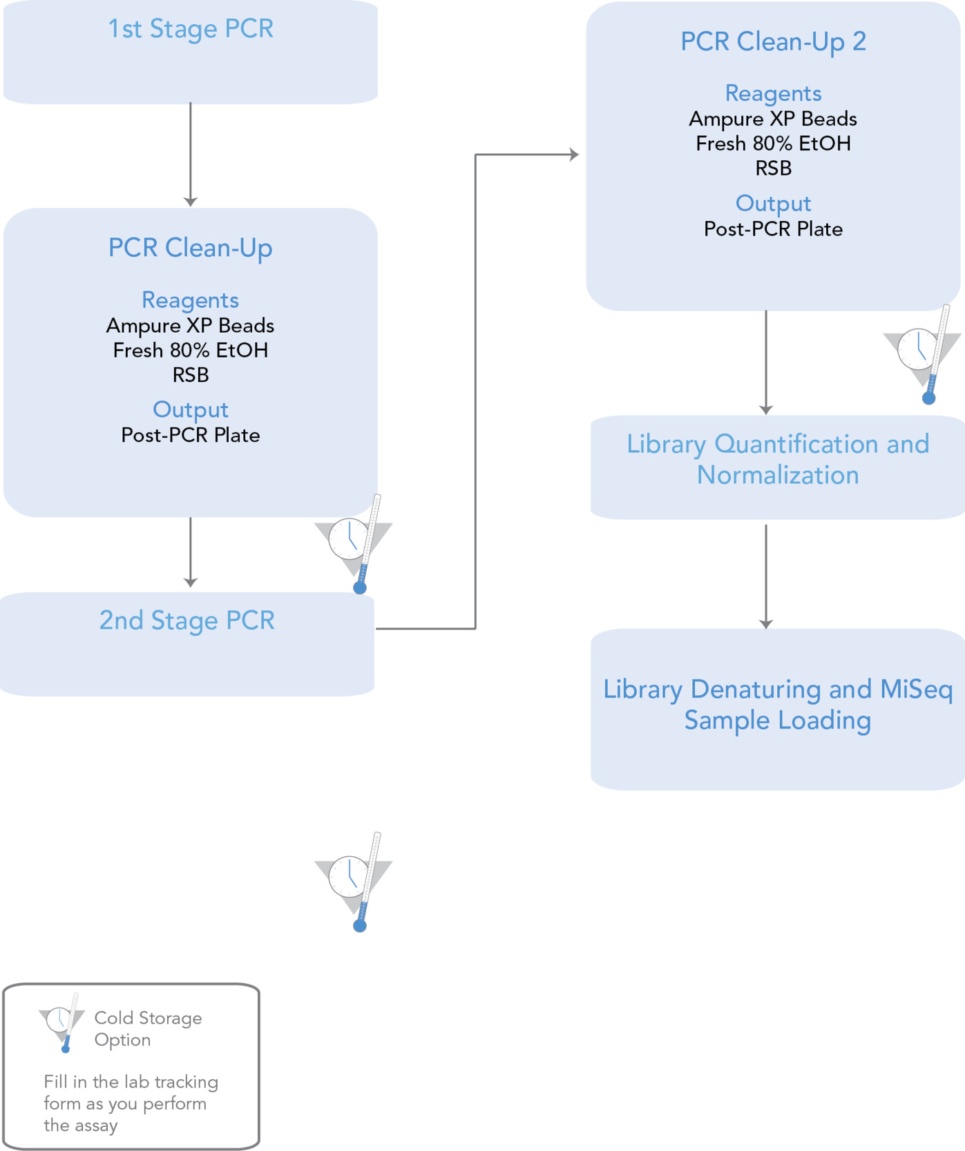
To generate 16S amplicons, 50 µg of DNA was used as a template for the V3-V4 base coded primers targeting 357F and 92GR of the bacterial 16S gene in Kappa HiFi Hot Start ready mix. The polymerase chain reaction (PCR) cycling was set at 95°C for 3 minutes, followed by 34 cycles at 98°C for 20 seconds, 70°C for 15 seconds, and a 5-minute extension at 72°C. Polymerase chain reaction (PCR) products (385bp) were verified by Agilent 2200 type station system with DIOCO severe Tape A and purified Agincourt AMPura xpDCR purification steps. Amplicons were quantified and concentrations were equalized for sequencing using the Illumina MiSeq2x300. Refer to supplement 2 for detailed mythology for PCR sequencing. After exact sequencing depth per sample, paired-end reads were analyzed using the IM-TORNADO bioinformatics pipeline. The following software packages were used: DADA2 (Callahan et al. 2016) and phyloseq (McMurdie and Holmes 2013). Both open and close-reference methods were used to generate operational taxonomic unit (OTU) tables at various phylogenetic levels for each sample, and OTUs were annotated using the Greengenes and HOMD databases. Sequences were tagged so that every participant sample was independently characterized. Controls included positive and negative control samples.
Data Analyses
Descriptive statistics were used to summarize patients’ demographic and clinical characteristics. Calculated α- diversity and β-diversity measures were summarized using univariate statistics. For α-diversity, a paired t-test was used to assess change between time points (T1 to T2, T1 to T3, and T1 to T4). The effect size of change was calculated and interpreted using Cohen’s d (Rosenthal et al. 1994). For β-diversity, de-trended correspondence analysis (DCA) ordination plots were generated for saliva and stool separately. Additional ordination plots are presented in the supplement file. Trends were described. A p-value <0.05 was considered statistically significant. All statistical analysis was performed using R Programing Language.
Results
Out of 104 patients screened for the study, 8 agreed to participate and provided consent. However, 1 patient withdrew after surgery, therefore 7 participants are included in the analysis. Table 1 provides patient demographics, clinical characteristics, and post-operative antibiotic regime. The sample was predominantly female (71.4%), with a mean age of 72.6 ± 9.2 years, and mean BMI of 25.6 ± 4.4 kg/m2. Additionally, only two patients (28.6%) were ever smokers and three patients (42.9%) reported ≥5 drinks per week. The end-stage hip and/or knee osteoarthritis was the primary diagnosis for all patients. All patients were on a regular diet, without special diet restrictions or regulations. All participants were followed for a minimum of 2 years and did not have a PJI or any disrupted gut microbiota-related complications.
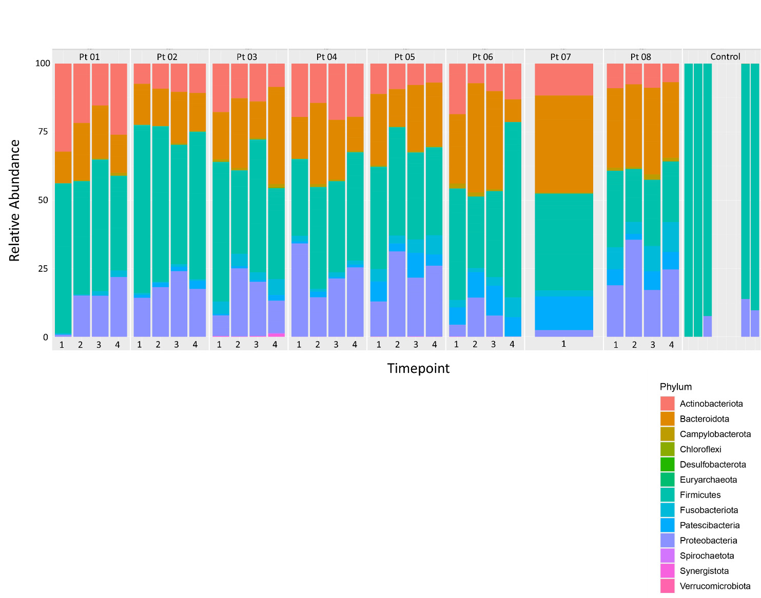
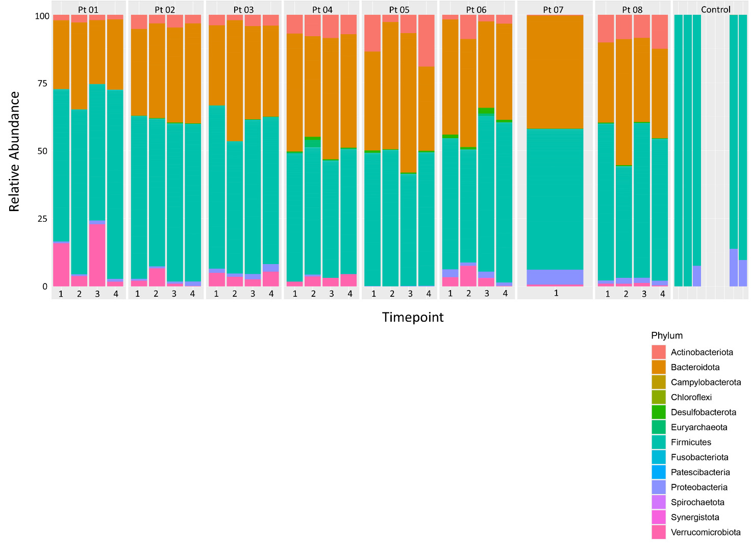
Figures 1 and 2 present the relative phylum abundance for saliva and stool, respectively. Figure e3 in the supplement file presents the relative class abundance of saliva and stool. Except for Pt04, all patients had an increase in Proteobacteria in their saliva at T2 compared to T1. Minimal differences were seen in the stool.
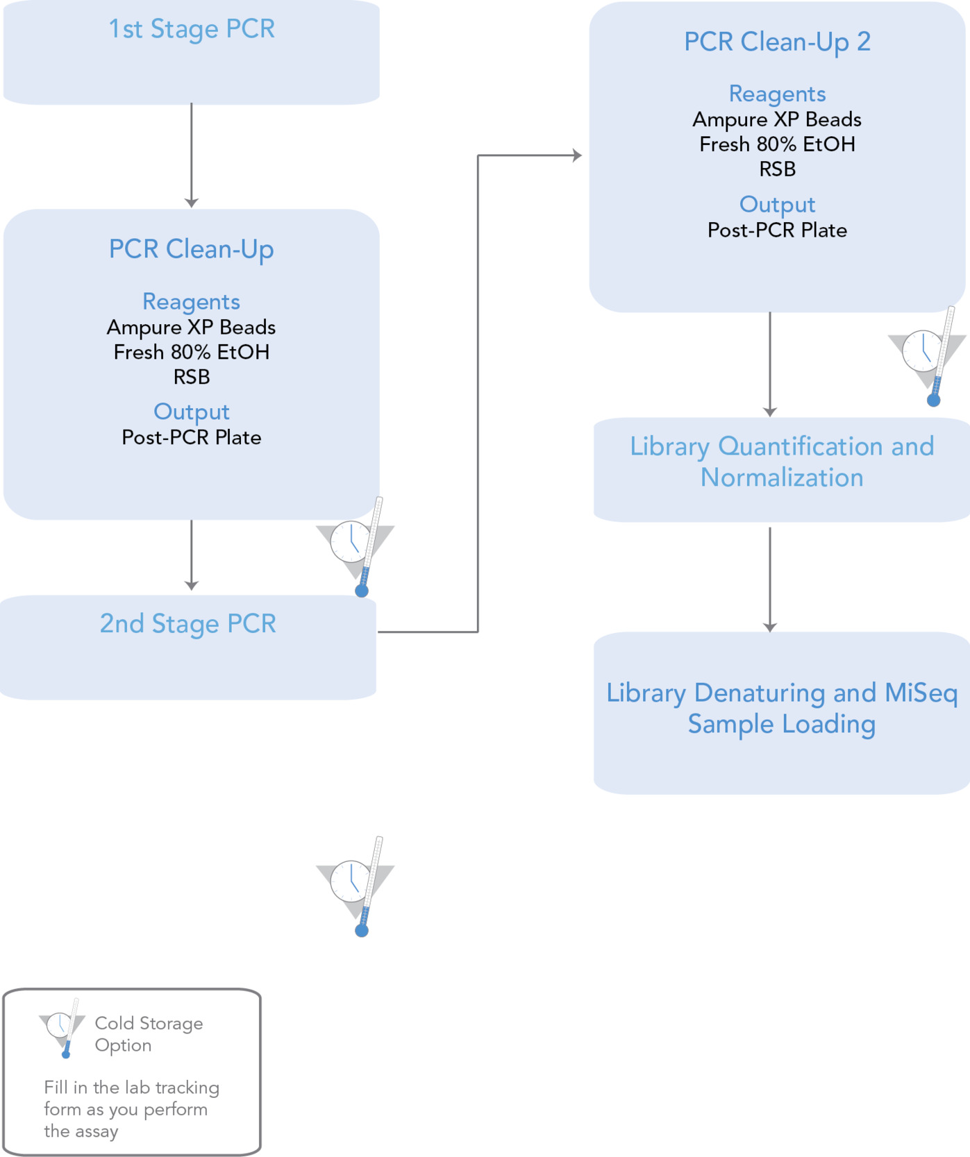
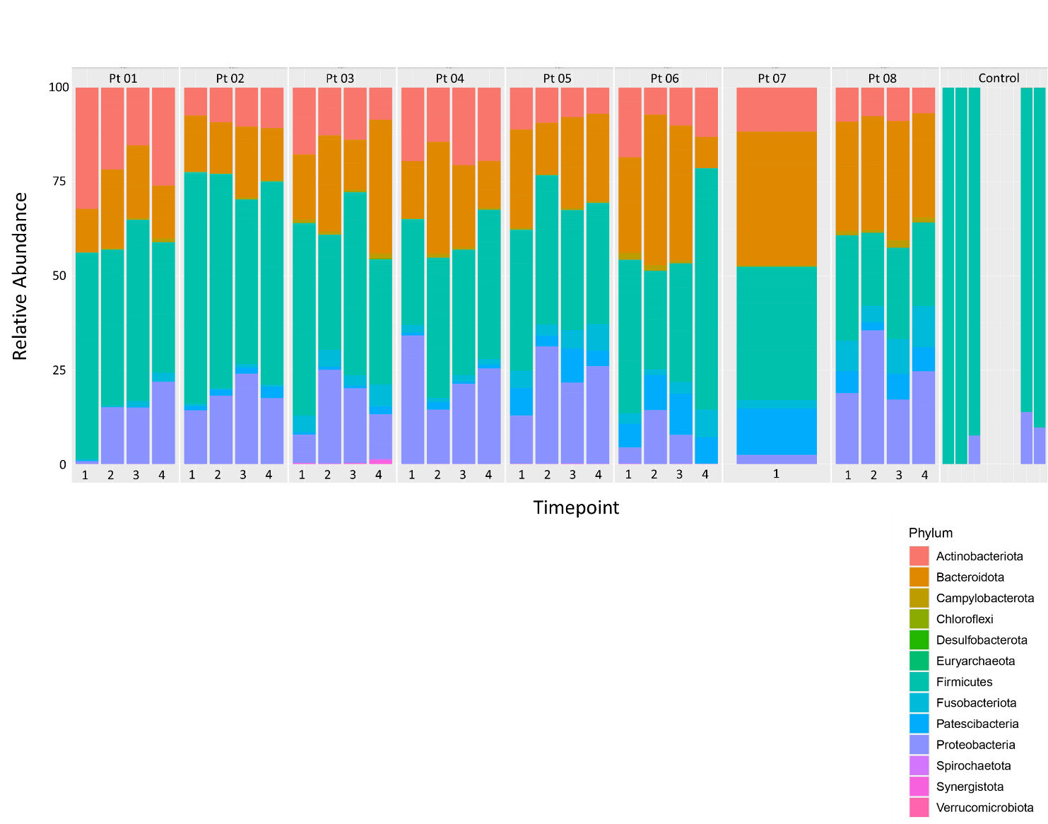
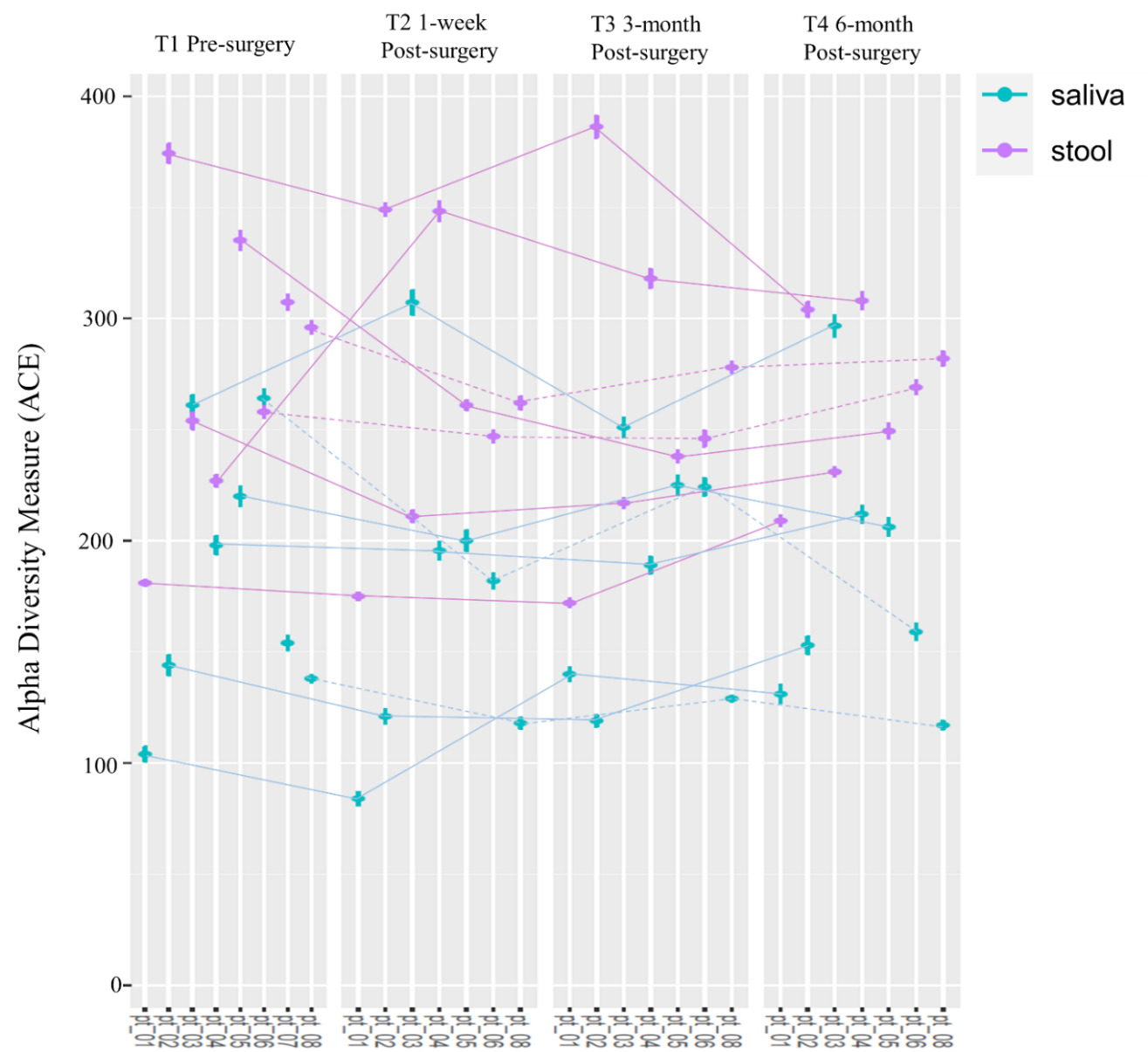
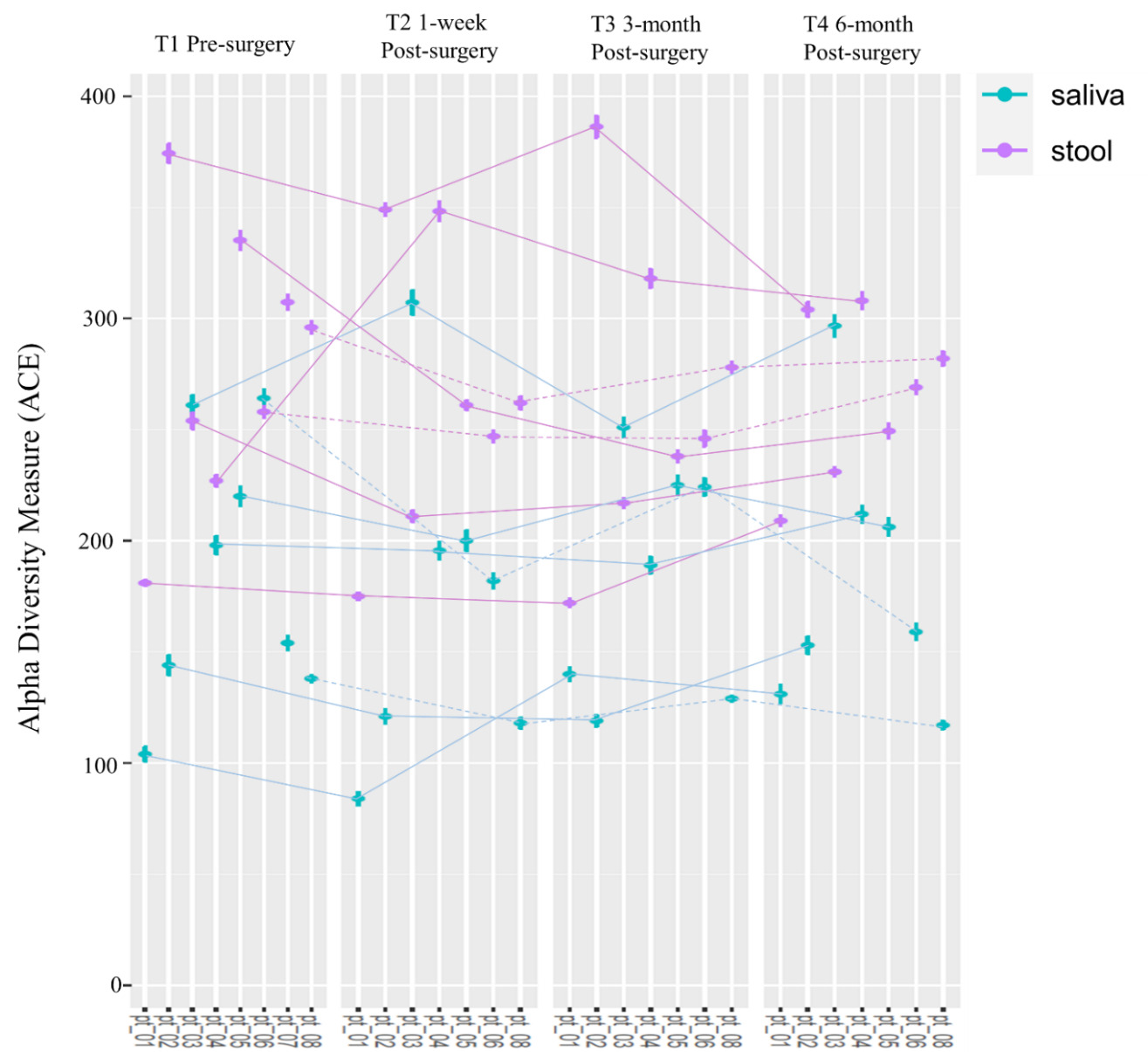
Figure 3 presents the absolute α-diversity of stool and saliva samples at all four time points. Stool samples, on average, had greater diversity than saliva samples. The mean values for each time point for saliva samples were the following: T1 = 190.14 ± 62.13; T2 = 173.14 ± 73.82; T3 = 179.86 ± 50.33; and T4 = 182.29 ± 61.32. The mean paired decrease in α-diversity between T1 and T2 was 17.00 (-17.48, 51.48, p = 0.273), between T1 and T3 was 10.28 (-13.98, 34.55, p = 0.340) and between T1 and T4 was 7.86 (-35.58, 51.29, p = 0.674). The effect size of change was 0.46 (-0.34, 1.22), 0.39 (-0.39, 1.15) and 0.17 (-0.56, 0.91), respectively. The mean values for each time point for stool samples were the following: T1 = 274.86 ± 65.59; T2 = 264.86 ± 64.73; T3 = 265.14 ± 70.67; and T4 = 264.14 ± 37.21. The mean decrease in α-diversity between T1 and T2 was 10.01 (-47.94, 67.94, p = 0.688), between T1 and T3 was 9.71 (-42.72, 62.15, p = 0.666) and between T1 and T4 was 10.71 (-42.44, 63.87, p = 0.639). The effect size (Cohen’s d) of change was 0.16 (-0.59, 0.90), 0.17 (-0.58, 0.91) and 0.19 (-0.57, 0.93), respectively.
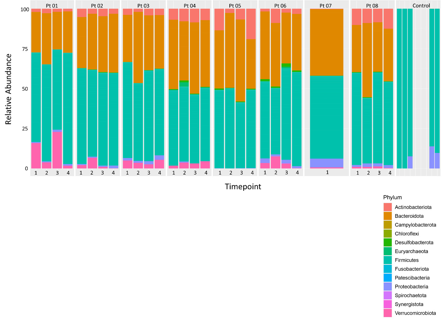
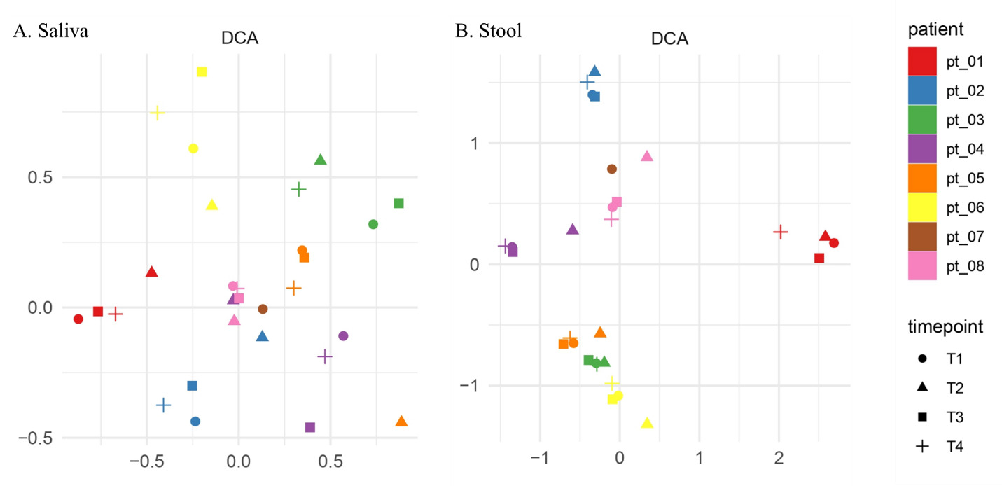
Figure 4 shows the DCA for saliva and stool. On visual assessment of clustering patterns, participants’ samples at different time points clustered around themselves. For saliva, the T2 sample (triangle) was the furthest from the participant’s individual centroid for Pt01, Pt02, Pt04, Pt05 and Pt06 (5/7 participants) suggesting that the T2 sample was the least similar sample from all other time points. For stool, a similar trend of T2 being the furthest from the participant’s individual centroid was observed for Pt04, Pt05, Pt06 and Pt08 (4/7 participants).
_and_stool_(b)_beta_diversity_dca_visualization_of_relationships_of_the_7_subjec.png)
Discussion
Our pilot case series investigated changes in oral and gut microbiome after patients underwent routine TJA and received perioperative antibiotics. Conducting this study has helped create a framework to investigate the effect of antibiotic use on oral and gut microbiomes and to plan for future trials. There has been some controversy regarding the overuse of preoperative antibiotics in orthopedics (Prokuski 2008). Since the study conducted by Ericson et al (Ericson et al. 1973). in 1973 that showed an infection rate of 0% and 13% between antibiotic prophylaxis and placebo, other researchers have replicated this study with less drastic results. Pavel et al (Pavel et al. 1974). conducted a double blinded trial which demonstrated an infection rate of 2.8% in patients given antibiotics and 5% in patients receiving placebo prior to undergoing general orthopedic procedures. Henley et al (Henley et al. 1986). also performed a double blinded trial and found an infection rate of 1.6% in patients treated with antibiotics and 4.2% in patients receiving placebo prior to surgery. Even though infection rates are lower, its impact on the healthcare system is burdensome. It is projected that more than 4 million TJAs will be performed per year in the UK and USA by 2030 (Kapadia et al. 2016). Even if we accept the infection rate of 1% for total hips and 1-2% for knees, it is clear that PJI will have a significant impact on the healthcare system, necessitating the use of antibiotic prophylaxis.
To negate the effects caused by dysbiosis, several research articles suggest the use of prebiotics and probiotics to replenish the microorganisms found in healthy gut microbiota (Gagliardi et al. 2018). Probiotics have the potential to help protect against infections of the surgical site and can be implemented in standard protocols after TJA to improve patient outcomes. However, studying this is difficult. If we only compare the outcome of developing PJI and use an estimated incidence of 1.74% (Zeng et al. 2023), we would need to perform a randomized trial of 2,671 participants per intervention arm to be able to identify a moderate (d = 0.5) effect. This sample size estimation is not practical, prompting us to measure changes in the microbiome instead.
Our study suggests that there is a small reduction in absolute α-diversity within a week of TJA and antibiotic use, but the effect was more prominent in the oral microbiome than the gut. We observed a moderate reduction (d = 0.46, moderate = 0.50) in saliva but only a small reduction (d = 0.16, small = 0.20) in stool. Comparing reduction in stool absolute diversity at one time point and expecting a moderate reduction in a small effect (d = 0.08) would require 2454 participants in each arm, which is also an impractical number. Instead, we propose collecting samples at multiple weekly time points over 3 months and base our sample size estimation using longitudinal analysis. This would only require a total of 101 participants, and we would be able to control one important confounder, such as read depth. Alongside side changes in microbiome diversity, we suggest collecting serological markers of inflammation, such as erythrocyte sedimentation rate and C-reactive protein (Longo et al. 2024).
Our other possible marker for reduction in α-diversity was comparing the increase of Proteobacteria after intervention, which is a genetic signature of dysbiosis (Shin et al. 2015). Although we observed changes in saliva relative Proteobacteria proportions over time, limited changes were observed in stool. At T2, 5/7 participants’ (Pt01-05) Proteobacteria did not change, 1 decreased (Pt06) and 1 increased (Pt08), which limits our ability to use this variable to calculate sample sizes. Similarly, for β-diversity, although an interesting observation was made that most participants’ T2 time point was furthest from the individual participant’s centroid, samples from the same participant clustered around themselves. This suggests there are major differences between person to person, and it will be difficult to identify a small change within the same participant at a group-wise level. Differences in β-diversity are due to the individual’s choice of diet, environmental or genetic factors, as well as medical comorbidities (Ousey et al. 2023).
Our pilot feasibility case series study has several limitations apart from its limited sample size. Our sample only includes patients who were given antibiotic prophylaxis so we cannot comment on how much the microbiome changed due to antibiotics alone. All patients did not receive the same antibiotic. Undergoing major surgery stresses the body and can also lead to changes in microbiome independent of antibiotics (Tsigalou et al. 2023). Possible generalizability bias should be mentioned, since all 7 patients are from a single institution and surgeon. Additionally, our other limitation is that none of our participants developed PJI or any other dysbiosis-related complications. It would have been interesting to see how the microbiome changes from pre-surgery baseline in those who develop PJI since current literature only reports how it changes when they have already developed a PJI (Levast et al. 2021). Lastly, since this was only feasibility pilot study with limited funding, we did not control for several relevant factors including participant’s diet, activity levels, or markers of systemic inflammation.. Future prospective trials should account for them.
Conclusion
Our pilot feasibility case series study has important findings that can help design future trials attempting to treat antibiotic prophylaxis-related dysbiosis in patients undergoing TJA. Although there is greater diversity in the gut than in the oral cavity, only small changes in α- or β-diversity within 2-years of post TJA follow-up were observed in the gut in patients undergoing pre-operative antibiotic prophylaxis. We suggest that comparing absolute α-diversity in stool over time is more feasible than measuring changes in Proteobacteria or β-diversity when performing large prospective study.
Funding
This study was, in part, supported by Orthopaedic Research and Education Foundation of Buffalo.