
Introduction
Total hip arthroplasty (THA) is established as one of the most successful orthopaedic procedures, providing excellent functional outcomes and quality of life improvements for patients with end-stage hip disease. However, the complexity of modern THA procedures requires considerations including prevention and treatment of intraoperative iatrogenic fractures, need for planned trochanteric osteotomies, and fixation of postoperative periprosthetic fractures. For these challenging clinical scenarios, cerclage fixation is often employed in isolation or in conjunction with other fixation constructs.
The incidence of intraoperative fractures during cementless THA remains significant, with reported rates ranging from 1.5% to 27.8% across multiple studies (K. R. Berend et al. 2004; Fishkin et al. 1999; Waligora et al. 2017). These fractures most commonly occur at the posteromedial calcar during broaching or implantation of the femoral component. Berend et al. demonstrated that “nondisplaced or minimally displaced proximal femur fractures can be secured with cerclage techniques and the final results of cementless THA are not compromised, as long as fracture and prosthetic stability are achieved at the time of surgery” (K. R. Berend et al. 2004).
The biomechanical rationale for cerclage fixation stems from the substantial forces transmitted through the proximal femur during normal activities. Press-fit cementless femoral prostheses generate loads exceeding 2-3 times body weight during routine activities such as stair climbing, rising from a chair, or brisk walking, creating significant hoop stresses within the proximal femur (Fishkin et al. 1999). These stresses predispose the bone to fracture, particularly in patients with compromised bone quality or abnormal anatomy. Cerclage fixation counteracts hoop stresses imparted by the femoral stem in the proximal femur by employing circumferential compression around the bone segment(s).
Prophylactic cerclage application has gained attention in high-risk populations, including patients with developmental dysplasia of the hip, who demonstrate a 5-fold increased relative risk of intraoperative fracture during THA (Zhang et al. 2016). Additionally, this technique has been described in treatment of femoral neck fracture with total hip arthroplasty given the poor bone quality in these patients. However, routine use of prophylactic cerclage remains debated, as current evidence lacks strong long-term clinical outcome data and raises concerns about complications such as soft tissue irritation, infection, and potential disruption of bone healing (Ponzio et al. 2015).
Despite their potential advantages, cerclage techniques carry inherent risks, particularly to the femoral neurovascular bundle and sciatic nerve during proximal femur applications (Devendra et al. 2018; Apivatthakakul et al. 2018; Grob et al. 2015). Understanding anatomical relationships and employing proper surgical techniques are essential for minimizing these complications while maximizing fixation effectiveness.
The purpose of this expert-driven review is to examine the current evidence regarding cerclage fixation in THA, including anatomical considerations, available implant options, surgical techniques, and clinical outcomes. By synthesizing the existing literature, we aim to provide guidance for the safe and effective application of cerclage techniques in both primary and revision hip arthroplasty procedures. We utilized PubMed to identify and obtain relevant clinical and biomechanical studies regarding this topic. This manuscript represents a narrative expert review based on the authors’ clinical experience and synthesis of the existing literature and is not intended to represent a systematic review.
Anatomy and Surgical Technique
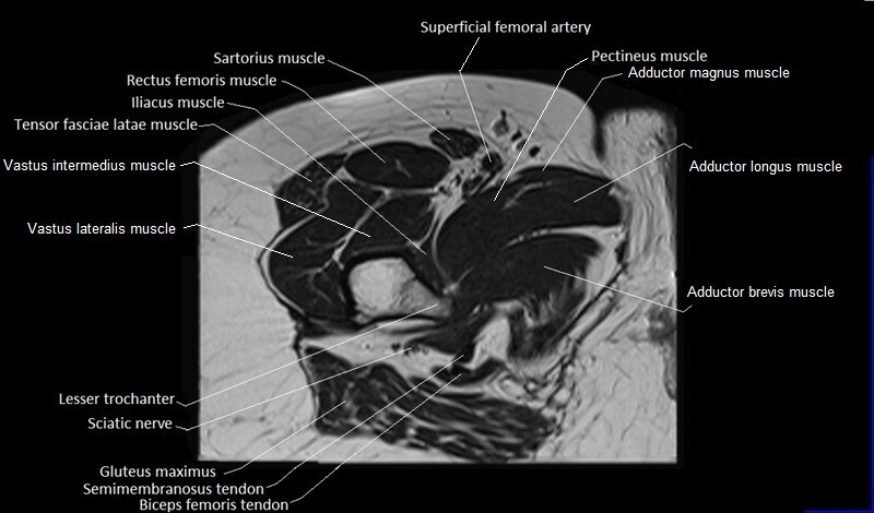
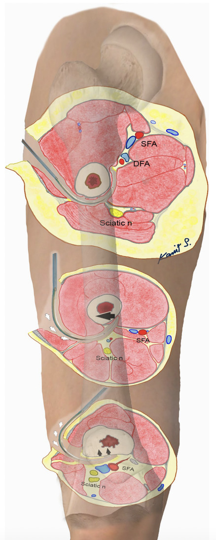
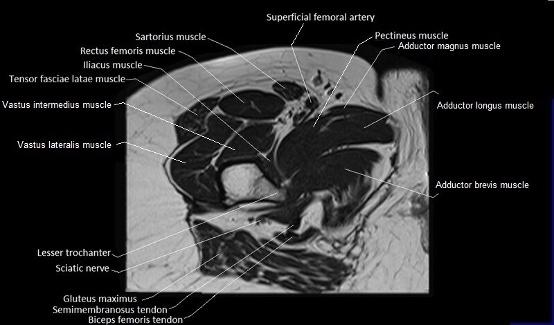
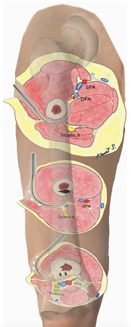
Application of cerclage wiring around the femur requires a thorough understanding of complex neurovascular anatomy to minimize iatrogenic injury. The femoral shaft receives vascular supply from three parallel arterial pathways: the superficial femoral artery (SFA), the profunda femoris artery, and collateral perforators, with additional supply from the descending branch of the lateral femoral circumflex artery (Apivatthakakul et al. 2018). The proximity of these vessels to the femoral cortex varies significantly along the shaft length, creating distinct zones of risk. Apivatthakakul et al. demonstrated through computed tomographic angiography that the SFA maintains safe distances of 26-38mm from the femoral cortex in the proximal half of the femur but approaches dangerously close distances of less than 15mm in the distal quarter (Apivatthakakul et al. 2018). The profunda femoris artery system poses particular risk in the subtrochanteric region where it lies within 9-27mm of the cortex (Apivatthakakul et al. 2018, 2013).
Previous literature has validated these anatomic concerns, with reported vascular injury rates of 1.59% for proximal femur shaft fractures and an alarming 7.14% for distal femur shaft fractures when using cerclage passer instruments (Devendra et al. 2018). Devendra et al. documented four cases of major vascular injury, including two SFA injuries requiring resection and end-to-end anastomosis, and two profunda femoris system injuries managed with vessel ligation (Devendra et al. 2018). The higher injury rate in distal femur procedures appears related to the instrument’s inner curvature mismatch with the femoral circumference and the closer proximity of the SFA to the bone in this region (Devendra et al. 2018). Additionally, surgical positioning factors significantly impact risk, with Yang et al. demonstrating that adduction of 20° reduces the SFA-bone distance from 20.2mm to 11.8mm, further decreasing to 9.4mm with additional 20° internal rotation (Yang et al. 2004).
The neurovascular risks also include critical nerve structures supplying the quadriceps muscle group. Grob et al. identified that the area immediately distal to the intertrochanteric line serves as a common entry point for multiple nerve branches, with some branches entering muscle bellies as close as 9mm distal from the intertrochanteric line (Grob et al. 2015). Distal extension of surgical approaches or cerclage application in this region inevitably compromises nerve supply to the anterolateral quadriceps musculature, including branches to the vastus lateralis and lateral portions of the vastus intermedius (Grob et al. 2015). Introduction of cerclage instruments through anterior approaches poses particular risk to the lateral femoral circumflex artery branches and the intramuscular nerve branches serving the articularis genus (Grob et al. 2015).
Safe Application Techniques for Cerclage Fixation
Based on comprehensive anatomic studies, safe cerclage application requires adherence to specific technical principles and anatomically-informed zone-specific approaches. The fundamental principle involves starting the cerclage passer insertion from the posterior intermuscular septum at the linea aspera, progressing from the side closest to structures at risk toward safer anatomical zones (Apivatthakakul et al. 2018, 2013). The proximal femoral region represents the safest zone for cerclage application, where both SFA and DFA maintain safe distances exceeding 26mm from the femoral cortex (Apivatthakakul et al. 2013). In this region, standard cerclage techniques may be employed with confidence, maintaining continuous posterior cortical contact throughout cerclage passage.
For the challenging mid-shaft to distal three-quarters region, meticulous technique becomes paramount. The cerclage passer tip must remain in close contact with the posteromedial cortex, with careful lateral translation of the instrument handle to move the tip away from the SFA before advancing past the mid-axis of the femur (Apivatthakakul et al. 2018, 2013). Below the distal quarter of the femur, where the SFA approaches within 10-14mm of the cortex, the cerclage passer must maintain strict contact with the posterior cortex to avoid both SFA injury and sciatic nerve damage (Apivatthakakul et al. 2018, 2013). Critical technical modifications include reducing traction force, limiting patient positioning in adduction and internal rotation, and minimizing the number of instrument passes (Devendra et al. 2018; Yang et al. 2004). When cerclage application is required in high-risk zones, alternative approaches should be considered such as lateral subvastus access, which allows better instrument control while protecting superficial nerves and major vessels (Grob et al. 2015). Vigilant intraoperative monitoring for unexpected bleeding during instrument clamping and disengagement enables immediate recognition of vascular injury, facilitating prompt intervention and optimal patient outcomes (Devendra et al. 2018).
Cerclage Options
The evolution of cerclage fixation in orthopaedic surgery reflects a shift from availability-based choices to evidence-based decision-making, driven by advances in biomechanics, clinical outcome data, and economic considerations. Modern cerclage systems, ranging from traditional monofilament wires and sutures to advanced polymer and plate combinations, are designed to address specific clinical challenges while optimizing bone healing and minimizing complications. Each fixation method offers distinct advantages and limitations in terms of strength, biocompatibility, ease of use, and cost. A thorough understanding of these factors is essential for selecting the most appropriate system for each patient and surgical scenario.
Wire
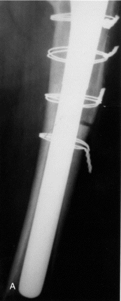
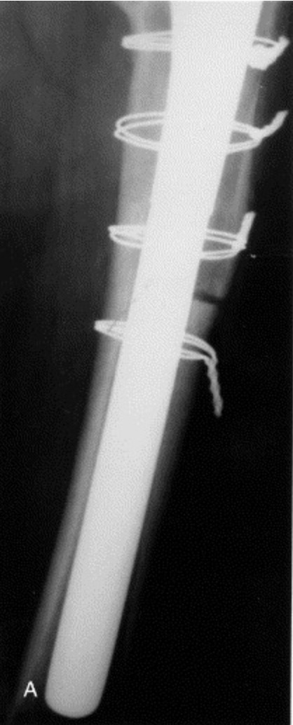
Monofilament stainless steel wire has been the traditional standard for cerclage fixation in orthopaedic surgery for over 50 years. Wire cerclage offers several advantages including biologic inertness, easy handling, good radiographic imaging, and relatively low cost (Ritter et al. 2006). Clinical studies have demonstrated acceptable outcomes with doubled wire configurations (i.e., using two unbraided wires rather than a single wire), achieving breakage rates as low as 5.12% when properly applied (Ritter et al. 2006). Monofilament wire cerclage presents limitations that have driven the search for alternative materials. Documented breakage rates range from 0.4% to 28%, with trochanteric nonunion rates between 0.4% and 21% (Oe et al. 2013). Wire failure typically occurs due to low cycle fatigue, with even a 1% notch potentially reducing fatigue resistance by 63% (Oe et al. 2013). Additional complications include unraveling of the twist, and the “cheese wire effect” in osteoporotic patients where the thin wire can cut through fragile bone (Oe et al. 2013; Kuruvalli et al. 2008). Biomechanical testing has shown that monofilament wires demonstrate the poorest fatigue performance among cerclage options, failing at approximately 10 lb (44.5 N) after 3 million cycles (Dickman et al. 1997).
Suture
High-performance sutures represent the newest frontier in cerclage fixation, offering potential advantages in terms of handling, cost, and imaging compatibility. Modern suture materials such as FiberWire (Arthrex, Naples, FL, USA), consisting of a UHMWPE core with braided polyester, and Ethibond (Ethicon, Raritan, NJ, USA), a braided polyester non-absorbable suture, have demonstrated superior biomechanical properties compared to traditional wire in laboratory testing [16]. FiberWire achieved mean failure loads of 232-254N compared to only 61.8N for wire, while Ethibond showed the best cyclic displacement resistance [16].
Biomechanical studies comparing suture cerclage to stainless steel wire in shoulder arthroplasty found that while wire achieved higher initial compression (618N vs. 131-137N for sutures), FiberWire configurations demonstrated equivalent performance in preventing prosthetic subsidence and superior ultimate failure strength (Wähnert et al. 2011). Additionally one study comparing biomechanical properties of FiberTape (Arthrex, Naples, FL, USA) found comparable subsidence and fragment translation compared to traditional metallic cables (Han et al. 2024). This suggests that sutures may be particularly suitable for non-displaced fractures requiring stabilization rather than active reduction.
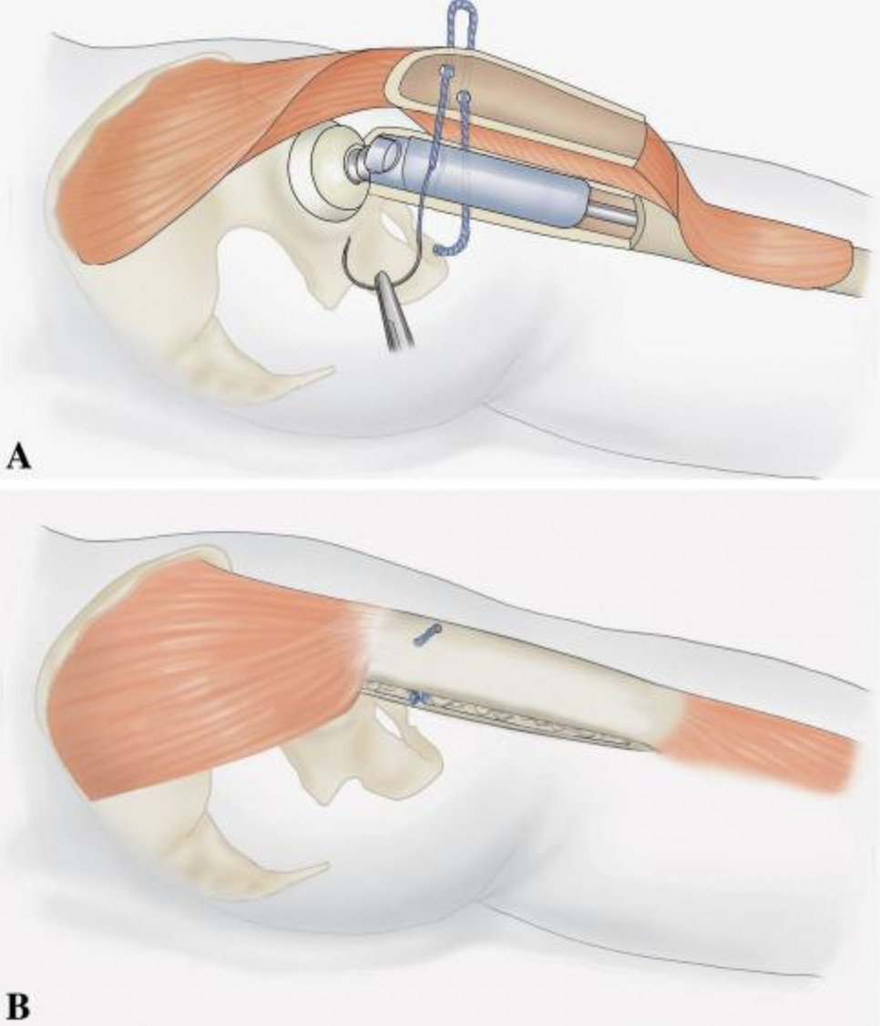
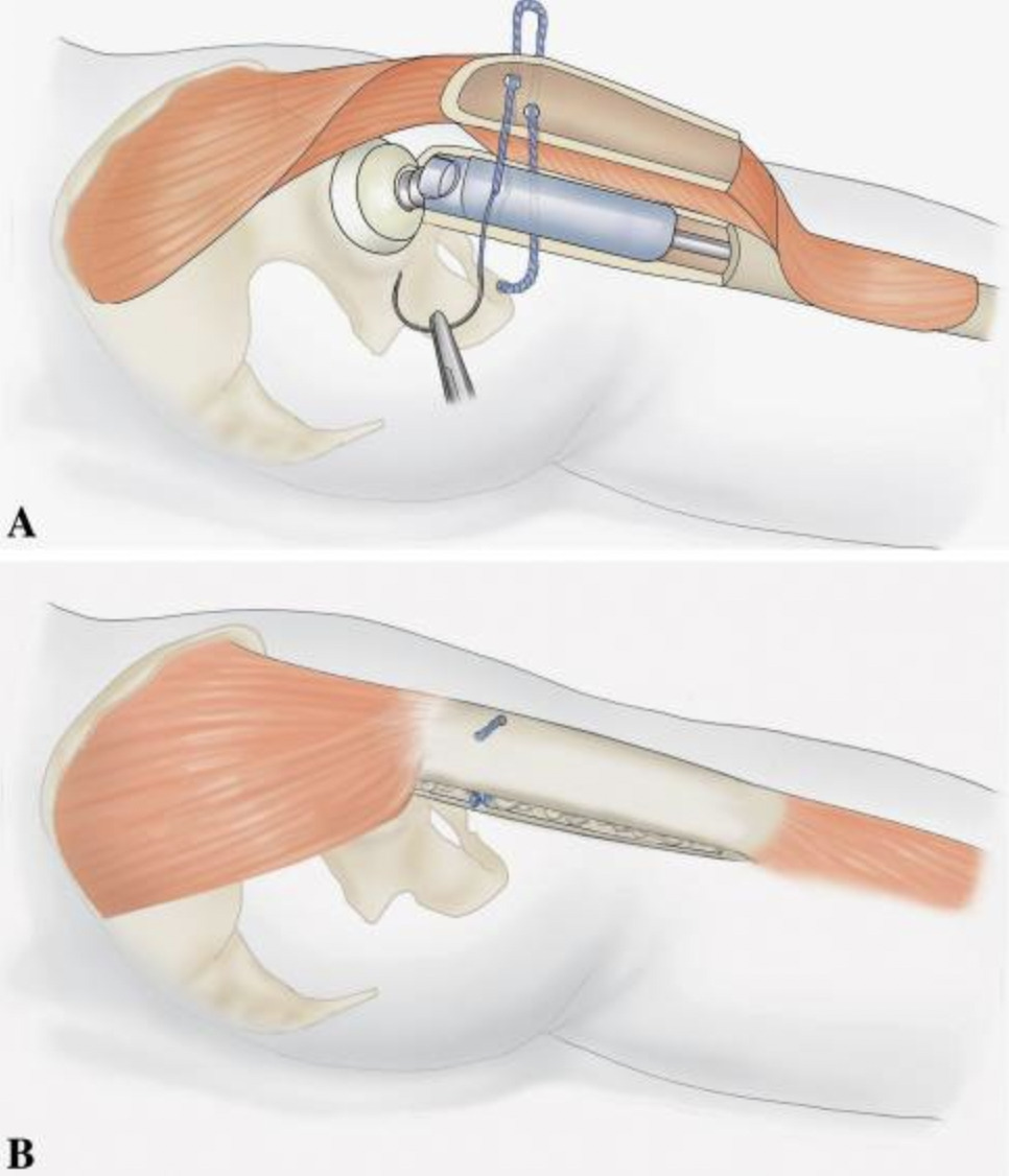
An innovative approach using braided absorbable suture cords has shown promising clinical results. Kuruvalli et al. reported 100% healing rate (95% bony union, 5% stable fibrous union) in 20 patients using cords made from eight strands of Vicryl suture (Ethicon, Raritan, NJ, USA) with breaking strength exceeding 1000 Newtons (Kuruvalli et al. 2008). This technique differs from traditional cerclage by passing the cord through drill holes in the proximal fragment and anchoring it directly around the prosthetic stem below its shoulder, rather than encircling the entire femur. This direct prosthetic anchoring offers unique advantages including preservation of blood supply to the bone, more secure fixation against abductor muscle forces, elimination of permanent foreign bodies, and significant cost savings, particularly valuable in resource-limited settings (Kuruvalli et al. 2008).
Metallic Cable
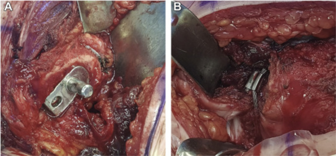
Metallic cable systems were developed to address mechanical limitations of monofilament wire. These multifilament constructs, typically made from stainless steel or titanium alloys in various configurations (7×7 or 19×7 bundles), offer superior tensile strength compared to single wires (Dickman et al. 1997). Stainless steel cables consistently demonstrate higher strength and fatigue resistance than their titanium counterparts, with some systems achieving fatigue strength of approximately 50 lb (222 N) at 3 million cycles (Dickman et al. 1997).
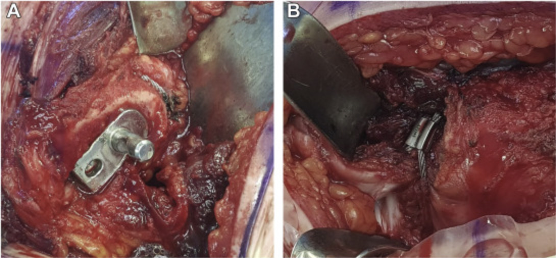
Metallic cable systems are available in both beaded and non-beaded configurations, with each design offering distinct technical advantages. Beaded cerclage cables are commonly recommended for use with single-sided tensioners and enable a more minimally invasive approach, particularly for supplemental cerclage fixation. Non-beaded cables are generally recommended for use with grip plates or for the proximal portion of fixation systems and are tensioned using double-sided tensioning devices that allow for sequential re-tensioning to optimize cable tension before final crimping.
Despite their mechanical advantages, cable systems have not eliminated complications. Clinical breakage rates range from 3.1% to 44%, with cable fraying and fragmentation observed in up to 50% of patients (Oe et al. 2013; Silverton et al. 1996). Trochanteric nonunion rates with cable fixation vary from 1.5% to 38% (Oe et al. 2013). Additionally, cables generate metallic debris through fretting and breakage, which has been implicated in accelerated polyethylene wear, third-body generation, and increased rates of acetabular component loosening (Lenz et al. 2012). Economically, cables are significantly more expensive than wires, costing 9.2 times more per unit with equipment costs 12.8 times higher, without demonstrating superior clinical outcomes (Ritter et al. 2006). An often-overlooked issue is the substantial tension loss during installation, with some systems losing up to 52% of applied tension once the tensioning device is removed (Ménard et al. 2013).
Polymer Cable
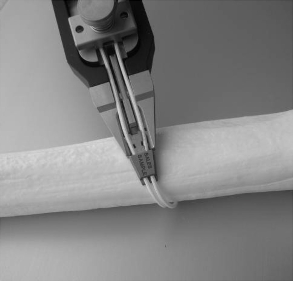
Polymer cables, particularly those made from ultra-high molecular weight polyethylene (UHMWPE), represent a significant advancement in cerclage technology. These nonmetallic systems offer several theoretical advantages including elimination of metallic debris or metal on metal irritation, no radiographic interference, and superior fatigue resistance compared to metallic alternatives. Biomechanical testing has demonstrated that polyethylene cables achieve remarkable fatigue strength of approximately 130 lb (578 N) at 3 million cycles, significantly superior to all metal systems tested (Dickman et al. 1997).
Early results with polymer cables have been encouraging. A study of 29 patients using nonmetallic cables (SuperCableTM; Kinamed Inc, Camarillo, CA), consisting of a nylon core with UHMWPE braided fiber jacket, reported zero cable failures and a 93% union rate for osteotomies and fractures (Ting, Wera, Levine, and Valle 2010). Polymer cables’ ability to conform to bone surfaces by flattening under load distributes forces over a greater contact area, potentially reducing the “cheese wire effect” in osteoporotic bone (Dickman et al. 1997). Additionally, UHMWPE fiber cables in tape form demonstrated superior fixation stability with no failures compared to a 25% failure rate with traditional soft wire in an animal model (Oe et al. 2013).
Polymer cables are not without limitations. Significant creep under constant load can lead to loosening over time, with some systems experiencing up to 46% tension loss during installation (Ménard et al. 2013). The radiolucent nature of these materials prevents radiographic assessment of cable integrity, and long-term degradation cannot be monitored without direct inspection (Lenz et al. 2012). Furthermore, while polymer cables do not abrade bone, they can be abraded by sharp bone edges, failing after 8,000-9,000 cycles in biomechanical testing (Dickman et al. 1997). Another consideration in use of polymer cables is increased propensity for biofilm formation compared to metallic cables or wires as demonstrated by Cichos et al. however the results of this in vitro study have yet to be corroborated by in vivo studies (Cichos et al. 2024). Lastly, there is currently lacking long-term data regarding the use of polymer cables in THA.
___591.png)
Cable-Plate
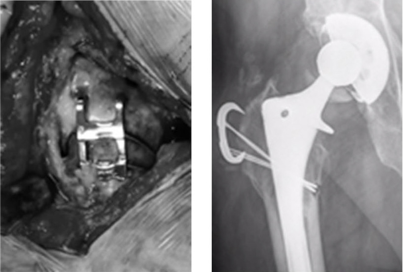
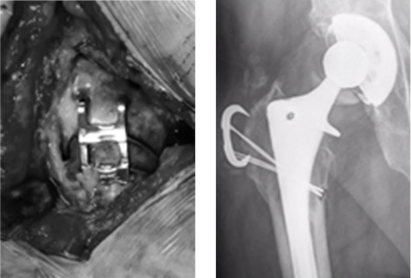
Cable-plate systems represent an evolution in cerclage technology. They combine the ability of cerclage fixation to create interfragmentary compression and dissipate hoop-stresses with a plate-screw fixation’s ability to neutralize fragments preventing axial and rotational displacement. These systems are designed to provide more robust fixation for complex reconstructions, particularly in cases with significant bone loss or challenging anatomy. The SuperCable System (Kinamed Inc., Camarillo, CA, USA), utilizing nonmetallic isoelastic cables with grip-plate configurations, demonstrated an 81% mechanical success rate in a clinical series of 27 revision THA procedures (K. Berend et al. 2014).
The versatility of cable-plate systems allows for customization based on defect size, with options ranging from single-hole (50 mm) grip-plates to 6-hole (245 mm) configurations (K. Berend et al. 2014). However, these systems are not without complications. Failures were particularly notable with shorter grip-plates used for trochanteric slides and intraoperative fractures, and soft-tissue irritation requiring hardware removal occurred in 11% of cases despite successful bony healing (K. Berend et al. 2014). The overall reoperation rate related to the grip-plate system was 30%, highlighting the need for careful patient selection and technique optimization (K. Berend et al. 2014).
Choice of Cerclage Option
The above options have all been described for fixation of periprosthetic fracture and trochanteric osteotomies. Choice of a particular cerclage construct should be based on goals of fixation, available bone stock, and surgeon preference. In fixation of most simple periprosthetic fractures and osteotomies the authors prefer cable fixation given proven biomechanical characteristics, ease of use, and wide availability. If using wires, consider doubling wires to enhance biomechanical stability. In the setting of poor bone stock a cable-plate construct may provide greater fixation and capture comminuted greater trochanteric fragments. Polymer cables are a promising option particularly for osteoporotic bone in which the “cheese-wire effect” is a concern. Additionally it is important to consider that polymer and suture constructs are radiolucent which limits postoperative monitoring of cerclage position and integrity with radiographs.
Prophylactic Cerclage
Indications
Prophylactic cerclage application has emerged as a strategy to minimize the risk of intraoperative and early postoperative periprosthetic femoral fractures during primary and revision THA. The clinical rationale for prophylactic cerclage is fundamentally different between cemented and cementless femoral fixation techniques. Cementless stems rely on press-fit fixation and generate significantly higher hoop stresses during insertion due to their wedge-shaped geometry and the need for cortical engagement to achieve initial stability. In contrast, cemented stems distribute loads more evenly through the cement mantle, which acts as a stress-distributing interface between the prosthesis and bone, substantially reducing peak cortical stresses during implantation. The biomechanical rationale centers on increasing resistance to hoop stresses generated during femoral canal preparation and stem insertion (Waligora et al. 2017). These circumferential stresses, which can increase by 400% with oversized stems, represent the primary mechanism of proximal femoral fracture during cementless implantation (Fishkin et al. 1999).
Current indications for prophylactic cerclage include poor bone quality (Dorr type C femurs), low trabecular indices on preoperative imaging, and subjectively identified poor bone quality intraoperatively (Waligora et al. 2017). The technique has gained attention in managing unstable femoral neck fractures in elderly patients, where the combination of osteoporotic bone and traumatic injury creates a high-risk scenario for periprosthetic fracture.
Clinical Outcomes: Prophylactic Cerclage
Recent evidence suggest potential benefit of prophylactic cerclage in preventing periprosthetic fractures in high-risk populations, however long-term data and broader validation remain limited. Iwasa et al. demonstrated dramatic reductions in intraoperative (6.9% to 0%, p<0.001) and postoperative (5.4% to 0.6%, p=0.016) periprosthetic fracture rates when prophylactic cerclage cabling was employed for cementless stems in unstable femoral neck fractures (Iwasa et al. 2024). Notably, no patients in the cerclage group required reoperation compared to 5.0% in the non-cerclage group (p=0.003) (Iwasa et al. 2024). This suggests that prophylactic cerclage significantly increased both the rotation and energy required for failure in cadaveric specimens, though fracture classification patterns remained largely unchanged. This increased torsional energy requirement may be particularly relevant during the critical period of stem insertion when most intraoperative fractures occur (Waligora et al. 2017).
Cost
The economic impact of periprosthetic fractures treated with cerclage techniques remains significant. While prophylactic cerclage adds minimal cost and operative time (approximately 2 minutes and $200-500 in materials), the prevention of even a single periprosthetic fracture requiring revision surgery (average cost $50,000-80,000) provides substantial cost savings (Iwasa et al. 2024). The cost-effectiveness of prophylactic cerclage likely depends on patient-specific fracture risk and institutional cost structures.
Technical Considerations
Optimal placement involves positioning cerclage cables just proximal to the lesser trochanter before final femoral preparation (Iwasa et al. 2024). Per Iwasa et al., this technique adds minimal surgical burden, with only 2 minutes of additional operative time and 16 mL of increased blood loss reported (Iwasa et al. 2024). For medial calcar cracks, Fishkin et al. demonstrated that cables placed normal to the crack direction provide superior biomechanical stability compared to those placed normal to the femoral neck axis, reducing stem subsidence by 3.17mm and crack opening by 1.55mm (Fishkin et al. 1999).
Trochanteric Osteotomies
_and_troc.png)
Types of Trochanteric Osteotomies
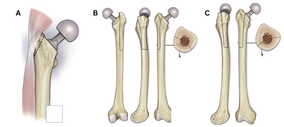
Trochanteric osteotomy has evolved from Charnley’s routine use in primary hip arthroplasty to become an essential technique for complex revision scenarios. Three primary osteotomy techniques have emerged, each offering specific advantages based on surgical approach and exposure requirement (Sundaram et al. 2020).
Trochanteric Slide Osteotomy (TSO)
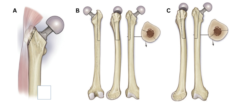
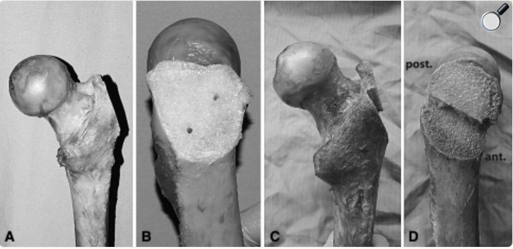
The TSO creates a bone fragment containing the greater trochanter with preserved soft tissue attachments. The osteotomy begins medial to the piriformis fossa, preserving the gluteus medius insertion proximally, and exits just distal to the vastus ridge, protecting the vastus lateralis attachment distally. This continuous soft tissue sleeve allows the fragment to be mobilized anteriorly or posteriorly while maintaining vascularity (Sundaram et al. 2020). TSO is particularly valuable in young adults undergoing hip resurfacing arthroplasty and in complex primary cases involving developmental dysplasia, severe protrusio, or conversion from prior hip arthrodesis. For revision scenarios with abductor deficiency, TSO offers the advantage of preserving the posterior capsule and short external rotators. Alternatively, the trochanteric step osteotomy, a modification to this technique described by Schoeniger et al, may provide a more stable construct than a trochanteric slide osteotomy by incorporating a step cut into the traditional TSO (Figure 9) (Schoeniger et al. 2009).
Extended Trochanteric Osteotomy (ETO)
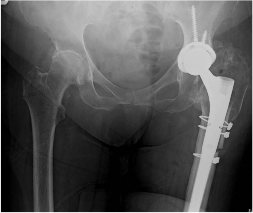
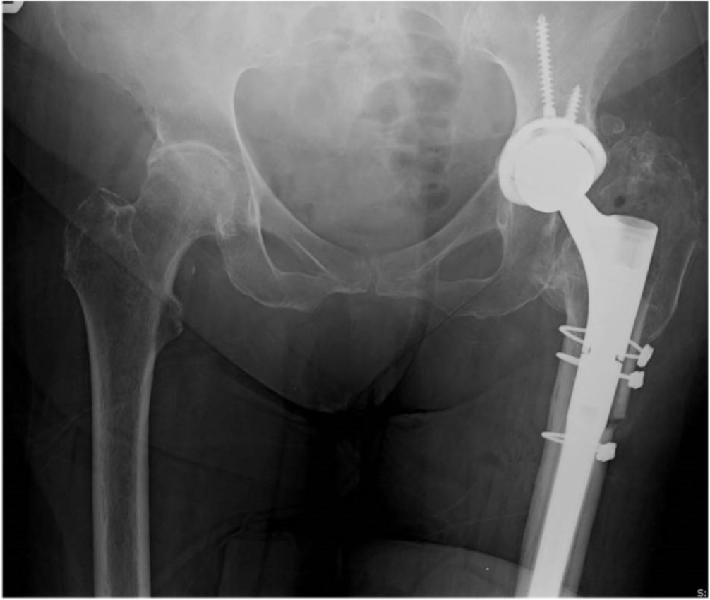
The ETO has become the workhorse for revision hip surgery requiring extensive femoral exposure. The osteotomy creates a lateral femoral sleeve with specific anatomical boundaries: the proximal pole at the greater trochanter, the posterior border along the linea aspera, the anterior border at the femoral midline, and a distal transverse cut at the surgeon’s discretion. Standard ETO length measures 12-15cm, with a minimum of 10cm recommended to prevent complications (Sundaram et al. 2020). The technique involves three phases: direct osteotomy of the posterior limb, a transverse cut encompassing no more than one-third of the femoral circumference, and anterior border completion using drill holes or osteotomes. ETO is indicated for removal of well-fixed stems, clearing significant cement mantles, addressing femoral remodeling, and managing periprosthetic infections (Sundaram et al. 2020). The ETO can then be repaired by reducing the fragment back to the femur and securing with 2-3 cerclage cables with or without a prophylactic cable distal to the osteotomy site (Figure 10) (Wronka et al. 2020).
Transfemoral (Wagner) Osteotomy
The transfemoral approach, also known as the Wagner osteotomy, differs from ETO by utilizing approximately half the femoral diameter compared to ETO’s one-third involvement. The cuts are made in the coronal plane rather than the sagittal plane used in ETO, creating an “open book” configuration. This approach is performed through a direct lateral incision with vastus lateralis splitting (Sundaram et al. 2020). The Wagner osteotomy is optimal for anterior-based approaches and particularly useful when addressing significant anterior femoral bowing, removing distal cement fragments, or extracting broken femoral components (Sundaram et al. 2020).
Clinical Outcomes: Trochanteric Osteotomies
Clinical outcomes following trochanteric osteotomies demonstrate consistently high union rates when appropriate cerclage fixation is employed. For TSO, reported union rates range from 84% to 98%, with Langlais et al. achieving 96% union (90/94 patients) and Lakstein et al. reporting 98% union (81/83 patients) in their series (Lakstein et al. 2010; Langlais et al. 2003). The lower union rate of 84% reported by León et al. in their modified TSO cohort highlights the importance of adequate osteotomy length, as fragments less than 10cm were associated with greater trochanteric migration risk (León et al. 2019). ETO demonstrates even more consistent results, with multiple series reporting union rates exceeding 98%: Miner et al. achieved 98.2% union (163/166) with 89.8% reoperation-free survival, while Mardones et al. and León et al. both reported greater than 98% healing rates (León et al. 2019; Mardones et al. 2005; Miner et al. 2001). The Wagner osteotomy shows similarly excellent outcomes, with Fink et al. achieving 98.5% union using their modified technique and De Menezes et al. reporting 95% union with significant functional improvement (mean Harris Hip Score improvement from 45.2 to 83.4 points) (Fink and Oremek 2016; de Menezes et al. 2012).
Periprosthetic Fracture
Classification
Periprosthetic femoral fractures represent a significant complication following THA, with classification systems evolving to guide treatment decisions. The Vancouver classification, originally developed by Duncan and Masri, remains the most widely utilized system for categorizing these fractures based on fracture location, implant stability, and surrounding bone stock quality (Brady et al. 1999). Vancouver Type A fractures involve the trochanteric region, Type B fractures occur around or just distal to the femoral stem, and Type C fractures occur well below the stem tip. Type B fractures are further subdivided based on stem stability: B1 (stable stem with good bone stock), B2 (unstable stem with good bone stock), and B3 (unstable stem with poor bone stock) (Brady et al. 1999).
An alternative classification specifically for intraoperative fractures was developed by Mallory et al., dividing fractures into three anatomical zones. Type I fractures are confined to the proximal zone including the lesser trochanteric area proximally (80% of cases), Type II fractures extend past the lesser trochanter but no further than 4cm distally (16% of cases), and Type III fractures extend more than 4cm distal to the lesser trochanter or within 4cm of the prosthesis tip (4% of cases) (Mallory et al. 1989). This zone-based system provides practical guidance for intraoperative management, as each zone requires different fixation strategies based on the fracture’s relationship to the implant.
Timing of fracture occurrence also influences classification and treatment. Intraoperative fractures typically occur during broaching or stem insertion due to excessive hoop stresses, while early postoperative fractures (within 90 days) often result from unrecognized intraoperative damage or inadequate initial fixation. Kheir et al. demonstrated that 50% of all periprosthetic fractures occur within 90 days postoperatively, highlighting the critical importance of the immediate perioperative period (Kheir et al. 2023).
Surgical Options Using Cerclage
The application of cerclage fixation for periprosthetic fractures depends on fracture classification, stem stability, and bone quality. For Vancouver B1 fractures with stable stems, cerclage wires or cables alone may provide adequate fixation. Berend et al. reported excellent outcomes treating intraoperative proximal femoral fractures with cerclage techniques, achieving 100% femoral component survival at up to 16 years follow-up when initial stability was achieved (K. R. Berend et al. 2004). This technique requires compression across the fracture site while maintaining implant stability.
Technical considerations for cerclage placement in fracture treatment differ from prophylactic applications. Fishkin et al. demonstrated that cerclage placed orthogonal to the fracture direction provide superior biomechanical stability compared to those placed perpendicular to the femoral neck axis (Fishkin et al. 1999). For a single medial calcar crack, cerclage placed orthogonal to the fracture reduced stem subsidence by 3.17mm and crack opening by 1.55mm compared to those placed normal to the neck (Fishkin et al. 1999). This orientation principle mirrors lag screw biomechanics, where optimal fixation occurs perpendicular to the fracture line.
The number of cerclage constructs also significantly impacts fracture stability. While a single wire placed normal to the crack provides adequate stability for loads up to 890N (representing normal walking), higher loads associated with stair climbing or rising from a chair (1780-2670N) require multiple cerclage rings. Fishkin et al. found that two wires eliminated fracture displacement under low loads and reduced it to less than 0.75mm at higher loads, while three wires provided a 50% reduction in subsidence and limited fracture displacement to less than 1mm (Fishkin et al. 1999).
For Vancouver B2 and B3 fractures with unstable stems, cerclage techniques serve as adjuncts to revision surgery. Extended trochanteric osteotomy may be performed to facilitate stem removal and fracture visualization, with subsequent cerclage fixation of both the osteotomy and fracture sites. The combination of revision to a longer, distally-fixed stem with proximal cerclage fixation addresses both implant stability and fracture healing requirements (K. R. Berend et al. 2004; Waligora et al. 2017).
Surgeons should also consider the indications for pure cerclage fixation constructs versus cerclage-plate combinations. Recall that cerclage fixation only counteracts hoops stresses in the femur imparted by forces transmitted through the hip and stem and do not directly prevent axial or rotational displacement of captured fragments. Muscular forces imparted by the hip abductors on the greater trochanter, iliopsoas on the lesser trochanter are common deforming forces, among others. Factors to consider when choosing the correct implant include fracture or osteotomy morphology and the principles surrounding the implanted stem’s fixation. Since cerclage fixation alone can only act to generate interfragmentary compression and dissipation of hoop stresses, situations will occur where addition of a plate and screw construct is warranted to counteract forces that will lead to inadequate fracture stability. Short/proximal fragments are often inadequately captured by cerclage techniques. Multifragmentary patterns also lack adequate bone stock to allow cerclage to provide compression and are at risk for displacement. Long ETOs and Vancouver B1, B2, and C periprosthetic fractures may be treated well with cerclage fixation alone if the implanted stem achieves adequate independent fixation distal to the fixed region in the femoral diaphysis. Plate fixation may still be required in this setting if required to bridge a short interprosthetic distance between the hip stem and distal femur implants/hardware.
Modern cerclage options for fracture treatment include traditional stainless steel wires, multi-strand cables, cable-plate systems, and non-metallic alternatives. Cable-plate systems such as the Dall-Miles device provide additional fixation points through bone while maintaining compression across the fracture site. Berend reported an 81% mechanical success rate using a polymer cable-grip plate system with locking screws for complex trochanteric fractures and osteotomies (K. Berend et al. 2014).
Clinical Outcomes: Cerclage for Periprosthetic Fracture
Clinical outcomes following cerclage treatment of periprosthetic fractures depend on multiple factors including fracture type, timing of recognition, adequacy of fixation, and patient characteristics. For intraoperative fractures recognized and treated immediately, outcomes can be excellent. Berend et al.'s long-term study of 50 intraoperative proximal femoral fractures treated with cerclage wires demonstrated 100% femoral component survival at up to 16 years, with no revisions, radiographic failures, or cases of severe thigh pain (K. R. Berend et al. 2004). This contradicts earlier concerns that intraoperative fractures compromise long-term implant survival.
The importance of achieving initial stability cannot be overstated. Mallory et al. found no long-term prognostic hazards for Type I and II fractures when properly managed with cerclage fixation, with functional outcomes comparable to matched controls without fractures (P > 0.1) (Mallory et al. 1989). However, inadequate fixation leading to fracture displacement or nonunion results in significantly worse outcomes, including chronic pain, implant loosening, and need for revision surgery.
Several patient factors have been found to influence outcomes of these fractures. Kheir et al. identified Dorr Type C bone as a major risk factor, with cementless stems in Type C bone demonstrating a 15.9% periprosthetic fracture rate compared to 2.3% in Type A bone (Kheir et al. 2023). Paradoxically, younger age was also associated with increased fracture risk (OR 0.96 per year, P = 0.02), possibly due to more aggressive broaching in patients with presumed better bone quality (Kheir et al. 2023).
Recent innovations in cerclage materials show promising results. Ting et al. reported 93% healing rates using SuperCable System (Kinamed Inc., Camarillo, CA, USA) for extended trochanteric osteotomies and fracture fixation, with no cable breakages compared to historical metallic cable breakage rates ranging from 27-44% (Ting, Wera, Levine, and Valle 2010). However long term data demonstrating the durability of this system remains limited.
Summary
Cerclage fixation has become an indispensable technique in hip arthroplasty, but its safe and effective use requires a thorough understanding of femoral anatomy and implant options. In particular, the close proximity of neurovascular structures, especially in the distal femur, demands zone-specific technical modifications to minimize the risk of iatrogenic injury. When performed with meticulous surgical technique and anatomical precision, cerclage fixation reliably achieves high union rates and preserves long-term implant survivorship.
Over time, cerclage systems have evolved significantly, progressing from traditional stainless steel wires to more advanced cable-based constructs, polymer loops, and suture-based technologies. These innovations aim to improve fatigue resistance, enhance biocompatibility, and eliminate complications associated with metallic debris. Modern fixation options offer a range of mechanical and handling characteristics that can be tailored to the clinical scenario, facilitating secure fixation while reducing procedural complications.
Clinically, cerclage fixation plays a critical role across a spectrum of indications—from stabilizing periprosthetic fractures and trochanteric osteotomies to preventing intraoperative calcar fractures during femoral broaching. Prophylactic application in high-risk patients has proven especially valuable, significantly reducing both intraoperative and early postoperative fracture rates with minimal added surgical time or cost. As THA expands to younger, more active populations and increasingly complex revision scenarios, surgeon mastery of cerclage techniques remains essential. Future research should focus on the long-term durability of non-metallic constructs, improved tensioning mechanisms to prevent intraoperative tension loss, and refinement of prophylactic usage guidelines to ensure optimal patient outcomes while avoiding overtreatment.