
INTRODUCTION
Antibiotic-loaded hip spacers are temporary implants used in two-stage revision arthroplasty for managing prosthetic joint infections (PJI) of the hip. These spacers serve a dual role: maintaining joint space and limb length while delivering high local concentrations of antibiotics to eradicate infection (Anagnostakos et al. 2006). Typically composed of antibiotic-loaded polymethylmethacrylate (PMMA) bone cement, they may be hand-crafted intraoperatively or prefabricated in standardized forms, with some incorporating a metal endoskeleton to enhance mechanical integrity (Anagnostakos et al. 2006; Leunig et al. 1998).
Despite their therapeutic benefits, antibiotic hip spacers are linked to local mechanical complications such as dislocation, mechanical failure, acetabular erosion, and femoral fractures. Contributing factors include spacer type (hand-crafted vs. prefabricated), head-to-neck ratio, bone loss, mismatch between spacer head size and acetabular dimensions, suboptimal femoral fixation, prior surgical revisions, poor bone quality, muscle weakness, and noncompliance with partial weight-bearing protocols [Jung et al. 2009; Sambri et al. 2023]. Among all, the biomechanical mismatch between the spacer head and native acetabulum is a major factor in spacer-related complications (Leunig et al. 1998; Jung et al. 2009; Sambri et al. 2023). Undersized or poorly shaped heads can cause instability, abnormal loading, dislocation, and acetabular damage, impairing mobility and complicating reimplantation. Optimizing spacer geometry and acetabular conformity is important to reduce these risks and improve outcomes (Sambri et al. 2023).
This technical note describes a reproducible, hand-crafted technique for assembling an anatomy-matched antibiotic cement hemi-articulating hip spacer using a standard cemented femoral stem, focusing exclusively on surgical technique and technical decision-making. Although similar techniques are likely to be in use, this report provides a detailed and reproducible step-by-step description of the technique to serve as a guide to young surgeons and a basis for more innovative methods.
Surgical technique (Hip spacer for native hip infection)
1. Surgical Approach and Femoral Head Removal
The procedure begins with a standard posterior hip approach. After femoral head dislocation, it is excised using an oscillating saw or osteotome with minimal bone and soft tissue disruption. Thorough debridement and irrigation follow to remove infected and devitalized tissue.
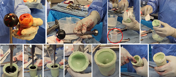
2. Femoral Head Sizing and Acetabular Fit Verification (Figure 1)
After femoral head removal, its size is measured with a calibrated device, and a corresponding trial head is inserted to assess acetabular fit. Achieving a proper suction fit is a sign of congruence, joint stability, and potential concentric loading during the interim period.
3. Femoral Canal Preparation
The femoral canal is broached to the maximal fitting size using a cemented stem broach system, followed by selection of a stem two sizes smaller. This downsizing ensures an adequate cement mantle, enhancing fixation and reducing mechanical failure risk.
4. Spacer Mold Preparation (Figure 1)
To create the spacer head, the selected femoral head trial is coated with sterile mineral oil and inserted into unset bone cement poured into a sterile plastic container slightly larger than the trial. Gentle circular motion forms a spherical mold matching the trial head. Cement overflow is avoided, and once partially set, the trial is removed, leaving a precise mold to harden fully.

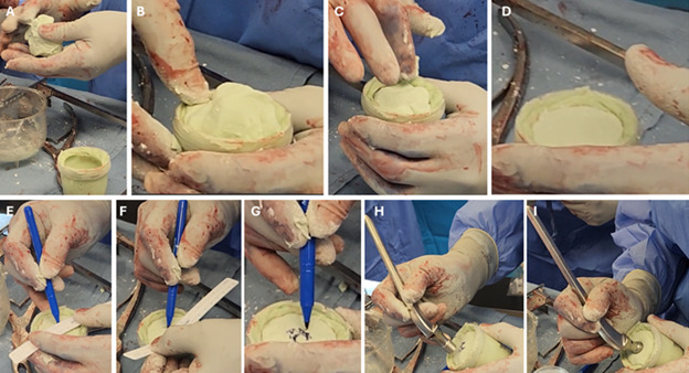
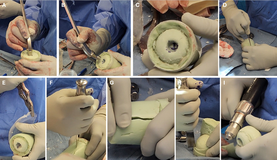
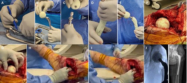
5. Formation of the Antibiotic Spacer Head (Figures 2, 3)
A second batch of antibiotic-loaded cement (2 g vancomycin and 2 g tobramycin per 40 g) is prepared. The hardened mold is lubricated with sterile mineral oil, and cement is hand-packed into the container. Excess is trimmed, and a central hole is created using a sterile ruler and the lubricated trial stem to ensure proper alignment. The stem is removed, and the cement is left to cure fully. It is important to note that the antibiotic combination described (e.g., 2 g vancomycin and 2 g tobramycin per 40 g cement) reflects the authors’ standard empiric protocol. Antibiotic selection and dosing should always be individualized based on the known culture results, patient allergies, and in consultation with infectious disease specialists.


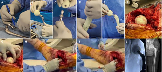
6. Final Assembly, Femoral Stem Preparation and Implantation (Figure 4)
Once cured, the cement head is extracted by breaking the mold, and its sphericity is refined with a surgical drill. As for the femoral stem, after canal preparation and broaching, a stem two sizes smaller than the final broach is selected to allow for an adequate cement mantle. Antibiotic-loaded cement is packed around the stem. The stem, coated with antibiotic-loaded cement, is then inserted while the cement is in its doughy phase to balance initial fixation with ease of later removal, with the goal of achieving axial and rotational stability without aggressive distal pressurization. Overly rigid fixation is avoided to reduce the risk of femoral fracture and to facilitate explantation at the second stage. Stem height is marked based on trial reduction to maintain leg length and soft-tissue tension. If a non-collared stem is used, a cement collar may be added at the level of the trial mark to enhance the stem stability. The custom head is then mounted, hip reduction is performed, and stability is confirmed mechanically and fluoroscopically.
Surgical technique (Hip spacer for prosthetic joint infection)
The surgical technique mirrors standard practice. After exposure, infected components are removed, and thorough debridement, including excision of devitalized tissue and femoral canal membranes, is performed. Femoral head or acetabular size is determined from the explanted prosthesis, followed by steps 2–5 as previously described.
Acetabular Preparation
In native hip infection, acetabular management consists of thorough synovectomy, debridement of infected tissue, and copious irrigation while preserving subchondral bone. In prosthetic joint infection, all components and cement are removed, followed by circumferential debridement of the acetabulum.
For cases involving acetabular preparation, such as revision total hip arthroplasty or primary septic hip arthroplasty with compromised acetabular cartilage, an optional step (Lausmann et al. 2018) can be incorporated to enhance joint stability and minimize erosion risk. This involves debriding and reaming the acetabulum to create a stable hemispherical bed, followed by the insertion of 40–80 g of antibiotic-loaded PMMA cement. A trial dual mobility acetabular liner component is then used to shape the cement into a smooth, concentric socket during the doughy phase, ensuring a stable articulation with the custom femoral spacer head.
Postoperative protocol
Postoperatively, patients are instructed to adhere to toe-touch weight-bearing (≤10% body weight) using assistive devices (e.g., walker or crutches) for the duration of spacer implantation. Standard hip precautions are enforced based on the surgical approach (e.g., avoidance of flexion >90°, adduction past midline, and internal rotation for posterior approaches). Early mobilization (e.g., seated transfers, gentle range-of-motion exercises) is encouraged to prevent stiffness and thromboembolic complications, while full weight-bearing, pivoting, or high-impact activities are strictly avoided to minimize mechanical stress on the spacer.
Construct duration
This construct serves as a temporary interim spacer, and its duration is dictated by infection eradication, host factors, and soft tissue healing rather than spacer integrity alone. Prolonged implantation (beyond 12 weeks) may increase the risk of acetabular erosion, cement fatigue, or mechanical failure (Erivan et al. 2018; Sambri et al. 2023). Typically, second-stage reconstruction is planned after 6–8 weeks, upon resolution of the clinical and laboratory signs of infection, as per the IDSA and ICM guidelines for PJI management (Osmon et al. 2013; Parvizi et al. 2013).
Discussion
Prosthetic joint infections are devastating complications after joint arthroplasty (Autorino et al. 2019; Mahmoud, Brule, et al. 2025; Mahmoud, Ordas-Bayon, et al. 2025). The management of periprosthetic joint infection (PJI) in hip arthroplasty often requires the use of antibiotic-loaded spacers during two-stage revision. Several spacer designs are currently available, each with distinct advantages and limitations (Table 1). This custom antibiotic-loaded hip spacer technique may offer some advantages by enhancing mechanical stability and reducing dislocation and acetabular erosion. The spacer head is molded to match native acetabular geometry using a trial head, promoting a suction fit that improves articulation and interim joint stability. Compared with prefabricated and metal endoskeleton spacers, this technique allows improved head–acetabulum congruency and flexibility in sizing of the cement head to match the native femoral head or explanted prosthetic head, aiming for a concentric and stable articulation within the acetabulum. Additionally, it does not rely on proprietary implants or molds, making it cost-conscious in resource-limited settings, especially when using low-cost cemented stems.
Despite its advantages, the technique has limitations, including the stem cost, prompting some centers to use alternative fixation methods. This technique, as with any manually fabricated molds, requires additional operative time and technical familiarity, and may yield variable results. Dependence on intraoperative materials (e.g., sterile containers, mineral oil) may limit feasibility. Risks of mechanical complications, such as acetabular erosion, dislocation, mechanical failure, cement fragmentation, stem subsidence, femoral fractures, and infection recurrence persist, especially with poor weight-bearing compliance due to the spacer’s mechanical limitations.
Acetabular erosion remains an inherent limitation of all hemi-articulating cement spacers, especially with prolonged implantation or undersized spacer heads. Acetabular erosion occurs particularly when the spacer head is undersized or lacks a smooth, spherical surface, leading to point loading and increased acetabular wear (Erivan et al. 2018). The anatomy-matched spherical head produced with this technique is intended to improve load distribution, minimize eccentric loading and distribute forces more evenly across the joint surface. While erosion cannot be fully eliminated, improved head–acetabulum congruency may mitigate the rate and severity of this process during the interim period.
Spacer dislocation is another limitation that is frequently reported as a complication after articulating hip spacers. Spacer dislocation rates were reported between 10% and 42% (Jung et al. 2009; Sambri et al. 2023; Erivan et al. 2018), often due to spacer-acetabulum mismatch, poor head–neck ratio, insufficient soft-tissue tension, or limb-length discrepancy (Leunig et al. 1998; Jung et al. 2009; Sambri et al. 2023). By deliberately sizing the cement head to match the native femoral head or the explanted prosthetic head, concentric and stable articulation within the acetabulum can be achieved with the described technique. Trial reduction before final spacer insertion allows for appropriate stem offset choice and adjustment of stem height to restore limb length and soft-tissue tension, which are critical contributors to spacer stability.
Femoral subsidence and fracture are additional concerns, particularly when spacer stems are either inadequately fixed or improperly sized. The current technique emphasizes controlled cementation using a downsized stem and insertion during the doughy phase of cement polymerization around the stem. This approach seeks to achieve sufficient axial and rotational stability while avoiding aggressive distal pressurization.
Among the inherent limitations of articulating spacers and this technique in particular is the risk of cement fracture and mechanical fatigue, particularly in patients with higher functional demands or noncompliance with weight-bearing restrictions. For this reason, the described construct is intended strictly as a temporary solution. The postoperative protocol should emphasize protected weight bearing and avoidance of high-impact activities to reduce mechanical stress on the spacer.
On the other hand, the described technique could offer significant cost savings compared to proprietary spacer systems, as it utilizes standard, widely available materials (e.g., antibiotic-loaded PMMA cement, trial components, and basic surgical instruments) rather than expensive prefabricated or modular implants. While the intraoperative time and technical demand are higher than with off-the-shelf spacers, the reduced material costs, estimated at a fraction of the price of commercial systems, make it a potentially cost-effective alternative, particularly in settings where budget constraints are a concern. The utilization of a cemented stem represents the most expensive item in the described technique, and this cost may be decreased by selecting low-cost cemented stems without compromising functionality.
While our patient-adapted, hand-crafted technique offers advantages in terms of anatomical fit and decreased cost, it is important to acknowledge that the literature presents mixed evidence regarding the superiority of surgeon-made spacers versus standardized preformed alternatives. For instance, prefabricated articulating spacers have been associated with ease of use, reproducibility, and standardized antibiotic loading, which may reduce variability in clinical outcomes (Citak et al. 2015; Li et al. 2023). Conversely, prefabricated spacers may suffer from limited sizing options, leading to potential mismatches with patient anatomy and increased risks of dislocation or acetabular erosion (Jung et al. 2009; Sambri et al. 2023).
Additionally, modular systems, such as the PROSTALAC, have demonstrated superior mechanical stability and lower dislocation rates, albeit at a higher cost and with limited availability (Scharfenberger et al. 2007; Cherubino et al. 2013). Our technique, while cost-conscious and customizable, requires technical skill and additional operative time, which may not be feasible in all clinical settings. The choice of spacer should ultimately be guided by patient-specific factors, institutional resources, and surgeon experience.
It is important to note that the antibiotic dosing used in our technique (2 g vancomycin and 2 g tobramycin per 40 g of cement) is based on empiric protocols aimed at achieving high local antibiotic concentrations to eradicate infection. However, it is essential to acknowledge that increasing antibiotic load in polymethylmethacrylate (PMMA) cement can compromise its mechanical properties, including compressive strength, fatigue resistance, and elasticity (Anagnostakos et al. 2006; Masri et al. 1998). Higher antibiotic concentrations may weaken the cement matrix, potentially increasing the risk of spacer fracture, acetabular erosion, or mechanical failure, particularly in load-bearing applications such as articulating hip spacers (Jung et al. 2009). In a trial to mitigate the risk of mechanical compromise from high antibiotic loads, our technique emphasizes a spherical, anatomy-matched head to optimize load distribution and reduce acetabular point loading. While this approach aims to preserve mechanical integrity, surgeons must tailor antibiotic dosing and spacer design to individual patient factors, such as bone quality and functional demands.
Comparative long-term data with prefabricated spacers are lacking. While similar methods are likely to be in use, this report provides a detailed, step-by-step documentation, aiming to standardize and support reproducibility, particularly in resource-limited settings. Importantly, this article is designed as a technical guide rather than an outcomes study. While the described steps are intended to address known failure mechanisms reported in the literature, clinical results and complication rates associated with this technique are being evaluated separately and shall be reported in a dedicated outcomes-focused investigation. This may limit the technique’s applicability, although similar techniques are already in use, and necessitate cautious interpretation of the evidence presented. Specifically, this technique remains limited by the inherent mechanical constraints of cement hemiarthroplasty spacers. Acetabular erosion, cement fracture, dislocation, and femoral subsidence may occur, particularly with prolonged implantation or noncompliance with weight-bearing restrictions.
Conclusion
In summary, this technique provides a practical and customizable approach to antibiotic spacer fabrication, with potential benefits in joint stability and acetabular preservation, provided that spacer duration and weight bearing precautions are followed.
Conflicts of interest
None.
Funding
No funding received