
Introduction
Ipsilateral hip and knee dislocations are rare, high-energy injuries that usually present in polytrauma and carry substantial neurovascular risk (Sharma et al. 2016; Falgons and Warner 2024). Early reports and small series describe frequent ligamentous and vascular damage at the knee, diagnostic difficulty, and wide variability in outcomes (Sharma et al. 2016; Gonzalez-Morgado et al. 2023; Jlidi et al. 2024). Larger knee-dislocation cohorts reinforce the limb-threat: popliteal artery injury occurs in 3–33%, mandating time-critical reduction and vascular assessment (Bernhoff et al. 2021; Naziri et al. 2018). Outcomes for “floating knee” patterns remain complication-prone, including amputation in severe cases (Nouraei et al. 2013). No consensus algorithm exists when a hip dislocation coexists with an unstable/open knee, where standard maneuvers may transmit harmful axial force across the knee; we present a case of ipsilateral closed hip and open knee dislocation with limb-threatening ischemia and a knee-sparing modification of the Allis maneuver performed in the trauma bay, adding to a literature that has documented only a handful of such combined injuries
Case Report
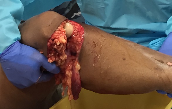
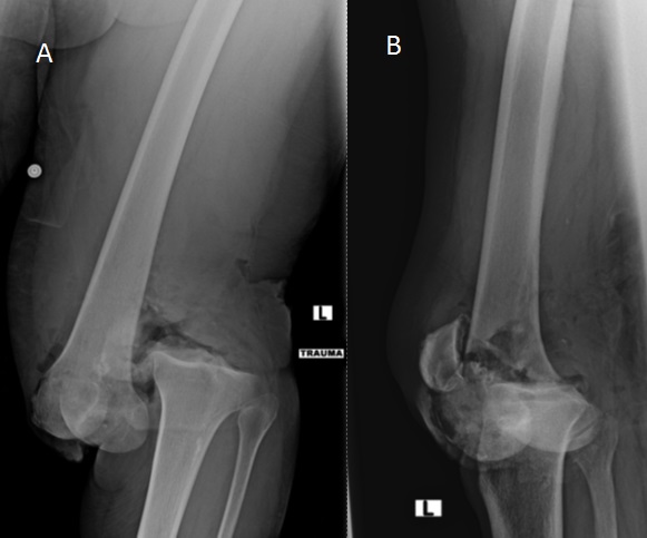
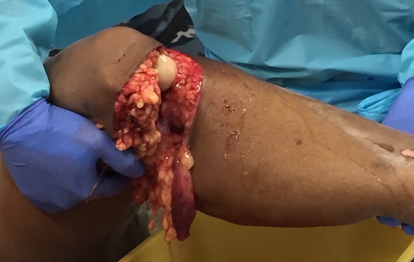
A 70-year-old woman presented after an auto–pedestrian collision. Examination showed a near-circumferential 25-cm posterior left knee laceration with a clear arthrotomy and extrusion of soft tissue (Figure 1). The foot was cold with no Doppler signals and no sensorimotor function below the knee. Because she was hypotensive, the open knee dislocation was provisionally reduced under direct visualization and the limb was placed in a rigid immobilizer; pre-reduction radiographs confirmed a complete dislocation (Figure 2A–B). Vascularity did not return after knee reduction. The femur appeared internally rotated relative to the pelvis. Given the unstable physiology and suspected multi-region injury, she proceeded immediately to whole-body CT with CT angiography of the left lower extremity.
_anterior-posterior_(b)_lateral_radiographs_of_the_left_knee_demonstrating_complete_dis.jpg)
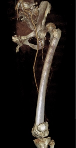
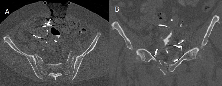
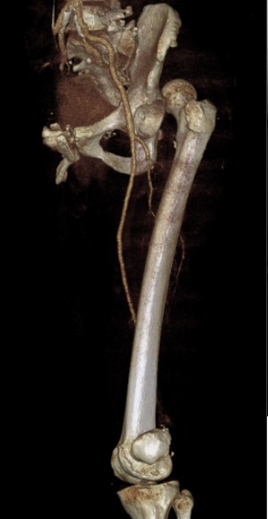
Imaging showed a left posterior–superior hip dislocation (Figure 3), fractures of the superior and inferior pubic rami with a zone-1 sacral fracture consistent with a floating hemipelvis (Figure 4A–B), a left posterior shoulder dislocation, right L1–L5 and left L5 transverse-process fractures, bilateral rib fractures with flail segments, pneumothoraxes, and a liver laceration. CT angiography demonstrated no flow at or distal to the popliteal artery.
_axial_cut_of_left_zone_1_sacral_fracture_and_(b)_coronal_cut_of_left_.jpg)
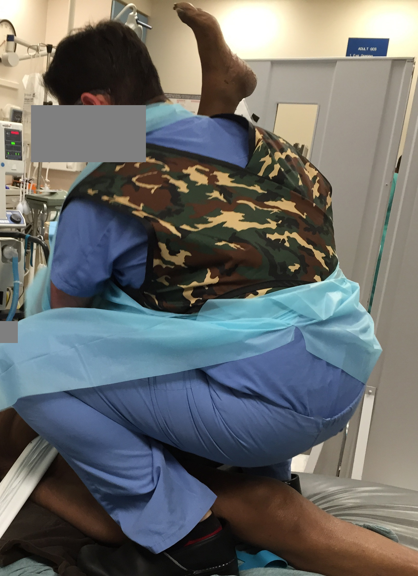
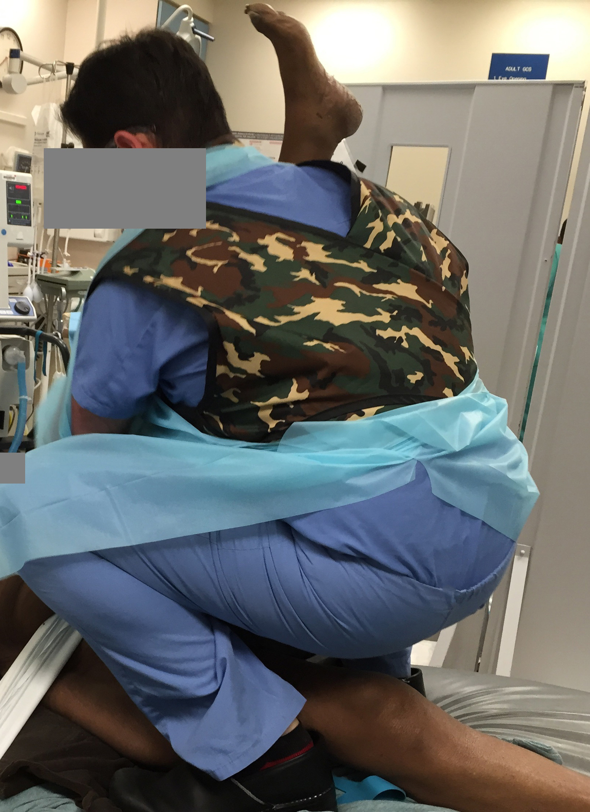
As her condition deteriorated, bilateral chest tubes were placed and she was intubated. Immediately after intubation, the hip was reduced in the trauma bay to take advantage of paralysis and to avoid further delay, using a modified Allis maneuver designed to avoid loading the injured knee (Figure 5). Post-reduction, the limb remained pulseless. She was taken emergently to the operating room for abdominal exploration and damage-control management. With a MESS of 9 and persistent neurovascular compromise, a guillotine left above-knee amputation was performed during laparotomy. No intra-abdominal hemorrhage was identified. A pelvic binder was applied and pelvic angiography was negative. The left shoulder was reduced in the ICU without difficulty. Her ICU course included abdominal compartment syndrome and ARDS; despite maximal support, the family elected to withdraw care approximately one month after injury.
Technique
The canonical Allis maneuver is performed with the patient supine: an assistant stabilizes the pelvis while the operator flexes the hip and applies in-line axial traction to the femur, with gentle adduction/rotation until reduction. In the setting of an open or unstable knee, flexion-based maneuvers and traction transmitted through the tibia/ankle are unsafe. We therefore use a knee-sparing modification that maintains rigid knee immobilization and applies traction directly to the distal femur while counter-pressure is maintained over the pelvis.
The patient is supine after intubation and paralysis. The pelvis is stabilized with a tightly secured sheet across both anterior superior iliac spines. The injured knee remains in a rigid immobilizer in full extension. The operator stands at the foot of the bed and rests the immobilized knee on the operator’s shoulder to unload the joint. Both hands grasp the distal thigh just proximal to the patella so traction is applied to the femur rather than through the tibia or ankle. Steady axial traction is applied in line with the femoral shaft while an assistant maintains firm counter-pressure over the pelvic sheet. With traction maintained, gentle adduction and small arcs of internal and external rotation are introduced until reduction occurs. Reduction is confirmed by a palpable clunk and immediate imaging. Distal perfusion as well as motor and sensory function are reassessed without delay (Figure 5).
Discussion
Simultaneous ipsilateral hip and knee dislocations are rare and time-critical; outcomes hinge on rapid hip reduction, protection of the unstable knee, and early, realistic appraisal of limb viability. Our patient differs from most published reports by combining an open knee dislocation with complete neurovascular compromise, a posterior–superior hip dislocation, and a floating hemipelvis in the setting of shock.
Comparable cases underline the central problems we faced. Motsis et al. described posterior hip dislocation with an open knee dislocation and popliteal artery disruption; despite vascular reconstruction, revascularization failed and the patient ultimately required amputation, highlighting the tenuous prognosis when the knee is open and ischemic (Motsis et al. 2006). DuBois et al. reported that reducing the hip often needs general anesthesia and adjuncts such as Schanz pins to avoid transmitting force across an unstable knee; their patient required pins to accomplish closed hip reduction, underscoring the technical challenge when the knee cannot safely bear traction (DuBois et al. 2006).
Recent summaries echo the vascular risk: in a 2024 review of ipsilateral hip–knee dislocations, popliteal injury occurred in about a fifth of cases and two of three patients with arterial injury ultimately underwent amputation, emphasizing the narrow window for salvage in contaminated, unstable knees (Jlidi et al. 2024).
Current gaps include: (i) no pragmatic guidance when hip dislocation coexists with an open/unstable knee, (ii) reliance on tibial/ankle traction that can aggravate a tenuous popliteal segment, and (iii) delays while waiting for skeletal traction or OR transfer in unstable patients. Our modification enables immediate, trauma-bay reduction after intubation using only a pelvic sheet and a knee immobilizer: traction is applied to the distal femur (not through the knee) while an assistant provides firm counter-pressure over the pelvis. This preserves soft-tissue protection, avoids loading the popliteal segment, and shortens time to a safe reduction.
Broader knee-dislocation series also document high rates of popliteal injury and limb loss, reinforcing the need for serial vascular checks and rapid imaging when pulses are absent (Vyas et al. 2025).
Our contribution is twofold. First, we provide a reproducible, knee-sparing modification of the Allis maneuver that enabled immediate hip reduction in the trauma bay after intubation and paralysis without loading the injured knee or resorting to skeletal traction or transfer to the operating room; this addresses the practical gap highlighted by DuBois et al. and others (DuBois et al. 2006; Millea et al. 1991).
Second, we clarify decision-making when persistent ischemia, massive soft-tissue injury, and shock coexist. With no distal flow after timely reductions and a high mangled-extremity score, early guillotine above-knee amputation during laparotomy represented appropriate damage-control care rather than a failure of sequencing, consistent with outcomes in similarly severe reports.
Overall, this case adds practical technique detail and a transparent rationale for early amputation in a pattern where vascular injury, contamination, and systemic instability make limb salvage unlikely.
Acknowledgements
We would like to thank the patient and their family for allowing this case to be reported for academic purposes.