
INTRODUCTION
The number of revision total ankle arthroplasties (rTAA) has increased in recent years, mirroring the rise in primary total ankle arthroplasties (TAA). From 2005 to 2017 there was a 155% increase in TAA, with 1,170 TAAs performed in 2017 (Shah et al. 2025). The number of TAA is projected to increase by another 110-796% by 2030 (Shah et al. 2025). As the use of TAA expands, so too does the complexity of handling implant failures. Issues such as infection, prosthesis malpositioning, loosening, and periprosthetic fractures can all compromise implant stability and patient outcomes (Jennison et al. 2022). Among these challenges, bone loss presents a particularly difficult and variable scenario as patient bone quality and quantity can require innovative implant solutions.
Given the increasing demand of TAA operative strategies, new implant systems with customized solutions have emerged as viable strategies to effectively manage bone loss and optimize functional outcomes. Small series have illustrated acceptable outcomes of patient-specific TAA component implantation in both primary and revision scenarios (Valan et al. 2024; Wagener et al. 2017) The integration of advanced customizable systems in complex revision scenarios with challenging bone defects holds the potential to significantly optimize patient outcomes.
Despite the rise in available off-the-shelf primary and revision implant systems, the literature on custom implant use in rTAA remains limited, especially in relation to the tibial component. Custom Short-Stemmed Tibial Trays (CSTT) match the patient’s tibial anatomy and incorporate a stem extension that projects into the tibial canal to enhance fixation. Customization is achieved through advanced imaging and allows the implant to precisely fit the patient’s unique bony morphology (Belvedere et al. 2019). This is particularly advantageous in rTAA patients who present with abnormal anatomy or compromised bone stock. Therefore, this study aims to share the safety and early experience of CSTT in rTAA.
METHODS
We performed a retrospective analysis of adult patients who underwent rTAA with a single fellowship-trained foot and ankle surgeon at a tertiary academic medical center between May 1st, 2023, and April 1st, 2024. Institutional Review Board approval was granted prior to study initiation. Inclusion criteria were patients who had undergone rTAA using an Apex [Zimmer, Warsaw, IN] CSTT and a minimum 1-year clinical follow-up. A standardized rehabilitation protocol from a single surgeon was utilized.
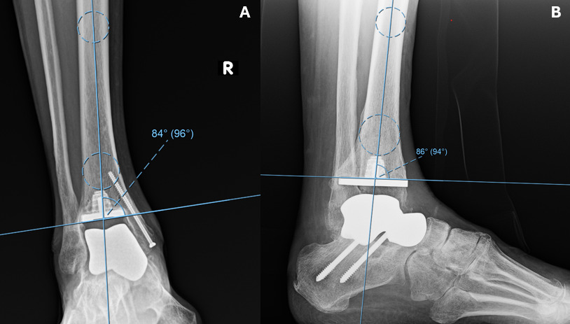
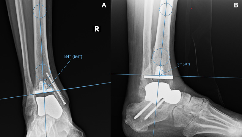
Data collected included age at the time of surgery, length of follow-up, time to weight bearing, adherence to postoperative physical therapy (PT), reported visual analog scale (VAS) pain scores at final follow-up, and reoperation. Radiographic parameters included lucency, subsidence, and the medial distal tibial angle (MDTA) and anterior distal tibial angle (ADTA). The MDTA and ADTA were measured as the angle between the tibial axis and the tibiotalar joint line on anteroposterior and lateral radiographic views, respectively (Figure 1). Lucency and subsidence were subjectively evaluated by a post-residency orthopaedic foot and ankle surgery fellow. Changes between immediate postoperative and most recent radiographs were calculated. Descriptive statistics were used to summarize patient characteristics and clinical outcomes. This study received no external funding.
For the total talus component, the contralateral talus and remaining native anatomy were used to guide implant sizing and shape. Similarly, for the CSTT, preoperative CT assessed tibial bone stock to identify regions of optimal bone quality and structural support for stem fixation. The implants were additively manufactured [Paragon28, Zimmer] using patient-specific imaging data. The total talus prostheses are available in cobalt chromium and titanium alloy. The lag time from implant design freeze to delivery was approximately 8-12 weeks and the cost for both implants’ ranges based on institutional contracts with the manufacturer.
RESULTS
Demographics and Baseline Characteristics
Six unique patients underwent rTAA using a 3D-printed CSTT. One patient underwent bilateral rTAA, resulting in 7 CSTT implants total with a minimum 1-year follow-up. Demographics and baseline characteristics of each case can be viewed in Table 1. The mean age at the time of surgery was 60.0 years (range, 36–80), and 3 patients (50%) were female. The mean preoperative body mass index (BMI) was 25.7 kg/m². No patients had diabetes, while 2 patients (28.6%) had a history of malignancy and 2 (28.6%) reported a history of tobacco use (both former smokers). The primary indication for the index TAA in 6 out of 7 cases was post-traumatic arthritis. The most common implant for primary TAA was the Cadence Total Ankle System [Smith+Nephew; Memphis, TN] (3), followed by the Scandinavian Total Ankle Replacement [Enovis; Wilmington, DE] (2), Agility Total Ankle System [DePuy Synthes, Warsaw, IN] (1), and INFINITY [Stryker; Kalamazoo, MI] (1).]
Surgical Characteristics
Indications and surgical characteristics regarding the CSTT revision procedure are displayed in Table 2. Failure was often driven by failure of the tibial component (4 cases, 57.1%). One surgeon performed all CSTT procedures, but 4 out of 7 primary TAAs were performed by other providers. Average time from primary TAA to CSTT implantation was 6.43 years (range 2.4 – 14.6 years). The most common concomitant procedure at time of CSTT implantation was total talus replacement (7/7 cases) followed by subtalar arthrodesis (6/7 cases). Other concomitant procedures included talonavicular fusions (4/7 cases) and prophylactic fixation of the medial malleolus (2/7 cases). Due to the high proportion of subtalar fusions, the average time to weight bearing was 69.14 days postoperatively. The single case that did not receive a subtalar fusion began weight-bearing as tolerated at 1-month postoperatively.
Outcomes
The mean medial distal tibial angle (MDTA) improved from 85.0° at first weightbearing to 87.9° at final follow-up, representing a mean change of +2.9°. The mean anterior distal tibial angle (ADTA) changed from 86.4° to 87.7°, for a mean change of +1.3°. One patient (14.3%) demonstrated radiographic lucency, while no cases of implant subsidence were observed. At a mean follow-up of 20.1 ± 5.6 months, no patients required reoperation. The mean postoperative VAS pain score was 3.4 ± 1.0.
Case Vignette – CSTT in Complex rTAA with Total Talus Replacement
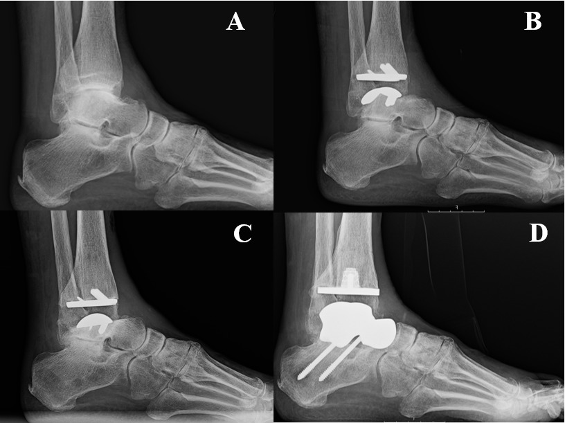
Patient demographics & background: A 78-year-old female with a history of degenerative joint disease and bladder cancer presented with recurrent right ankle pain following multiple prior surgeries [Figure 2A].
_preoperative_x-ray_26_days_prior_to_primary_taa__b)_postoperative_x-ray_31_days_after_p.png)
Initial surgical history: She underwent primary TAA on February 2020 with an outside provider [Figure 2B]. Approximately 21 months later, she required revision for loosening of the talar component. The implant failure was addressed with a polyethylene insert exchange and revision of the talar component [Figure 2C].
Presentation prior to CSTT revision: In early 2023, the patient developed worsening pain, reporting 8–9/10 severity. Further imaging was obtained (CT scan) demonstrating subsidence of the talar component with fracture of a posterior peg and absence of bony ingrowth at the implant–bone interface. Given the extent of bone loss and implant instability, she was referred to the current surgeon for further management.
Surgical intervention: In May 2023, the failed arthroplasty including all components were removed. Using patient-specific guides, the tibia was prepared and residual bone removed to create a stemmed socket for the custom short-stemmed 3D-printed tibial tray. A custom nickel-free total talus prosthesis was implanted with a stemmed design. Two screws were inserted across the subtalar joint for fusion [Figure 2D].
Postoperative course: The patient remained in a bulky Jones splint and was non-weight bearing for 10.8 weeks. The patient was able to progress to weight bearing in a cam boot and begin physical therapy 76 days post-revision. In the early postoperative period she experienced improvement with some stiffness, numbness, tingling, and minimal aching dull pain.
Follow-up & outcomes: At most recent follow-up, 30.3-months postoperatively, radiographs demonstrated stable implant alignment without evidence of progressive lucency or subsidence. Pain improved to 3/10 intermittent pain. Residual symptoms included swelling, numbness, and lack of proprioception.
DISCUSSION
The findings of the current study indicate that custom short-stem 3D-printed tibial trays serve as a viable option for managing complex rTAA procedures. In our retrospective case series, we show acceptable pain, radiographic outcomes, and no reoperations at short term follow-up.
Tibial component failure remains a challenging complication in TAA, particularly with the advent of modern low-profile implant designs (Cody et al. 2019; McKenna et al. 2020). McKenna et al. conducted a meta-analysis showing aseptic loosening accounted for 6.3% of all rTAA. Modern TAA implants are designed to be low profile and use smaller pegs or posts to fixate the tibia, aimed at allowing for minimal bone resection. Additionally, these smaller implants are thought to be more surgeon friendly, more straightforward to implant, and with reduced operative time and fluoroscopy use (Saito et al. 2019). A study by Cody et al. attempted to characterize tibial lucency following primary TAA with a modern “low-profile” implant (Cody et al. 2025). With a mean follow-up of 29 months, they found that 6.5% of primary TAAs had global tibial lucency evidenced by radiographic analysis. This suggests a lack of sufficient osseointegration around the tibial component and underscores the need for revision options to specifically address this issue.
Furthermore, several studies have shown that TAA patients with hindfoot fusion have significantly higher rates of TAA failure (Lewis et al. 2014; Teehan et al. 2025). Henry et al. reviewed 731 primary TAA cases, including 71 with hindfoot arthrodesis, and found that fusion was associated with 2.7-fold greater odds of failure (Henry et al. 2024). Similarly, Teehan et al. reported that patients with hindfoot arthrodesis were more likely to receive stemmed implants, suggesting a higher risk of tibial-sided failure in this population (Teehan et al. 2025). Interestingly, in Henry’s cohort only 1 of the 33 mechanical failures involved a stemmed component, and the use of a stem or keel was associated with 95% lower odds of tibial failure, despite their use in patients with higher bodyweight and greater coronal deformity.
These findings may be supported by the underlying biomechanics. The loss of subtalar motion transfers increased stress through the ankle joint and concentrates force across the tibial implant. Without robust fixation, this added stress may predispose to loosening or tibial-sided failure. Tibial stems provide greater stability and load distribution which makes them particularly advantageous in patients with prior or concurrent subtalar arthrodesis. Our cohort showed all but one patient undergoing rTAA with a CSTT received subtalar arthrodesis. Additionally, we observed no patients whose primary implant contained a tibial stem. These observations emphasize the importance of tailoring implant choice to patient-specific risk factors to mitigate tibial-sided failure.
In addition to CSTTs, custom TAA talar components have demonstrated promising results. A study by Ketz et al. looked at thirty-three patients with custom long-stemmed talar components for salvage of total ankle replacement with a minimum 4-year follow-up (Ketz et al. 2012). The authors observed acceptable postoperative range of motion, radiographic evaluation, clinical outcomes, and functional outcome scores. Similarly, Wagener et al. examined the outcomes of a custom-made talar component in 12 patients for 6.9 years, and found acceptable results in terms of range of motion and patient satisfaction with no instances of radiographic loosening (Wang et al. 2024). A number of other small studies involving custom total talar prostheses have also shown promising outcomes with little to no complications (Jennison et al. 2022; Akoh et al. 2020; Strand et al. 2022; Wang et al. 2024; Yano et al. 2023).
Regarding the tibial component, to our knowledge scant literature exists on custom-made components for rTAA. An investigation by Cook et al. examined the technique of antegrade tibial reaming in rTAA while retaining the original talar component. Both CSTTs and the antegrade tibial reaming technique aim to optimize stem alignment and fixation by maximizing engagement with the tibial canal, particularly in complex revision settings. In the single patient described by Cook et al., the antegrade tibial reaming approach allowed secure tibial component placement while preserving the talar implant and resulted in maintained function at follow-up (Cook et al. 2023). A key finding of this study is the minimal change observed in both MDTA and ADTA over the follow-up period which suggests preservation of coronal and sagittal alignment after revision TAA with talar reconstruction. From a clinical perspective, stable MDTA and ADTA measurements may suggest maintenance of alignment, although correlation with long-term fixation is unknown. Progressive changes in these angles have been associated with component loosening, malalignment, or subsidence in prior arthroplasty literature; thus, their stability in this series aligns with the absence of observed subsidence and low complication rates. Importantly, these findings suggest that even in complex revision scenarios, restoration and maintenance of alignment can be reliably achieved.
At the time these procedures were performed, no commercially available short-stemmed tibial trays existed for revision TAA, and available stemmed options required long stems that were inserted through the calcaneus, often necessitating more extensive bone resection and requiring talar component removal. The use of custom short stems provides greater versatility, particularly in cases where the talar component remains well-fixed and does not require revision. This was especially relevant in the current cohort, as all patients underwent total talus replacement, which was not compatible with any available tibial components and would have otherwise required explantation of a well-fixed talar implant. In addition, custom components can be designed for patients with metal allergies or hypersensitivities, such as nickel allergy, which occurred in three cases in our series. By incorporating preoperative CT imaging and CAD-based planning, the stem trajectory and fixation was optimized to achieve maximal engagement with the remaining bone stock thus expanding the surgical options we had in our series of complex rTAA scenarios.
Short stem implants are increasingly being favored in both primary and revision arthroplasty cases as they preserve proximal bone stock. In the case of rTAA, custom short stems maintain the structural integrity necessary to support revision components by minimizing diaphyseal preparation and conserving metaphyseal bone. This approach is particularly advantageous in younger or more active patients, where the likelihood of future revision is higher and maximizing remaining bone stock is essential for long-term reconstructive options. We observed an average time from index TAA to CSTT implantation of 77.1 months. This reflects its utility for patients who have undergone prior revision procedures, such as in the representative case example. Nonetheless, patients in this series with CSTT implantation occurring up to 15 years after the index arthroplasty and no subsequent revisions receive comparable results.
The use of a CSTT, and other patient-specific implants, introduces several important practical considerations. First, these implants require advanced preoperative imaging and CT-based templating, which increases both the cost and complexity of surgical planning. In addition, the manufacturing process for these implants is associated with approximately 8–12 weeks between final implant design approval and delivery. Implant costs may also vary substantially depending on institutional agreements. This can potentially limit widespread accessibility. Consequently, these patient-specific reconstructive options may be best suited for complex revision scenarios, as presented in this series, in which patients are willing to accept potential increased cost and longer preoperative wait times in exchange for an improved quality of life.
While the CSTT holds potential for challenging revisions, there are clear limitations of the current study. Most importantly, the sample size was limited and length of follow-up was up to only 21 months on average. Larger studies with longer follow-up are required in order to evaluate the efficacy of these implants over time. In addition, the study was descriptive in nature and did not compare the device against other modes of revision total ankle arthroplasty. This study also lacked multifaceted outcome data including function patient reported outcomes, range of motion, or other relevant functional outcome parameters. Lastly, all procedures were performed by a single surgeon at a tertiary care center, which may limit generalizability to other settings. Nonetheless, the study is valuable in the novelty it offers by presenting early evidence supporting the use of a 3D-printed custom tibial tray for complex rTAA.
CONCLUSION
This case series provides early experience supporting the feasibility of using a custom short-stem 3D-printed tibial tray in rTAA. All patients demonstrated stable radiographic alignment, satisfactory pain control, and no reoperations at short-term follow-up. These results suggest that CSTTs may provide an additional reconstructive option in select patients. While encouraging, these findings must be interpreted in the context of the study’s limitations, including its small sample size, lack of a control group, and limited follow-up duration. Outcomes of this series reflect the combined reconstructive strategy involving multiple concomitant procedures rather than the isolated effect of CSTT. Future research with larger cohorts, longer-term outcomes, and comparative analyses is warranted to fully assess the long-term performance and potential advantages of custom tibial implants in complex revision scenarios. Additionally, as barriers to patient-specific implant use continue to recede, including cost and manufacturing time, further investigation may extend to evaluating these implants in a primary TAA setting.