Loading [Contrib]/a11y/accessibility-menu.js

This website uses cookies

We use cookies to enhance your experience and support COUNTER Metrics for transparent reporting of readership statistics. Cookie data is not sold to third parties or used for marketing purposes.

Skip to main content
null
J Orthopaedic Experience & Innovation
  • Menu
  • Articles
    • Brief Report
    • Case Report
    • Case Series
    • Conference Proceedings
    • Data Paper
    • Editorial
    • Meeting Reports/Abstracts
    • Methods Article
    • Product Review
    • Research Article
    • Review Article
    • Review Articles
    • Systematic Review
    • All
  • For Authors
  • Editorial Board
  • About
  • Issues
  • Blog
  • "Open Mic" Topic Sessions
  • Advertisers
  • Recorded Content
  • CME
  • JOEI KOL Connect
  • Resident Research League
  • search
  • RSS feed (opens a modal with a link to feed)

RSS Feed

Enter the URL below into your favorite RSS reader.

https://journaloei.scholasticahq.com/feed
ISSN 2691-6541
Methods Article
Vol. 7, Issue 2, 2026October 02, 2026 EDT

On How I Do It™…Attachment Preserving Hinge Arthroplasty vs. ORIF for Complex Periarticular Knee Fractures

Michael Langworthy, MD,
HingeTraumaplastyComplex TKADistal femur fracture
Copyright Logoccby-nc-nd-4.0 • https://doi.org/10.60118/001c.168111

Articles in Vol. 7, Issue 2, 2026

Vol. 7, Issue 2, 2026
  • A Human Factors Analysis of Personal Electronic Device Use and Cognitive Distractions in Orthopedic Surgery
    Asfand KhanAlbert Boquet
  • Does Preoperative Suzetrigine Impact ASC Opioid Consumption For Total Joint Arthroplasty?
    Louis BattistaAndrew Wickline
  • Pickleball Pains: A 10-year Epidemiologic Analysis of Rising Upper Extremity Injuries
    Kevin ValdesAghdas MovassaghiJehad Feras AlSamhoriXiomara OrtizJocelyn LubertVani J. Sabesan
  • From Innovation to Inaccuracy: The Impact of ChatGPT on Orthopaedic Surgery Research Citations in Sports Medicine
    Calista StevensAlexander HahnGregory ConnorsShiraz MumtazMartinus MegallaZachary GraceJohn CorviMatthew PartanKatherine Coyner
  • Does CMS Hate Specialists?
    Benjamin Schwartz, MD
  • Voices in Orthopaedics™...The Residency Programs: The Unionized Orthopod: Apprenticeship, Labor, and the Changing Identity of Orthopedic Residency at Jefferson
    Eric R. TecceJalen N. BroomeTyler W. HenryGabriel I. Onor Jr.Daniel A. NemirovDaniel E. DavisJames J. Purtill
  • Cerclage Fixation in Total Hip Arthroplasty: Anatomy, Surgical Options and Clinical Outcomes
    Zuhdi AbdoZachary FullerThomas ChristensenAhmed Siddiqi
  • In My Experience™...Orthopaedics: Then, Now, and Tomorrow: Reflections on a Half Century of Change
    Richard Conn, MD
  • Genicular artery embolization for symptomatic knee osteoarthritis: A narrative review
    Junaid MakdaAhmed SiddiqiKhalid YousufOsman Ahmed
  • Simplifying Orthopedic Patient Education: A ChatGPT-Based Readability Analysis
    Mendel ShloushKlaudia GreerJonathan BruttiBrayden TolmanJacob RosenthalRafael Aldaya BourricaudyMarcia VarellaFernando Aran
  • Digital Fatigue and Orthopedic Injuries: The Postural Burden of the Digital Era - A Narrative Review
    Ambrose Loc NgoNiki Gharavi AlkhansariTruong HoRachana TadakamallaLinda NguyenJared Nichols
  • Concurrent Floating Hip and Open Knee Dislocation: Damage-Control Management
    Sara LowVladislav MuldiiarovMark AyzenbergGermanuel LandfairGene Shaffer
  • Higher Pain Catastrophizing Scores are Associated with Increased Pre-operative Anxiety in Ambulatory Hand Surgery that is not Impacted by Watching a High-Quality Pre-Operative Video
    Christopher G. LarsenMichael J. SayeghAmr TawfikCaroline ApriglianoChloe HeitingKate W. Nellans
  • MOTIV™ and the Next Frontier of Orthopaedic Evidence Generation: A New Model for Physician-Led Clinical Research
    John Mercuri, MDAndrew Wickline, MD
  • Evidence on Demand: OpenEvidence versus Established LLMs on the Orthopaedic In-Training Examination
    Asim A. KhanShaan S. LalvaniSam PourarbabLord J. HyeamangRyan M. LewZaamin B. HussainGregory P. NicholsonGrant E. Garrigues
  • Trends in Orthopedic Surgeons Signing Medicare Opt-Out Affidavits
    Thriaksh RajanAndre RevnewJoshua PortoMonish LavuComron SaifiAtul Kamath
  • Voices in Orthopaedics™...The Residency Programs: Training for the Future of Orthopaedic Surgery: Residents’ Perspective on the UT Austin Dell Medical School Orthopaedic Surgery Residency
    Cassidy ShieldsSemran ThamerAmanda SeymourAlec Giron
  • Preoperative GLP-1 Receptor Agonist Use Improves Thromboembolic, Infectious, and Wound-Related Outcomes Following Unicompartmental Knee Arthroplasty
    Zachary FullerManjot SinghAbhiram DawarJeremiah ThomasAlan H DanielsZuhdi E Abdo
  • Beyond the Breaking Point: Solutions for Burnout in Orthopaedic Surgery
    Aghdas MovassaghiCamryn McIntyreSamir SakariaMitchell J. ChristiansenJocelyn LubertMary MulcaheyVani Sabesan
  • Feasibility and Early Experience of Custom Stemmed Tibial Trays in Revision Total Ankle Arthroplasty: A Case Series
    Grant M. ThomasKush S. ModyJoydeep BaidyaCorinne SommiDavid I. PedowitzSelene G. Parekh
  • Postoperative Outcomes by Timing of Total Hip and Knee Arthroplasty Following Coronary Revascularization
    Zachary FullerManjot SinghThomas ChristensenJeremiah J ThomasAlan H DanielsZuhdi E Abdo
  • Tibial Plafond Fractures and the Impact of Social Media Support Groups on Patient Perceptions
    Alexandra F. FlahertyDana PerimAnnie WaiteAlvarho GuzmanErnest N. Chisena
  • Retained Male Component After Intra-operative Dissociation of a Tibial Magnetic Lengthening Nail: A Case Report covering Technical Challenges and Management Strategies
    Eunice Anastasia WiliantoNeeraj MishraDerrick Jun Liang LamKenneth Pak Leung WongBenny Kai Guo LooAshik Mohammad
  • A physician led consensus building intervention at a multi-specialty practice results in meaningful reduction of opioid prescriptions for post-operative patients
    Jenna M. GodfreyJohn Paul BigouetteConnor FitzpatrickJohn W. OverHeather A. CampionErin C. Owen
  • Voices in Orthopaedics™...The Residency Programs...The Role of Networking and Interconnectivity in Building an Orthopedic Career
    Gabriel I. Onor Jr.Terence L. Thomas, MDDaniel Nemirov, MDJalen Broome, MDDaniel E. Davis, MDChristopher C. Dodson, MDJames J. Purtill, MD
  • From My Perspective… The Consent Discussion: The Robot Demands
    Stephen Howell
  • How to Assemble a Well-Fitting, Patient-Specific Antibiotic Cement Hip Spacer for Infection Management
    Ahmed Nageeb MahmoudNicholas BruleCatherine Mary DoyleGabriel MakarDaniel Horwitz
  • At The Conference™...How to Handle Severe Bone Defects in Knee Arthroplasty
    Christopher Grayson, MD
  • From My Perspective...Triple-Tapered Collared Stems: Are They All Equal—and How Have They Changed Primary Hip Arthroplasty?
    David Rodriguez-Quintana, MD
  • On How I Do It™...Attachment Preserving Hinge Arthroplasty vs. ORIF for Complex Periarticular Knee Fractures
    Michael Langworthy, MD
  • About The Innovation™...Arthrolense: Where the Idea Began
    Russell Nevins, MD
  • In My Experience™...Biointegrative Collagen Implant Use in Complex Hip and Knee Arthroplasty: Early Clinical Experience and Second-Look Observations
    Chris Hoedt, MD
  • Force-Modulating Tissue Bridges in Sports Medicine Incision Management: A First 100-Case Experience Evaluating Allergic Reactions and Postoperative Complications
    John A GrottingEge Karadag
J Orthopaedic Experience & Innovation
Langworthy, MD, Michael. 2026. “On How I Do ItTM…Attachment Preserving Hinge  Arthroplasty vs. ORIF for Complex Periarticular Knee Fractures.” Journal of Orthopaedic Experience & Innovation 7 (2). https://doi.org/10.60118/001c.168111.
Save article as...▾
Download all (3)
  • Figure 1. Intraoperative attachment-preserving preparation. A short epicondylar cut retains the collateral-ligament-bearing bone while creating space for the distal femoral replacement. Image provided in the accompanying pilot manuscript.
    Download
  • Figure 2. Representative native distal femur fracture and attachment-preserving hinge reconstruction. The sequence shows the injury, three-dimensional fracture pattern, early reconstruction, and later remodeling around the metal metaphysis. Images provided in the accompanying pilot manuscript.
    Download
  • Figure 3. Illustration of vascular perforation zones around the distal femur and proximal tibia—areas being investigated as stereotyped stress risers in fracture propagation. Artwork supplied with the pilot manuscript.
    Download

Error

Sorry, something went wrong. Please try again.

If this problem reoccurs, please contact Scholastica Support

Error message:

undefined

View more stats

Abstract

The author reviews his experience with this novel technique of tissue preservation in complex knee arthroplasty.

My goal is not simply to replace a shattered distal femur. It is to create a stable metal metaphysis while preserving the patient’s living soft-tissue envelope—bone fragments, collateral ligament attachments, posterior structures, and the biologic capacity to heal around the implant.

The question is not simply: Can I fix the joint surface?

When I look at a complex distal femur fracture, particularly in an older patient or around a total knee arthroplasty, I start with a different question than the one many of us were taught to ask. The articular surface may be reconstructable, but can the metaphysis support that reconstruction? In elderly, osteoporotic bone, the metaphysis is often the weak link. A technically accurate open reduction and internal fixation (ORIF) can still fail if the bone cannot hold the construct and heal while carrying the demands placed on it.

ORIF remains an excellent operation when I believe I can obtain stable fixation and preserve a biologically favorable environment. I am not arguing that every complex fracture needs a hinge. But when the metaphysis is profoundly compromised—when comminution, bone loss, osteoporosis, or a loose prosthesis makes durable fixation doubtful—I consider replacement early rather than waiting for fixation to fail.

The problem with a traditional distal femoral replacement is that it was largely conceived as a resection operation. The fractured distal femur is removed, the hinge is implanted, and the radiograph can look impressive. Biologically, however, the soft tissues have lost much of the native scaffold to which they attach. The implant becomes a purely load-bearing device surrounded by a diminished envelope.

I want the implant to provide immediate mechanical stability, but I want the patient’s own tissues to provide the envelope, proprioception, vascularity, and eventual biologic reinforcement.

Why I preserve the envelope

My approach grew out of two parts of my training. I was a trauma surgeon in the Navy and later completed a joint fellowship. In trauma, you learn to respect every viable fragment with a soft-tissue attachment. In arthroplasty, you learn how to establish joint line, rotation, fixation, and immediate stability. The envelope hinge brings those two ways of thinking together.

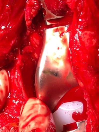
If a fragment is viable and carries a meaningful attachment, I leave it. That includes the epicondylar origins of the collateral ligaments, capsular attachments, posterior cortex, metaphyseal fragments, and—when the fracture pattern permits—the extensor mechanism. I do not expect every fragment to unite anatomically as it would after plate fixation. I expect the retained bone and soft tissue to organize around the implant. Bone remodels; gaps fill; and the maturing fibrous tissue links the fragments like the tension wires on an old biplane. The metal supplies the new metaphysis, while the envelope supplies biologic continuity.

The preserved epicondyles do more than retain ligament attachments. They are also reliable landmarks for restoring the joint line. Instead of estimating the level from a segmental prosthesis or from a resected field, I can place the reconstruction where the patient’s own epicondyles tell me it belongs.
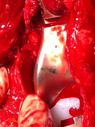
Figure 1
Figure 1.Intraoperative attachment-preserving preparation. A short epicondylar cut retains the collateral-ligament-bearing bone while creating space for the distal femoral replacement. Image provided in the accompanying pilot manuscript.

How I decide: ORIF or an attachment-preserving hinge

My decision is patient-specific, but the following comparison captures the way I think about the problem. It is a decision framework, not a rigid algorithm.

Table 1.Practical decision factors used in choosing fixation versus attachment-preserving hinge arthroplasty.
Question Favors ORIF Favors envelope hinge
Metaphyseal support Bone can securely hold fixation Severe osteoporosis, comminution, or bone loss makes fixation unreliable
Implant status Stable existing components; fracture can be spanned or fixed Loose or unsalvageable component, or fixation would depend on poor bone
Weight bearing Patient can safely comply with the required protection Immediate weight bearing is a major clinical priority
Biologic envelope Fixation can preserve blood supply and attachments Fragments can be retained around a stable prosthesis instead of excised
Failure consequence Revision remains manageable A failed ORIF would impose a major second operation on a frail patient

The operation: three cuts and a stable metal metaphysis

The technique is intentionally simple. I expose the fracture sufficiently to understand the pattern, but I avoid stripping the fragments. I preserve the posterior bone and soft tissues, which keeps me away from the neurovascular structures and maintains the envelope behind the prosthesis.

1. Define what must be removed. I remove the failed or loose prosthesis when present and clear only what prevents stable preparation and implantation. I do not excise fractured bone simply because it looks untidy.

2. Make the diaphyseal cut. I create a clean transverse level in the femoral shaft that will accept the stem and establish the proximal end of the reconstruction.

3. Preserve the epicondyles. I make two short, approximately 15-mm epicondylar cuts. Conceptually, the preparation resembles an upside-down U: one diaphyseal cut and two epicondylar cuts. The epicondylar fragments—and their ligament attachments—remain in place.

4. Protect the posterior envelope. I leave the posterior cortical and soft-tissue structures whenever they do not interfere with safe implantation. This protects the neurovascular bundle and avoids unnecessary devascularization.

5. Pot and cement the prosthesis. I prepare and cement a trauma-oriented rotating hinge/distal femoral replacement to create immediate mechanical stability. The retained fragments are laid back against the metal metaphysis rather than being stripped away.

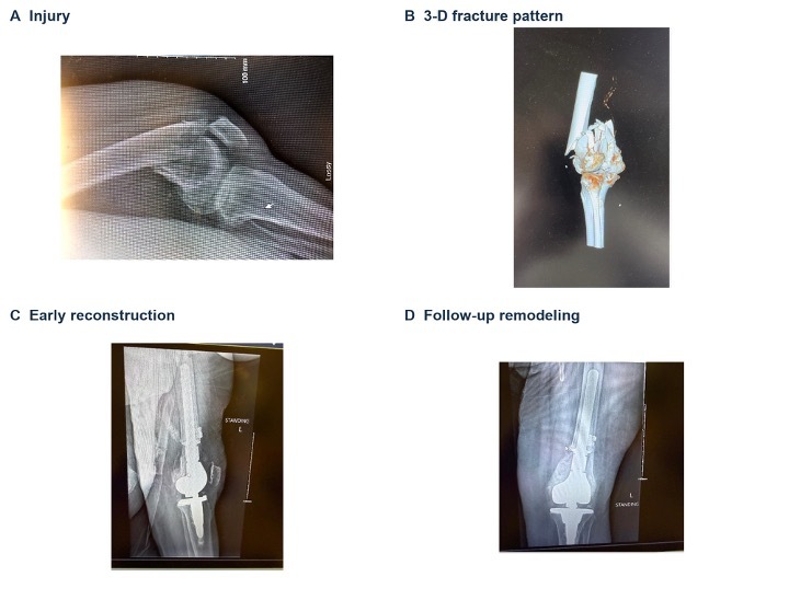
6. Let biology complete the construct. On the femoral side, I generally do not try to turn every fragment into a formal fixation problem. The retained envelope scars and remodels around the prosthesis. Over six months to a year, radiographs can show striking consolidation and incorporation.
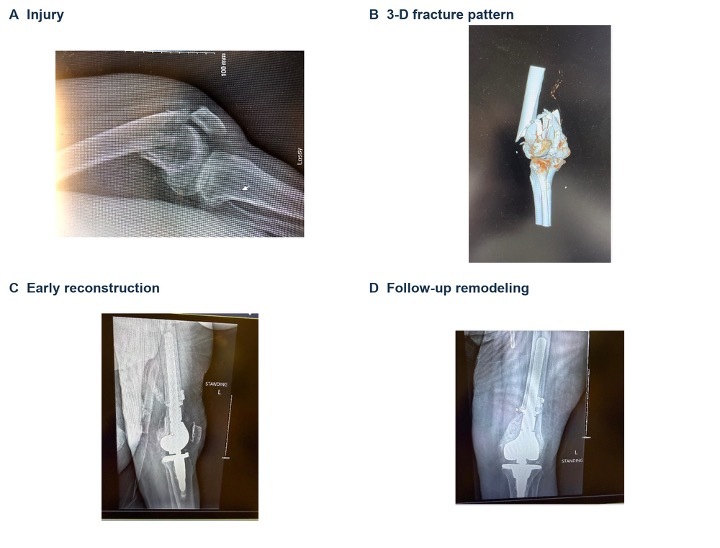
Figure 2
Figure 2.Representative native distal femur fracture and attachment-preserving hinge reconstruction. The sequence shows the injury, three-dimensional fracture pattern, early reconstruction, and later remodeling around the metal metaphysis. Images provided in the accompanying pilot manuscript.

What makes surgeons uncomfortable

The greatest technical pitfall is often psychological: the broken bone is still there. Surgeons are accustomed to believing that every fragment left beside a prosthesis will remain painful or become a nonunion problem. In my experience, once the environment is stable, that concern has not played out the way one might expect. The retained fragments organize within scar, and the pain associated with an unstable fracture diminishes as stability and soft callus develop.

A second pitfall is overworking the envelope. If I strip fragments to make them look anatomically perfect, I defeat the biologic purpose of the operation. If I chase the posterior cortex, I increase risk around the neurovascular structures. The prosthesis must be correctly aligned, securely fixed, and appropriately positioned; the fragments do not all need to be converted into a plate-and-cable reconstruction.

I also resist adding attachment features merely because they are available. On the tibial side I have used cables. On the femoral side, however, a smooth implant surrounded by the retained tissues has produced good motion and a stable-feeling knee. A highly porous or aggressively ingrowth-oriented surface may increase attachment, but it may also encourage stiffness. That balance deserves further study.

Postoperative priorities

The value of this operation is that it changes the early postoperative problem. My patients are permitted to bear weight immediately as tolerated. In a frail older adult, avoiding weeks of protected weight bearing can be as important as the reconstruction itself. Early mobility reduces the cascade of deconditioning, dependence, and medical complications that often follows these fractures.

I still watch the wound and soft-tissue envelope closely, manage swelling, and begin functional rehabilitation with the same respect I would give any major revision knee reconstruction. The hinge provides coronal-plane stability; the preserved extensor mechanism and collateral attachments help the limb behave more like a knee than a resected segment.

What our experience has taught me

In the clinical experience discussed in the interview, we had performed well over 60 cases, with at least two years of follow-up in the reported cohort and an increasing referral volume. Patients commonly regained motion from full extension to roughly 115 degrees, and gait videos could make it difficult to identify which knee carried the hinge. The accompanying pilot manuscript reports a defined consecutive series of 53 hinge arthroplasties—39 distal femoral and 14 proximal tibial replacements—with immediate weight bearing in all patients, average motion of 110 degrees, one infection, and three revisions. Those numbers are encouraging, but they should not be mistaken for a comparative trial.

The larger lesson is conceptual. A hinge need not mean abandoning biology. If I preserve living, attachment-bearing tissue around a stable prosthesis, I can combine the immediate reliability of arthroplasty with the envelope principles of trauma surgery. That may help explain the low infection burden, useful motion, and radiographic remodeling we have observed.

I also believe there is an economic argument for getting the index operation right. A hinge construct costs more than an initial ORIF, but a failed fixation followed by hardware removal, reconstruction, and prolonged hospitalization can be far more expensive—financially and physiologically. The choice should never be made on implant price alone; it should account for the probability and consequence of failure.
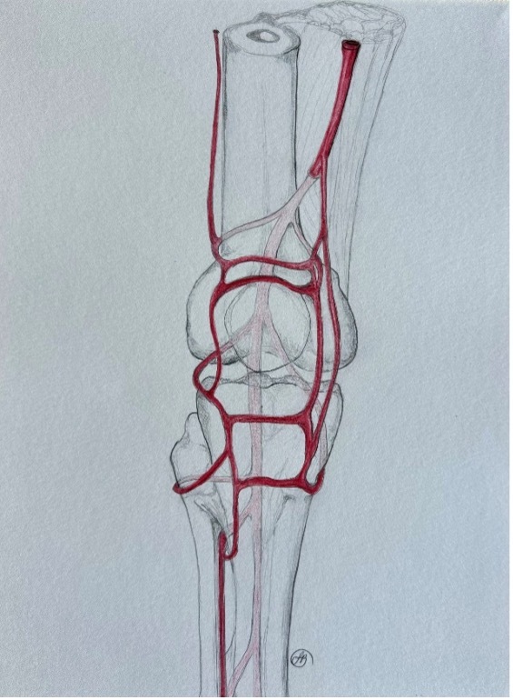
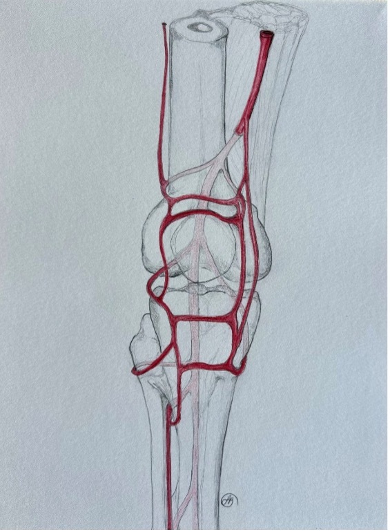
Figure 3
Figure 3.Illustration of vascular perforation zones around the distal femur and proximal tibia—areas being investigated as stereotyped stress risers in fracture propagation. Artwork supplied with the pilot manuscript.

Where I think this goes next

This technique will only become legitimate if other surgeons can reproduce it. The operation is straightforward, but the indications, implant design, degree of fragment preservation, and role of surface finish all need independent evaluation. Trauma-specific hinges should be designed around fracture anatomy rather than inherited wholesale from tumor reconstruction. Our work with engineers has focused on where fractures propagate and where vascular channels create recurring stress risers, with the goal of making the implants easier and safer to use in trauma. We also preserve the tibial bone in the same manor for native proximal tibia and peri-prosthetic fractures.

For now, my message is simple: do not discard viable bone merely because you have decided to replace the joint. When fixation is unlikely to survive poor metaphyseal bone, a hinge may be the better mechanical answer. But the best hinge, in my view, is one that keeps the patient’s own envelope. Preserve the attachments, restore the joint line, create a stable metal metaphysis, allow immediate mobilization, and let the biology work around the implant.

Submitted: August 21, 2026 EDT

Accepted: August 21, 2026 EDT

Attachments

Powered by Scholastica, the modern academic journal management system