
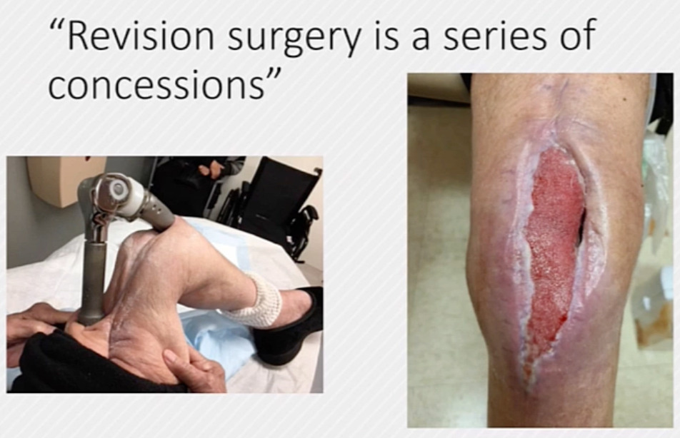
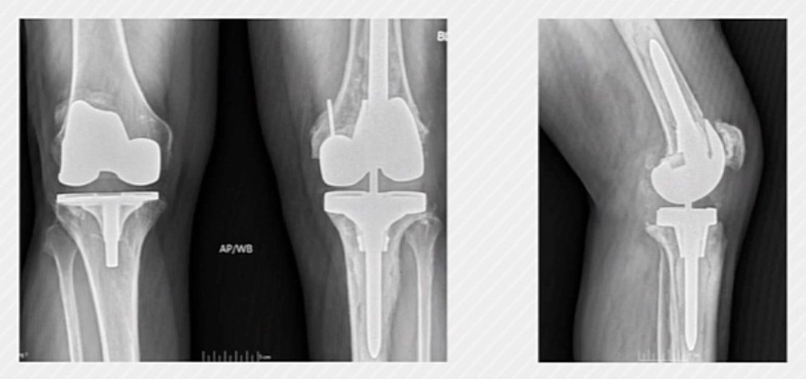
This article focuses primarily on metaphyseal fixation, but we will also discuss how to manage the bone defects encountered in revision surgery. When I trained with Dr. Meneghini, one of his well-known sayings was, “Revision surgery is a series of concessions.” I like to joke that clinic can sometimes feel like “Russian roulette”—you never know what you are going to encounter when you open the door. (Figure 1)
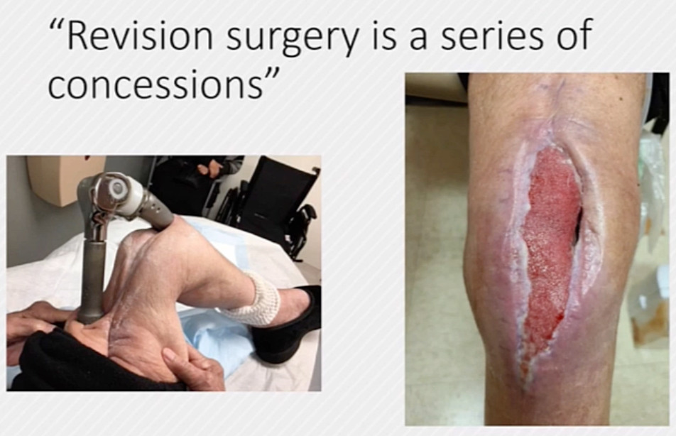
The Anderson classification system (Engh and Ammeen 1998) is familiar to most of us. Although I do not place too much emphasis on any single classification system, it is important to understand the progression from smaller defects to larger defects and, in the most severe cases, to compromise of the ligamentous and extensor mechanisms.
Anderson Classification System
1. Intact metaphyseal bone
2a. Metaphyseal bone defect involving only one femoral condyle or tibial plateau
2b. Metaphyseal bone defect involving both femoral condyles or tibial plateaus
3. Deficient metaphyseal bone involving the femur and tibia
I try to keep my approach to these cases simple. In revision total knee arthroplasty, our general goals are to provide good initial implant stability—particularly when using metaphyseal fixation to permit bony ingrowth—facilitate long-term implant survival through durable fixation, and provide stability to the knee joint.
Zonal fixation is an extremely important concept. We need fixation in at least two of the three zones; as bone loss becomes more severe, we often need to extend into zone three to establish durable fixation for a long-lasting construct.
I think it is important to start by asking: What do we have left to work with? This assessment begins in the clinic with the initial radiographs. We evaluate the extent of bone loss and perform a clinical examination to assess collateral ligament integrity and the extensor mechanism. Beyond zonal fixation, we also need to determine how we will provide rotational and axial stability to the components. That framework is what ultimately leads to a durable reconstruction.
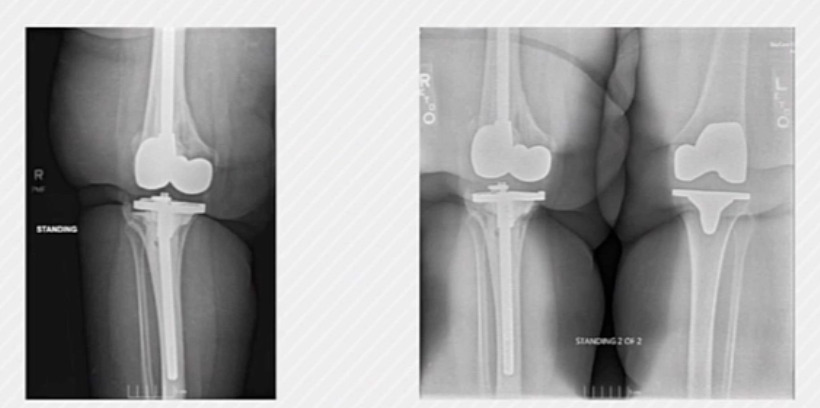
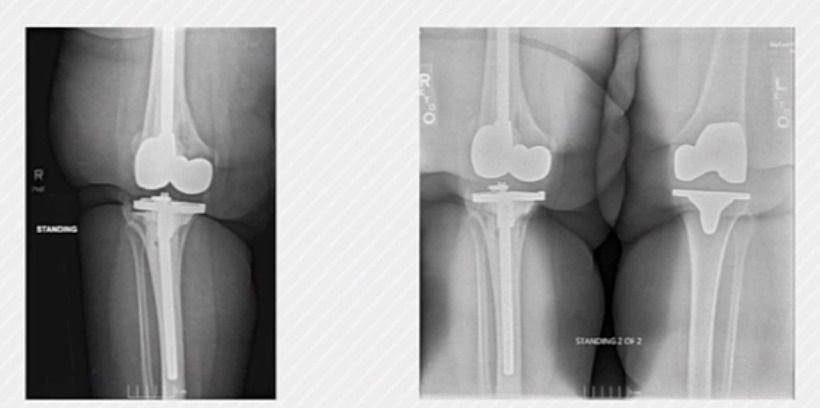
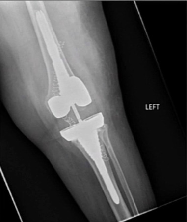
When these principles are not followed, cases like this show up in my clinic. This patient, who had a BMI of approximately 60 kg/m2, had a very long, well-cemented tibial stem but no proximal fixation, and the component ultimately fractured. (Figure 2) This was a difficult day in the operating room trying to remove it. With infection, severe obesity, and numerous prior surgeries, she now has a fusion nail functioning as a spacer two to three years later. Sometimes in revision surgery, you get one chance to get it right; when the basic principles are not followed, the consequences can be disastrous.
Starting with the most minimal defects, we still need axial and rotational stability. When bone loss is limited, that stability can often be obtained from the femoral box and keel and the tibial keel, without the need for additional adjunctive fixation.
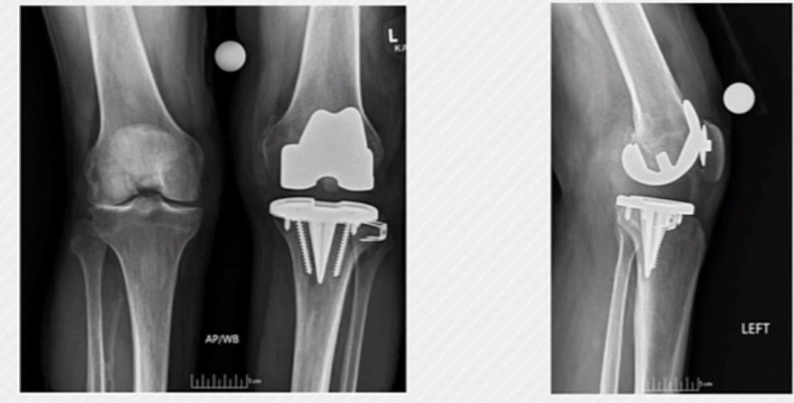
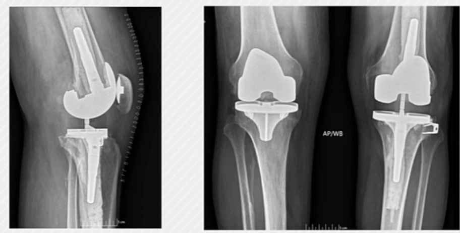
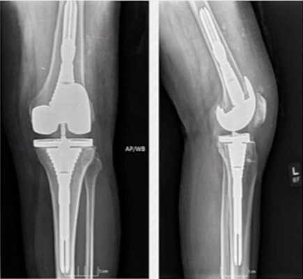
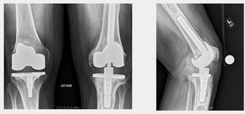
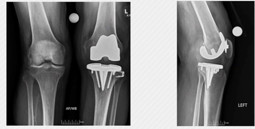
This case is an example of a patient who developed instability over time in a primary knee. (Figure 3) We did not need to add extensive metaphyseal fixation—just short cemented stems. We achieved good zone-one fixation and used augments to improve stability, without the need for more complex reconstructive techniques. (Figure 4)
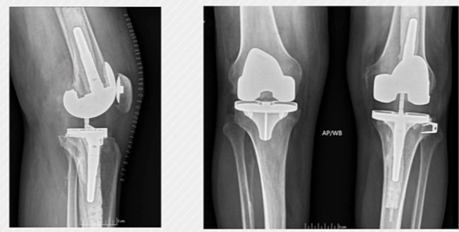
I think the indication for metaphyseal fixation has too often become, “We are doing a knee revision, so we are going to add metaphyseal fixation.” When there is good-quality bone, however, we do not need to remove healthy bone simply to add more metal.
For small, contained peripheral defects, cement alone can be used to fill the defect. For larger defects, screw-and-cement constructs can work very well. Ritter and colleagues demonstrated durable results with this technique (Berend et al. 2015). In their series, only one of 93 revision knees treated with screws failed at 17 years. Notably, these were patients with larger defects—worse problems, but excellent long-term outcomes.
Augments are useful for larger uncontained or unilateral defects and for restoring the joint line. Some augment systems also allow us to mate a smaller tibial tray with a larger femoral component. As tibial bone loss increases, component-size mismatches can become a problem. A wedge augment that allows a smaller tibial baseplate to accommodate the femoral size while still filling the flexion gap can be a very helpful tool. (Figure 5)
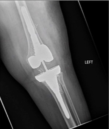
Posterior condylar bone loss is another important consideration. It is useful to have implants that can be posteriorized, or to use offset stems when needed, to appropriately fill the flexion gap. I was trained to be particularly mindful of flexion instability, so in revision cases I am always trying to restore the flexion gap and provide stability, including by posteriorizing the femoral component when appropriate.
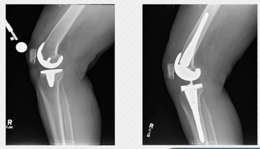
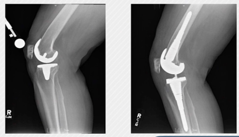
Here is a case in which we did exactly that. (Figure 6) The patient had instability and a suboptimal posterior condylar offset with the primary implant. We used posterior augments and upsized the femoral component, which restored excellent flexion-gap stability. The patient had a successful outcome.
Metaphyseal fixation has become very common, and I am often struck by how fortunate we are to be practicing orthopedics today. We have many tools that have made revision surgery much easier than it was 20 years ago. When we train fellows in Tampa, one of the questions they frequently ask is, “How do we know when to use these?” This is the framework I use. Large central defects are clearly situations in which cones and sleeves can be very beneficial. It is also important to understand the revision system you are working with. Implants with more minimal keels provide less rotational stability, particularly in larger defects. In those cases, metaphyseal fixation can be very helpful. Similarly, as we add tibial augments, we reduce the amount of keel-bone interface available, and adjunctive metaphyseal fixation may again be needed to provide rotational stability.
I view cones and sleeves as largely interchangeable tools. Metaphyseal fixation has been a game changer in revision surgery, and I have used it more frequently over time. The ability to obtain long-term bony ingrowth in zone two is a major advantage. Both cones and sleeves can provide axial and rotational stability and contribute to long-term construct durability.
Sleeves often provide a large surface area for ingrowth but generally require more bone removal or a larger defect to accommodate them. They use a tapered junction that connects the stem to the implant. Unfortunately, sleeves are usually implant-specific, which commits you to the associated revision system. In my practice, I use mostly cementless stems with sleeves, which can allow for a fully cementless construct.
Bloch et al. reported on 319 revision knees with more than two years of follow-up and demonstrated excellent survivorship through 10 years, with minimal radiolucent lines and subsidence (Bloch et al. 2020). When I use sleeves, one technical pearl is to prepare with a longer stem trial. (Figure 7) We can often use a shorter stem in the final construct, but sleeve preparation still needs to be referenced from the diaphysis. I generally use at least a 110-mm stem trial during sleeve preparation to ensure appropriate alignment. I also prefer to undersize the stem trial by one size so that as much stability as possible comes from the sleeve itself. Once the sleeve is prepared, a shorter stem can be used in the final construct when appropriate.
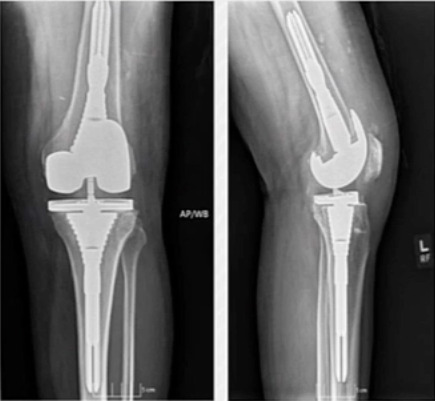
Moving on to cones, these provide another method of obtaining reliable metaphyseal ingrowth. Unlike sleeves, cones require cement for the implant construct. One advantage is that they can be implant-agnostic, allowing you to use the cone you prefer with the revision implant system you prefer.Cones can also fit into smaller defects and may require less bone removal. The stem passes through the cone, and the implant is cemented into the cone. In my practice, I have never been entirely comfortable with hybrid fixation because I want reliable cement pressurization into the cone while also obtaining secure stem fixation. Therefore, I tend to use a short cemented stem when I use cones. The Mayo group reported on 228 revision knees treated with porous tantalum tibial metaphyseal cones, with 97% 10-year survivorship free of cone removal, although only 58% were free of any reoperation (Hadley et al. 2024). These results highlight how difficult revision knee surgery can be. Even when we have excellent tools for obtaining fixation, reoperation rates remain high in this complex patient population. I personally find milling or burring systems easier to use than broaching, particularly in sclerotic bone. I also think we should not be afraid to stack cones in very large defects when doing so improves fixation. There is no need to feel obligated to use the same manufacturer’s cone and implant if another option works better.Another point worth considering is bone quality. In the United States, we have tended to move away from cement, despite how useful cemented fixation can be in hip arthroplasty. As more companies offer both sleeves and cones—and as cones allow more implant-system flexibility—a cone-and-cement construct may make particular sense in an osteoporotic patient. I become cautious when considering large sleeves and press-fit stems in an 85-year-old patient with osteoporotic bone.For stems, anything I can do to avoid a long cemented stem is beneficial. I have never been happy when I have had to revise a long, well-cemented stem. In my hands, short cemented stems are preferable to long press-fit stems in the appropriate construct. My first case using the LINK OptiStem (Waldemar Link GmbH & Co. KG, Hamburg, Germany) is in about two weeks, and I am excited to have it as another option for these challenging reconstructions. One final case brings these concepts together. This patient had a loose implant with severe bone loss, and we used several of the reconstructive options discussed above. (Figure 8)
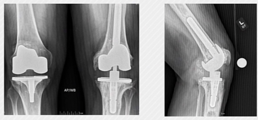
We used cones, screw-and-cement fixation, and the largest augments available to obtain stability. The patient had an excellent outcome and is now five to six years out from revision. (Figure 9)
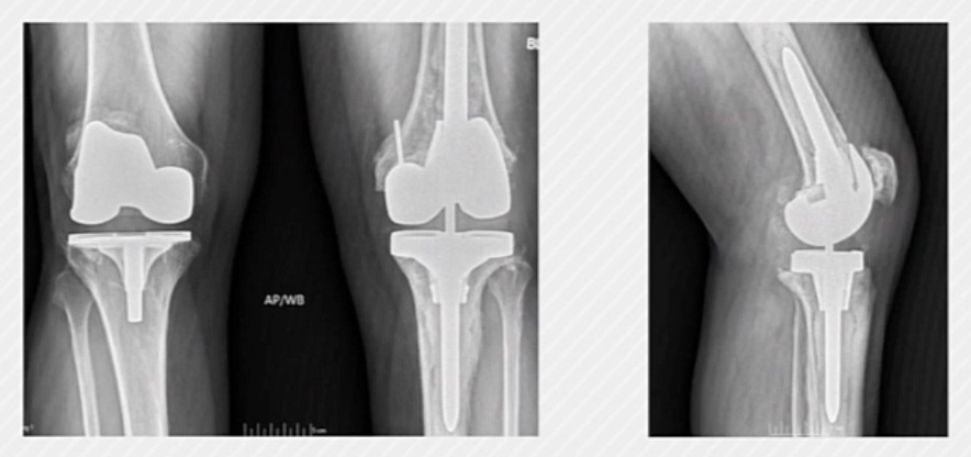
With type 3 defects—particularly massive bone loss or loss of a collateral ligament insertion—we may need to move to larger, more structural reconstructive options to compensate for the deficient bone.
In summary, both sleeves and cones provide additional fixation and an opportunity for bony ingrowth in revision knee arthroplasty. The fundamental goals remain the same: obtain fixation in at least two of the three zones and achieve both axial and rotational stability.