Loading [Contrib]/a11y/accessibility-menu.js

This website uses cookies

We use cookies to enhance your experience and support COUNTER Metrics for transparent reporting of readership statistics. Cookie data is not sold to third parties or used for marketing purposes.

Skip to main content
null
J Orthopaedic Experience & Innovation
  • Menu
  • Articles
    • Brief Report
    • Case Report
    • Case Series
    • Conference Proceedings
    • Data Paper
    • Editorial
    • Meeting Reports/Abstracts
    • Methods Article
    • Product Review
    • Research Article
    • Review Article
    • Review Articles
    • Systematic Review
    • All
  • For Authors
  • Editorial Board
  • About
  • Issues
  • Blog
  • "Open Mic" Topic Sessions
  • Advertisers
  • Recorded Content
  • CME
  • JOEI KOL Connect
  • Resident Research League
  • search
  • RSS feed (opens a modal with a link to feed)

RSS Feed

Enter the URL below into your favorite RSS reader.

https://journaloei.scholasticahq.com/feed
ISSN 2691-6541
Research Article
Vol. 7, Issue 2, 2026November 09, 2026 EDT

A Hyaluronic Acid-Based Patch for Use in Rotator Cuff Repair: Short-Term Clinical Exam-Based Outcomes

David A. Porter, M.D., William S. Pietrzak, Ph.D.,
rotator cuff tearrepairaugmentscaffoldhyaluronic acid
Copyright Logoccby-nc-nd-4.0

Articles in Vol. 7, Issue 2, 2026

Vol. 7, Issue 2, 2026
  • A Human Factors Analysis of Personal Electronic Device Use and Cognitive Distractions in Orthopedic Surgery
    Asfand KhanAlbert Boquet
  • Does Preoperative Suzetrigine Impact ASC Opioid Consumption For Total Joint Arthroplasty?
    Louis BattistaAndrew Wickline
  • Pickleball Pains: A 10-year Epidemiologic Analysis of Rising Upper Extremity Injuries
    Kevin ValdesAghdas MovassaghiJehad Feras AlSamhoriXiomara OrtizJocelyn LubertVani J. Sabesan
  • From Innovation to Inaccuracy: The Impact of ChatGPT on Orthopaedic Surgery Research Citations in Sports Medicine
    Calista StevensAlexander HahnGregory ConnorsShiraz MumtazMartinus MegallaZachary GraceJohn CorviMatthew PartanKatherine Coyner
  • Does CMS Hate Specialists?
    Benjamin Schwartz, MD
  • Voices in Orthopaedics™...The Residency Programs: The Unionized Orthopod: Apprenticeship, Labor, and the Changing Identity of Orthopedic Residency at Jefferson
    Eric R. TecceJalen N. BroomeTyler W. HenryGabriel I. Onor Jr.Daniel A. NemirovDaniel E. DavisJames J. Purtill
  • Cerclage Fixation in Total Hip Arthroplasty: Anatomy, Surgical Options and Clinical Outcomes
    Zuhdi AbdoZachary FullerThomas ChristensenAhmed Siddiqi
  • In My Experience™...Orthopaedics: Then, Now, and Tomorrow: Reflections on a Half Century of Change
    Richard Conn, MD
  • Genicular artery embolization for symptomatic knee osteoarthritis: A narrative review
    Junaid MakdaAhmed SiddiqiKhalid YousufOsman Ahmed
  • Simplifying Orthopedic Patient Education: A ChatGPT-Based Readability Analysis
    Mendel ShloushKlaudia GreerJonathan BruttiBrayden TolmanJacob RosenthalRafael Aldaya BourricaudyMarcia VarellaFernando Aran
  • Digital Fatigue and Orthopedic Injuries: The Postural Burden of the Digital Era - A Narrative Review
    Ambrose Loc NgoNiki Gharavi AlkhansariTruong HoRachana TadakamallaLinda NguyenJared Nichols
  • Concurrent Floating Hip and Open Knee Dislocation: Damage-Control Management
    Sara LowVladislav MuldiiarovMark AyzenbergGermanuel LandfairGene Shaffer
  • Higher Pain Catastrophizing Scores are Associated with Increased Pre-operative Anxiety in Ambulatory Hand Surgery that is not Impacted by Watching a High-Quality Pre-Operative Video
    Christopher G. LarsenMichael J. SayeghAmr TawfikCaroline ApriglianoChloe HeitingKate W. Nellans
  • MOTIV™ and the Next Frontier of Orthopaedic Evidence Generation: A New Model for Physician-Led Clinical Research
    John Mercuri, MDAndrew Wickline, MD
  • Evidence on Demand: OpenEvidence versus Established LLMs on the Orthopaedic In-Training Examination
    Asim A. KhanShaan S. LalvaniSam PourarbabLord J. HyeamangRyan M. LewZaamin B. HussainGregory P. NicholsonGrant E. Garrigues
  • Beyond BMI: Surgical Readiness in the Age of GLP-1 Therapy
    Thomas Meade, MDReza Jazayeri
  • From My Perspective… Medial or Lateral? The Native Knee Pivot Controversy and Its Implant Consequences
    Stephen Howell
  • Trends in Orthopedic Surgeons Signing Medicare Opt-Out Affidavits
    Thriaksh RajanAndre RevnewJoshua PortoMonish LavuComron SaifiAtul Kamath
  • Voices in Orthopaedics™...The Residency Programs: Training for the Future of Orthopaedic Surgery: Residents’ Perspective on the UT Austin Dell Medical School Orthopaedic Surgery Residency
    Cassidy ShieldsSemran ThamerAmanda SeymourAlec Giron
  • Preoperative GLP-1 Receptor Agonist Use Improves Thromboembolic, Infectious, and Wound-Related Outcomes Following Unicompartmental Knee Arthroplasty
    Zachary FullerManjot SinghAbhiram DawarJeremiah ThomasAlan H DanielsZuhdi E Abdo
  • Beyond the Breaking Point: Solutions for Burnout in Orthopaedic Surgery
    Aghdas MovassaghiCamryn McIntyreSamir SakariaMitchell J. ChristiansenJocelyn LubertMary MulcaheyVani Sabesan
  • Utilization of a 3D Printed Customized Knee Extender Device and An At-Home Video Rehabilitation Program on Patient Outcomes in Rural Areas Following ACL Surgery
    Kevin P. FeltzBrooklyn VanDerWoldeKyle CochranEleanor GibbonsBenjamin C. NoonanNathan Wm. Skelley
  • Reclaiming the Continuum of Care: A Physician-Owned Capital Platform for Aligning Care Delivery, Risk, and Financial Infrastructure
    Marty NicholsJohn MilesThomas Chambers
  • Feasibility and Early Experience of Custom Stemmed Tibial Trays in Revision Total Ankle Arthroplasty: A Case Series
    Grant M. ThomasKush S. ModyJoydeep BaidyaCorinne SommiDavid I. PedowitzSelene G. Parekh
  • Postoperative Outcomes by Timing of Total Hip and Knee Arthroplasty Following Coronary Revascularization
    Zachary FullerManjot SinghThomas ChristensenJeremiah J ThomasAlan H DanielsZuhdi E Abdo
  • Tibial Plafond Fractures and the Impact of Social Media Support Groups on Patient Perceptions
    Alexandra F. FlahertyDana PerimAnnie WaiteAlvarho GuzmanErnest N. Chisena
  • Retained Male Component After Intra-operative Dissociation of a Tibial Magnetic Lengthening Nail: A Case Report covering Technical Challenges and Management Strategies
    Eunice Anastasia WiliantoNeeraj MishraDerrick Jun Liang LamKenneth Pak Leung WongBenny Kai Guo LooAshik Mohammad
  • A physician led consensus building intervention at a multi-specialty practice results in meaningful reduction of opioid prescriptions for post-operative patients
    Jenna M. GodfreyJohn Paul BigouetteConnor FitzpatrickJohn W. OverHeather A. CampionErin C. Owen
  • Voices in Orthopaedics™...The Residency Programs...The Role of Networking and Interconnectivity in Building an Orthopedic Career
    Gabriel I. Onor Jr.Terence L. Thomas, MDDaniel Nemirov, MDJalen Broome, MDDaniel E. Davis, MDChristopher C. Dodson, MDJames J. Purtill, MD
  • From My Perspective… The Consent Discussion: The Robot Demands
    Stephen Howell
  • Changes in Oral and Gut Microbiome Diversity in Patients undergoing Total Joint Arthroplasty and Receiving Perioperative Antibiotics, a Pilot Case Series
    Mary Bayers-TheringNadir HaiderSonja PavlesenKaren FalknerMatthew Phillips
  • How to Assemble a Well-Fitting, Patient-Specific Antibiotic Cement Hip Spacer for Infection Management
    Ahmed Nageeb MahmoudNicholas BruleCatherine Mary DoyleGabriel MakarDaniel Horwitz
  • At The Conference™...How to Handle Severe Bone Defects in Knee Arthroplasty
    Christopher Grayson, MD
  • From My Perspective...Triple-Tapered Collared Stems: Are They All Equal—and How Have They Changed Primary Hip Arthroplasty?
    David Rodriguez-Quintana, MD
  • On How I Do It™...Attachment Preserving Hinge Arthroplasty vs. ORIF for Complex Periarticular Knee Fractures
    Michael Langworthy, MD
  • About The Innovation™...Arthrolense: Where the Idea Began
    Russell Nevins, MD
  • Voices in Orthopaedics™...The Residency Programs: From the University of Washington- The Paradox of Healing
    Sean PirkleKevin ZhaiSarah PirkleJessi ForeHoward Chansky
  • Subscapularis Repair in Reverse Shoulder Arthroplasty: Does Implant Design Matter?
    Nathan GilmoreAghdas MovassaghiAntonio Da CostaElisheva KnopfJacob CalpeyJocelyn LubertVani J. Sabesan
  • In My Experience™...Biointegrative Collagen Implant Use in Complex Hip and Knee Arthroplasty: Early Clinical Experience and Second-Look Observations
    Chris Hoedt, MD
  • The Process of Customization in Shoulder Arthroplasty: Advancements in 3D Design and Manufacturing
    Benjamin R. WesorickAghdas MovassaghiLana SmithLexi LewisVani J. Sabesan
  • From My Perspective... The Quadriceps Malalignment Question: No Evidence to Externally Rotate the Femoral Component in Kinematic Alignment
    Stephen Howell
  • The Case for Reforming VSLO Eligibility and ERAS Screening to Address the Declining Representation of US Citizen International Medical Graduates in Orthopaedic Surgery
    Jaxon S. BoleyRobert B. AtkinsonGarrett R. Jackson
  • Specialist-Led Care in Hand Surgery: Matching Clinical Complexity to the Future of Care Delivery
    Marney ReidBrian ChristieDamon AdamanyDana Jacoby
  • A Novel System for Validating 3D Mapping for Patients with Rotator Cuff Arthropathy
    Ashley IsenbergAman ChopraEliana S. LeMasterAnand M. MurthiMelissa A. WrightTyler Hwang
  • Value-Based Care: Orthopaedic Surgeons Are a Paradigm Shift Away From Enabling a Specialist-Led Care Model
    Marney ReidJason ScaliseRina C. VertesDana Jacoby
  • Force-Modulating Tissue Bridges in Sports Medicine Incision Management: A First 100-Case Experience Evaluating Allergic Reactions and Postoperative Complications
    John A GrottingEge Karadag
  • Concordance of ChatGPT with a Fellowship-Trained Orthopaedic Surgeon in Detecting Periprosthetic Osteolysis Following Total Shoulder Arthroplasty
    Colton C. MowersJustin T. ChildersAlexander L. HornungAmelia HummelArden ShenGregory P. NicholsonGrant E. Garrigues
  • A Hyaluronic Acid-Based Patch for Use in Rotator Cuff Repair: Short-Term Clinical Exam-Based Outcomes
    David A. PorterWilliam S. Pietrzak
  • Beyond MIC: Biofilm Eradication, MBEC, and Sustained Intra-articular Antibiotic Delivery in Periprosthetic Joint Infection
    Jared R.H. ForanPeter NoymerCharles De CookSpencer Albert
  • In My Experience™...Limb Salvage- What is Possible? From Total Femur Replacement to Rescue Sleeves
    John Stammers, MD
  • Zero by Zero: The Patient Supply Chain- Delivering Every Surgical Patient Prepared, Supported, and On Time
    Charles DeCook, MDAnuj Gupta, MDThomas Bradbury, MD
J Orthopaedic Experience & Innovation
Porter, David A., and William S. Pietrzak. 2026. “A Hyaluronic Acid-Based Patch for Use in Rotator Cuff Repair: Short-Term Clinical Exam-Based Outcomes.” Journal of Orthopaedic Experience & Innovation 7 (2).
Save article as...▾
Download all (12)
  • Figure 1. Integrity™ Implant System showing the patch, tissue tack, and bone staple (images not to scale).
    Download
  • Figure 2. Bone staple inserted through the patch using the caddy guide.
    Download
  • Figure 3. Deployment of tissue tack onto the rotator cuff by light mallet impaction.
    Download
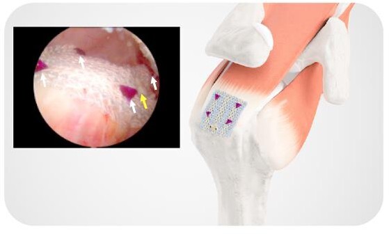
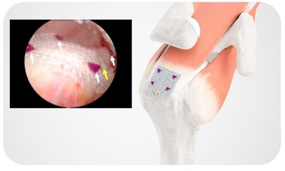
  • Figure 4. Completed repair with patch, bone staple, and four tissue tacks. Inset: Intraoperative photo of completed repair (white arrows showing tissue tacks and yellow arrow showing staple).
    Download
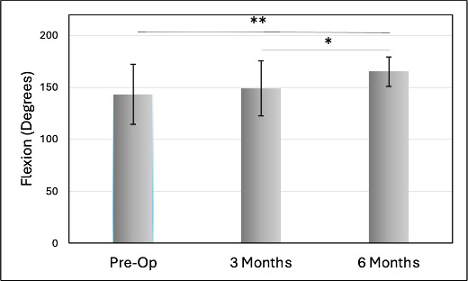
  • Figure 5. Flexion vs. interval for overall population. * P = 0.039, ** P = 0.0042
    Download
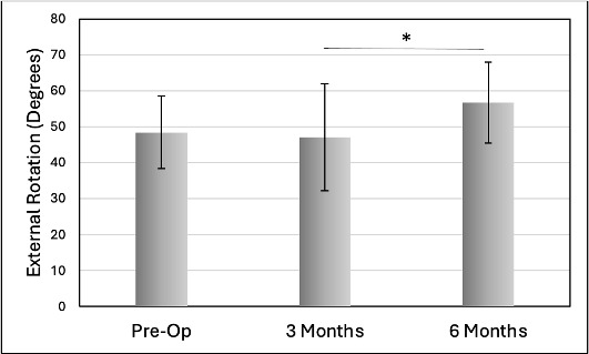
  • Figure 6. External rotation vs. interval for overall population. * P = 0.017
    Download
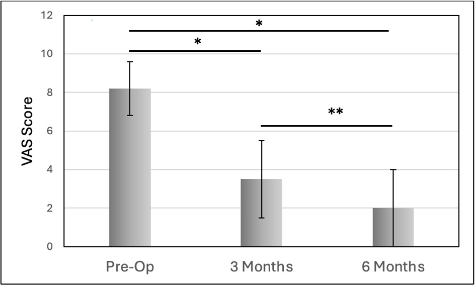
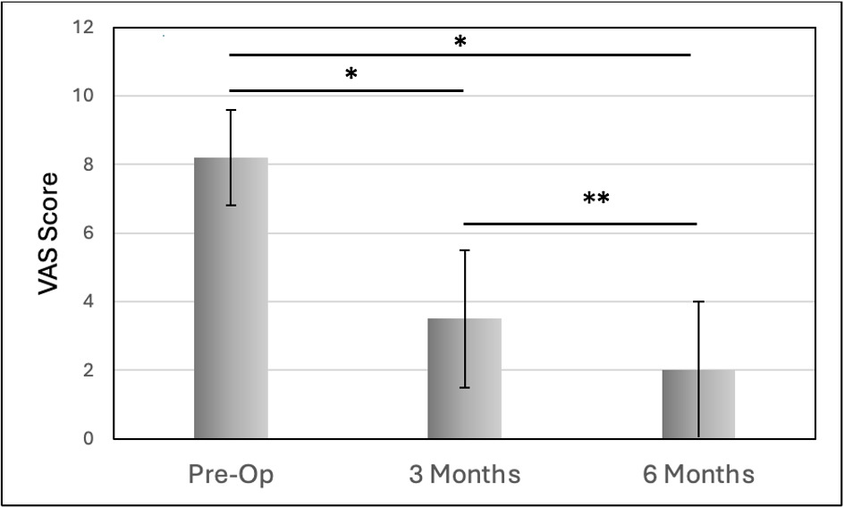
  • Figure 7. VAS pain score vs. interval for overall population. * P < 0.001, ** P = 0.015
    Download
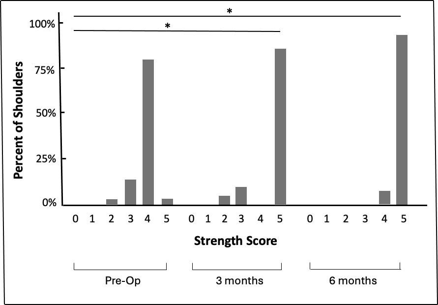
  • Figure 8. Percent of shoulders grouped by strength score and time interval. *P < 0.001.
    Download
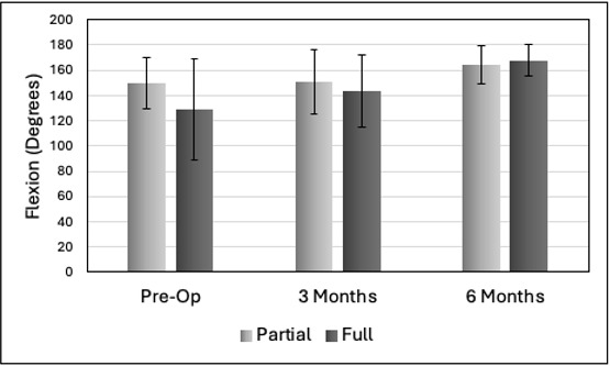
  • Figure 9. Flexion vs. interval for partial thickness tear and full thickness tear populations. No significant differences between the PTT and FTT groups.
    Download
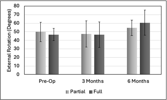
  • Figure 10. External rotation vs. interval for partial tear and full tear groups. No significant differences between the PTT and FTT groups.
    Download
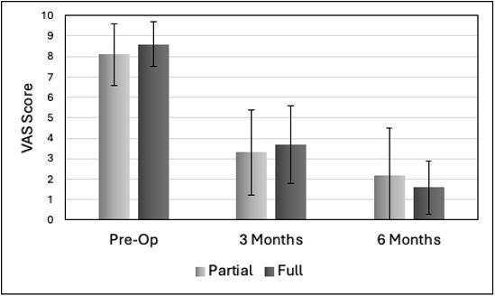
  • Figure 11. VAS pain score for partial tear and full tear groups. No significant differences between the PTT and FTT groups.
    Download
  • Figure 12. Percent of shoulders grouped by partial and full thickness tears, strength score, and time interval. *P < 0.001. **P = 0.014
    Download

Error

Sorry, something went wrong. Please try again.

If this problem reoccurs, please contact Scholastica Support

Error message:

undefined

View more stats

Abstract

Background

Rotator cuff tears are difficult to repair due to poor intrinsic healing ability and subsequent high retear rate. As such, patches have been developed to augment surgical repair and produce a local environment that is supportive of healing. While many have been clinically evaluated, the ideal patch has yet to be identified. Hyaluronic acid is ubiquitous in the musculoskeletal system and plays a significant role in the healing response through its cell signaling properties. The purpose of this study was to investigate the short-term clinical exam-based outcomes following rotator cuff repair utilizing a hyaluronic acid-based patch.

Methods

Twenty-nine patients with rotator cuff tears (mean age: 45.7 years - range 18-71 years; 14 male, 15 female) were treated with the Integrity™ Implant (Anika Therapeutics, Inc.). These patients were further divided into two subgroups by type of tear, i.e., partial tear (n=20) and full tear (n=9) which were statistically matched for gender (p = 0.782), BMI (p = 0.279), diabetic status (p = 0.124), and smoking status (p = 0.183) but with the full tear group (mean age 57.2 years) significantly older than the partial tear group (mean age 40.6 years) (p < 0.001). Thirteen partial tears were bursal side while 7 were articular side. Flexion, external rotation, VAS pain, and strength were evaluated at pre-op and at 3-month and 6-month post-op.

Results

Significant improvements in pain and function were obtained within 3-6 months. There were no significant differences in flexion, external rotation, or VAS pain between the partial and full tear groups from pre-op to 6 months. However, strength of the full tear group at pre-op and 3-month post-op was less than that of the partial tear group until reaching equivalence at 6 months. There was no clinical exam-based evidence of retear nor were any patch-related complications identified.

Conclusions

Rotator cuff repair utilizing augmentation with a hyaluronic acid-based patch demonstrated significant clinical improvement with no patch-associated complications or clinical evidence of retear. Furthermore, partial thickness and full thickness tear groups exhibited equivalent outcomes by 6 months. Longer term studies with MRI follow-up will further enhance understanding of the role of this patch in rotator cuff repair.

Introduction

Rotator cuff (RC) tears (RCT) are very common and often asymptomatic but over time can result in pain, disability, loss of shoulder function, and significant economic burden (Parikh et al. 2021). They are associated with age-related degenerative processes and other factors (Zhao et al. 2022). While partial thickness tears (PTT) and small full thickness tears (FTT) may benefit from conservative treatment, surgery to reattach the injured tendon to its enthesis is often required due to the limited intrinsic healing ability of the rotator cuff tendons (Tashjian 2012; Chalmers and Tashjian 2020).

The use of patches or scaffolds to mechanically strengthen the rotator cuff repair (RCR) and improve healing and minimize risk of retear is an active area of research (Cobb et al. 2022; Avanzi et al. 2019; Barber et al. 2012; Bryant et al. 2016; Cai et al. 2018; Iannotti et al. 2006). RC patches derived from autograft, allograft, xenograft, and synthetic materials have been used clinically with varying degrees of success (Cobb et al. 2022; Avanzi et al. 2019; Barber et al. 2012; Bryant et al. 2016; Cai et al. 2018; Iannotti et al. 2006). Currently, the ideal RC patch has not yet been identified.

A novel patch, the Integrity™ Implant System, (Anika Therapeutics, Bedford, MA), has recently been introduced for tendon repair procedures. This synthetic patch is a porous, flexible knitted construct of an absorbable benzyl ester of hyaluronic acid (HA) that increases the residence time of HA, reinforced with nonabsorbable polyethylene terephthalate (PET). The HA derivative takes advantage of the known wound healing properties of HA while PET enhances mechanical strength (Chang et al. 2024; Chen and Abatangelo 1999; Oliva et al. 2021; Yu et al. 2023). Moore et al (Moore et al. 2026) reported a short-term (6-month) clinical study utilizing this patch for onlay RC repair of PTT in 4 patients and augmentation repair of FTT in 6 patients finding significant improvements in the American Shoulder and Elbow Surgeons (ASES) score, the Single Assessment Numeric Evaluation (SANE) score, and the PROMIS Global Physical Health scores at 6 months. MRI evidence showed successful integration of the patch and healing of the repaired tendon. There were no complications or reoperations performed.

Our primary objective was to examine the short-term (6-month) clinical exam-based outcomes of this patch in the treatment of rotator cuff tears on a larger patient population than has previously been reported (Moore et al. 2026). Our secondary objective was to compare the outcomes of PTT and FTT subgroups due to little direct comparison in the literature with or without patch augmentation (Park et al. 2004; Warren et al. 2024).

Methods

This study was conducted according to principles outlined in the Declaration of Helsinki and Good Clinical Practice guidelines. This retrospective chart review clinical study protocol was approved by the WCG-IRB (Cary, NC USA) which waived consent to participate in accordance with the exclusions listed in US 45 CFR § 46. 104(d) (Chalmers and Tashjian 2020). All surgeries were performed by the lead author after an initial course of six weeks of formal physical therapy and anti-inflammatories.

Integrity™ implant system

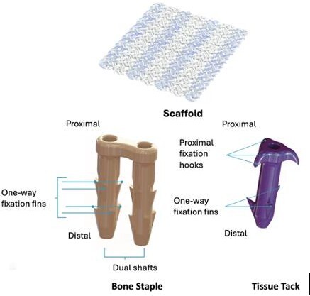
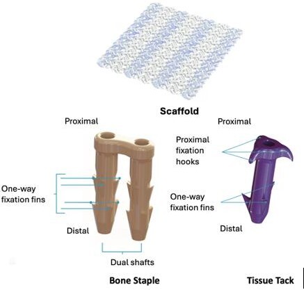
The Integrity™ Implant System includes the patch which is a porous, knitted composite of a benzyl ester derivative of hyaluronic acid that increases the residence time of HA and nonabsorbable PET that enhances the mechanical properties – Figure 1. The patch is available in multiple sizes with the 20 × 25 mm and 25 × 30 mm sizes used in this study for RCR. Also included are nonabsorbable poly ether ether ketone (PEEK) bone staples and absorbable 82% poly-L-lactic acid:18% poly glycolic acid copolymer tacks (7 mm and 8 mm) for fixing the patch to bone and tendon, respectively – Figure 1. The Integrity implant system obtained United States 510(k) clearance in 2023.
A diagram of a scaffolding AI-generated content may be incorrect.
Figure 1.Integrity™ Implant System showing the patch, tissue tack, and bone staple (images not to scale).

Surgical technique

Standard rotator cuff suturing was only performed for FTT cases and not PTT cases. Full-thickness tears were repaired arthroscopically using a standardized double-row repair construct. Anchor number and placement were selected according to tear size and tissue quality at the discretion of the operating surgeon while maintaining consistent repair principles across all cases. Following completion of the tendon repair, the HA implant was applied as an onlay augmentation in the manner described below.

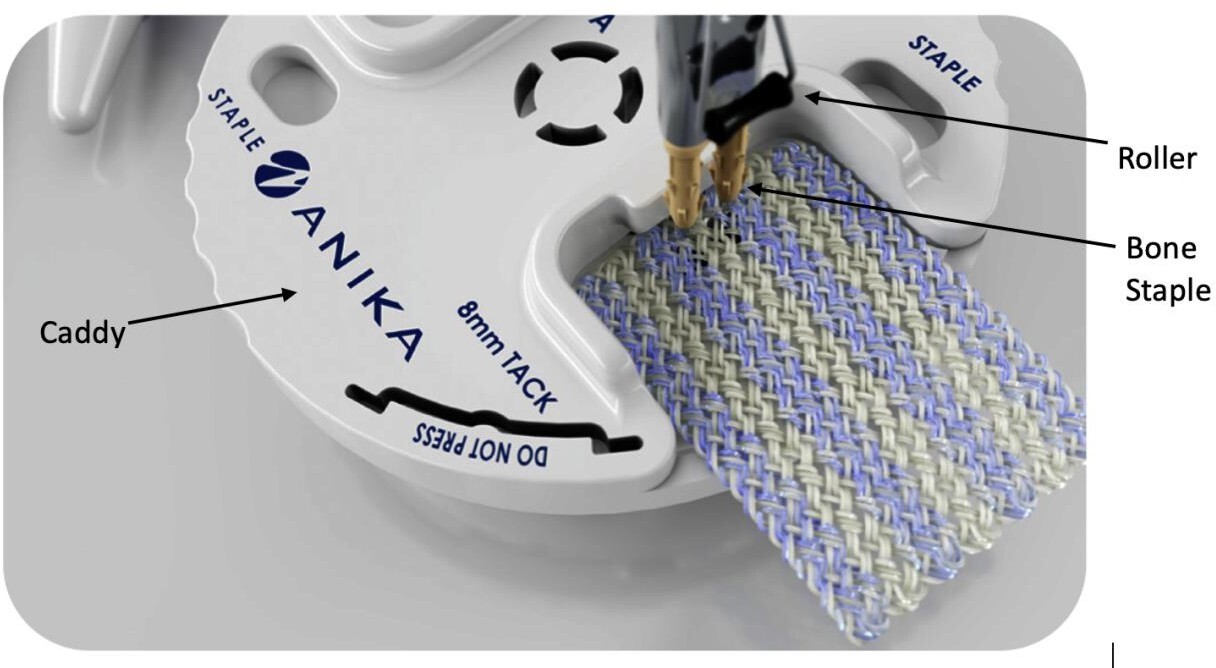
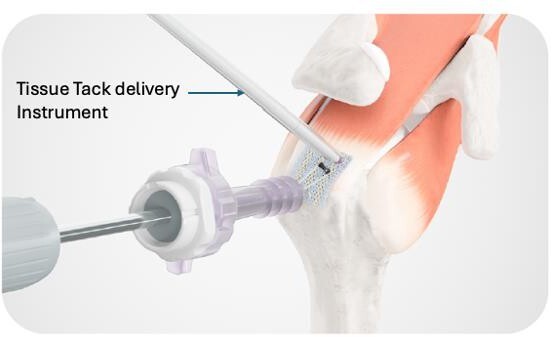
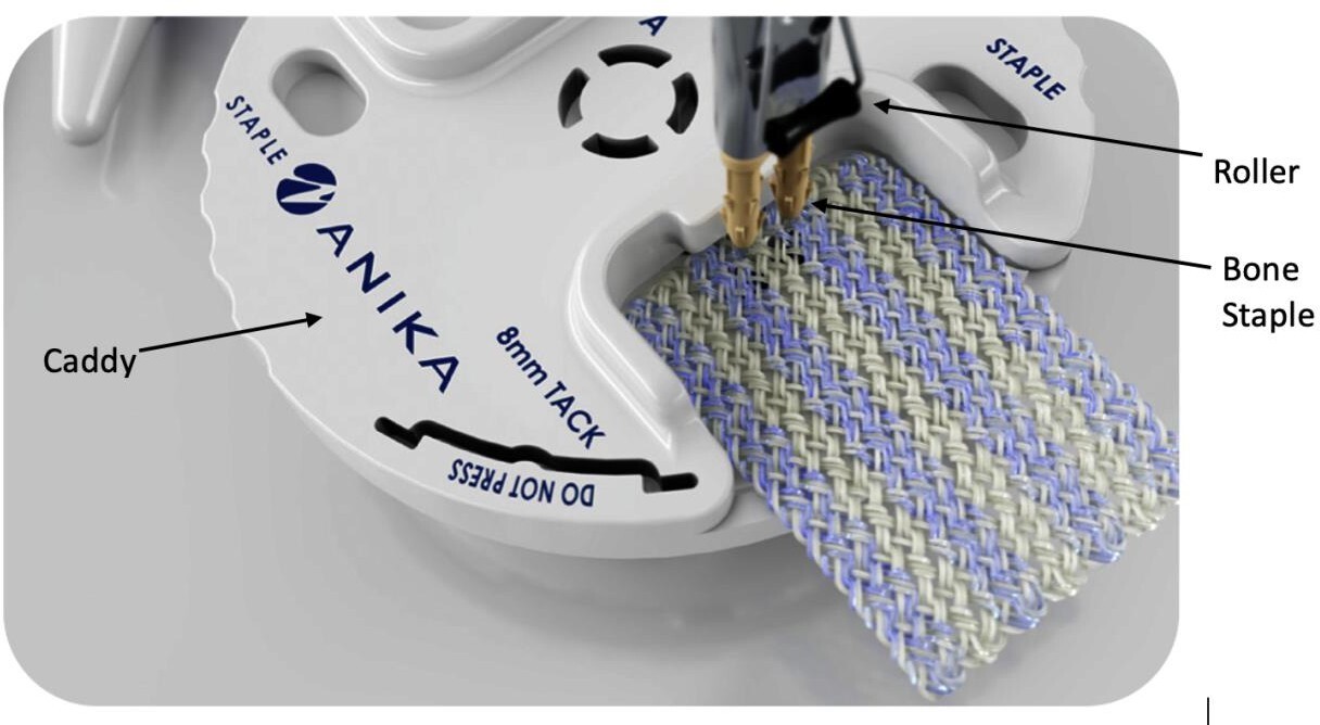
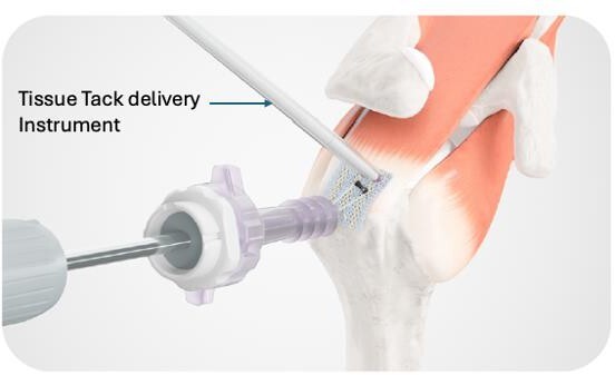
The patch was applied in identical fashion for both tear types. Viewing from the posterior portal, the subacromial space is entered. A lateral portal is established 2 cm distal to the lateral acromial tip and the 8.25 mm cannula is placed into the subacromial space. An accessory portal is established just lateral to the acromion. The distal tines of the bone staple delivery instrument are engaged with the bone staple (Figure 2). The patch is placed in the size-appropriate caddy guide with the staple inserted through the patch by manual pressure applied to the instrument, centrally positioning it approximately 4 mm from the short edge. Note that the bone staple orientation is perpendicular to the white and blue striations of the patch. The bone staple and patch assembly is then inserted into the lateral portal cannula and malleted to seat the staple at the appropriate lateral position on the greater tuberosity. A slider on the instrument handle controls longitudinal movement of a distal roller while rotation of the proximal end of the handle controls its transverse position to flatten the entire patch medially over the rotator cuff. Through the accessory portal, the tissue tacks are inserted through a protective sheath and delivered to the desired location on the patch. Gentle mallet taps are performed to fix the patch to the rotator cuff (Figure 3). Four tacks are deployed in this manner to secure the patch and complete the repair (Figure 4). Finally, the roller is retracted and the instrumentation removed. The operative site is then closed per standard protocol.
A close-up of a sewing machine AI-generated content may be incorrect.
Figure 2.Bone staple inserted through the patch using the caddy guide.
A close-up of a medical device AI-generated content may be incorrect.
Figure 3.Deployment of tissue tack onto the rotator cuff by light mallet impaction.
A close-up of a bone AI-generated content may be incorrect.
Figure 4.Completed repair with patch, bone staple, and four tissue tacks. Inset: Intraoperative photo of completed repair (white arrows showing tissue tacks and yellow arrow showing staple).

Postoperative protocol

Postoperatively after PTT repair there was 1) an initial 1 to 2-day period of immobilization, 2) passive and active-assisted range of motion (ROM) exercises to 2 weeks, 3) early strengthening exercises from 2 to 6 weeks to restore full, pain-free ROM and reactivate the rotator cuff and scapular stabilizers, and 4) advanced strengthening and functional rehabilitation (weeks 6–12), to enhance shoulder strength, endurance, and neuromuscular control, progressing toward sport- or work-specific activities.

The postoperative protocol following FTT repair included 1) a period of 4 weeks of mobilization in a sling, 2) beginning physical therapy at 2 weeks for passive range of motion, and 3) initiating strength, strengthening, and active-assisted and active motion at 6 weeks.

Clinical study

The goals of this retrospective study were 1) to investigate the short-term clinical exam-based outcomes of rotator cuff repair using the Integrity™ Implant System and 2) to compare outcomes between PTT and FTT cohorts. A consecutive series of patients underwent surgery to repair PTT and FTT of the rotator cuff from November 2023 to May 2024 with follow-up performed at 3 months and 6 months, postoperative. Follow-up consisted of measurement of shoulder flexion, external rotation, visual analog score (VAS) pain (0-10 scale), and strength. Strength was measured per Kleyweg et al (Kleyweg et al. 1991) as follows: 0 – No muscle contraction or movement, 1 – a trace of movement or flicker, 2 – movement at the joint, but not against gravity, 3 – movement against gravity, but not against added resistance, 4 – movement against gravity with some resistance, and 5 – normal strength or power.

Statistical analysis

Comparison of two groups of continuous data was performed with the 2-tailed Student t test. One-way analysis of variance (ANOVA) was used for multiple continuous data comparisons followed by a post hoc Tukey test. Categorical data was compared with the Fisher exact test with multiple comparisons utilizing the Bonferroni correction. Significance was taken for p < 0.05.

Results

The overall patient population was nearly evenly distributed among males (n=14) and females (n=15) with no significant difference between the PTT and FTT groups of 20 and 9 shoulders, respectively (p = 0.782) – Table 1. The overall patient age was 45.7±13.5 years, with the FTT group (57.2±8.6 years) significantly older than the PTT group (40.6±12.1 years) (p < 0.001). Overall BMI was 28.1±4.9 kg/m2 with no significant difference between the two groups (PTT: 27.4±5.0 kg/m2, FTT: 29.6±4.6 kg/m2, p = 0.279). Likewise, there were no significant differences between the distribution of patients having diabetes (p = 0.124) or their smoking status (p = 0183) between the two groups.

Table 1.Patient Demographics
Parameter Value (mean ± SD)
No. Patients 29
  • Partial thickness tear
20
  • Full thickness tear
9
Age (years), average (range): All patients 45.7±13.5 (range: 18-71)
  • Partial thickness
40.6±12.1 (range: 18-69) P < 0.001
  • Full thickness tear
57.2±8.6 (range: 43-71)
Sex, Male (Female): All patients 14 (15)
  • Partial thickness tear
10 (10) P = 0.782
  • Full thickness tear
4 (5)
BMI (kg/m2): All patients 28.1±4.9 (range: 19-39)
  • Partial thickness tear
27.4±5.0 (range: 19-39) P = 0.279
  • Full thickness tear
29.6±4.6 (range: 24-37)
Diabetes: All patients Yes: 5, No: 24
  • Partial thickness tear
Yes: 2, No: 18 P = 0.124
  • Full thickness tear
Yes: 3, No: 6
Smoking Status: All patients Yes: 8, No: 21
  • Partial thickness tear
Yes: 7, No: 13 P = 0.183
  • Full thickness tear
Yes: 1, No: 8

PTT vary widely but in this study were moderate and high-grade partial tears with greater than 50% articular or bursal surface tearing. Supraspinatus FTT were 1-2 cm in width and were classified as Goutallier grade 1 or 2 based on preoperative MRI. No patients demonstrated significant atrophy of the muscle.

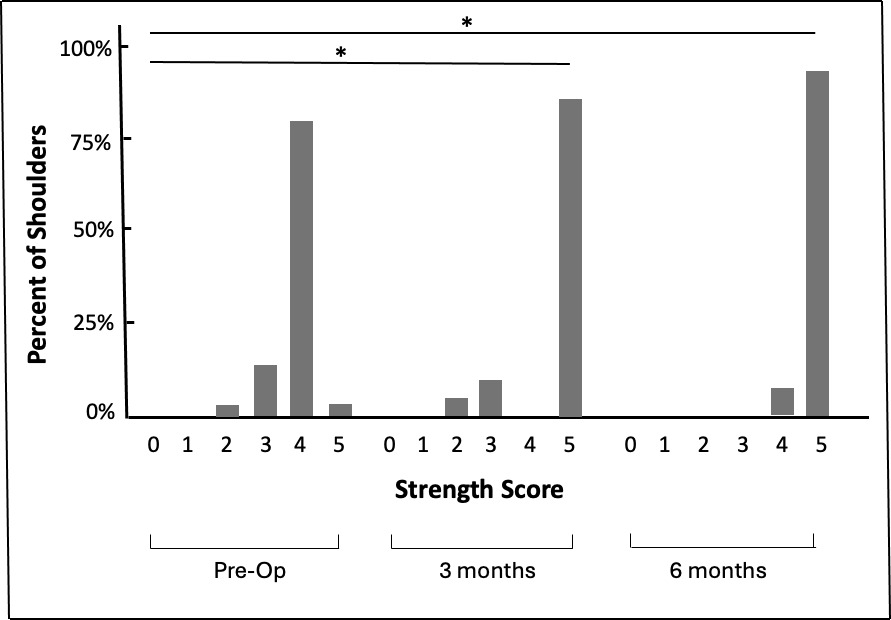
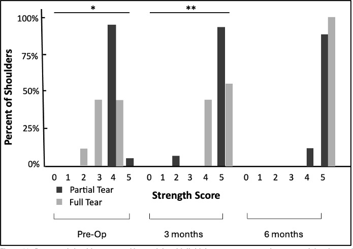
Table 2 lists the numerical results and Figures 5-12 illustrate the outcomes. Among the overall group of 29 patients there was no significant increase in flexion from pre-op to 3 months (∆=5.4°, p = 0.702), but the increases from pre-op to 6 months (∆=21.8°, p=0.0042) and from 3 months to 6 months (∆=16.4°, p=0.039) were significant (Figure 5). While external rotation was greater at 6 months than at pre-op (∆=7.9°, p=0.057), this difference was not significant, however the increase from 3 months to 6 months was significant (∆=9.5°, p=0.017) (Figure 6). There was a significant decrease in VAS score from pre-op to 3 months (∆=-4.7, p < 0.001) and pre-op to 6 months (∆=-6.2, p < 0.001) as well as from 3 months to 6 months (∆=-1.5, p < 0.015) (Figure 7). There was significant improvement in strength from pre-op to 3 months (p < 0.001) and pre-op to 6 months (p < 0.001) but no significant change from 3 months to 6 months (p = 0.853) with the percentage of patients exhibiting normal strength or power being 3.4%, 85.7%, and 92.6% at pre-op, 3 months, and 6 months, respectively (Figure 8)
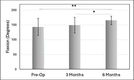
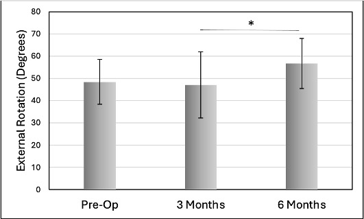
Table 2.Patient outcomes numerical outcomes.
Patient group Outcome Pre-Op 3 Months 6 Months
All 29 patients Flexion (°) 143.4±28.9 148.8±26.3 165.2±14.1
External rotation
(°)
48.8±10.1 47.1±14.9 56.7±11.3
VAS Pain (0-10) 8.2±1.4 3.5±2.0 2.0±2.0
Strength Score 0 0.0% 0.0% 0.0%
1 0.0% 0.0% 0.0%
2 3.4% 4.8% 0%
3 13.8% 9.5% 0%
4 79.3% 0% 7.4%
5 3.4% 85.7% 92.6%
Partial thickness tear
20 patients
Flexion (°) 150.0±20.1 150.9±25.9 163.9±15.1
External rotation
(°)
49.8±11.3 47.5±15.4 54.7±9.0
VAS Pain (0-10) 8.1±1.5 3.3±2.1 2.2±2.3
Strength Score 0 0.0% 0.0% 0.0%
1 0.0% 0.0% 0.0%
2 0.0% 7.1% 0.0%
3 0.0% 0.0% 0.0%
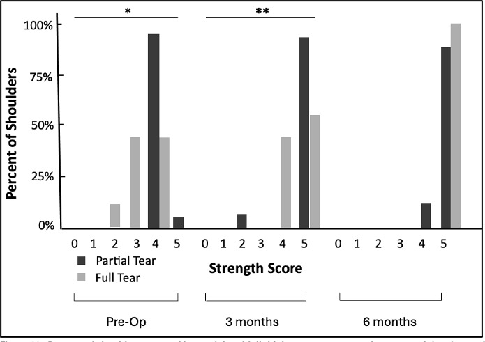
4 95.0% 0.0% 11.1%
5 5.0% 92.9% 88.9%
Full thickness tear 9 patients Flexion (°) 128.9±40.1 143.6±28.5 167.8±12.3
External rotation
(°)
46.7±7.1 46.4±14.9 60.6±14.7
VAS Pain (0-10) 8.6±1.1 3.7±1.9 1.6±1.3
Strength Score 0 0.0% 0.0% 0.0%
1 0.0% 0.0% 0.0%
2 11.1% 0.0% 0.0%
3 44.4% 0.0% 0.0%
4 44.4% 44.4% 0.0%
5 0.0% 55.6% 100.0%
A graph of a graph showing different stages of growth AI-generated content may be incorrect.
Figure 5.Flexion vs. interval for overall population. * P = 0.039, ** P = 0.0042
A graph of a graph showing different stages of growth AI-generated content may be incorrect.
Figure 6.External rotation vs. interval for overall population. * P = 0.017
A graph of a graph showing different stages of growth Description automatically generated with medium confidence
Figure 7.VAS pain score vs. interval for overall population. * P < 0.001, ** P = 0.015
A graph of strength and growth AI-generated content may be incorrect.
Figure 8.Percent of shoulders grouped by strength score and time interval. *P < 0.001.

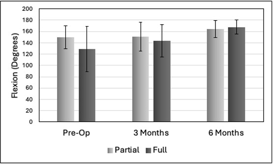
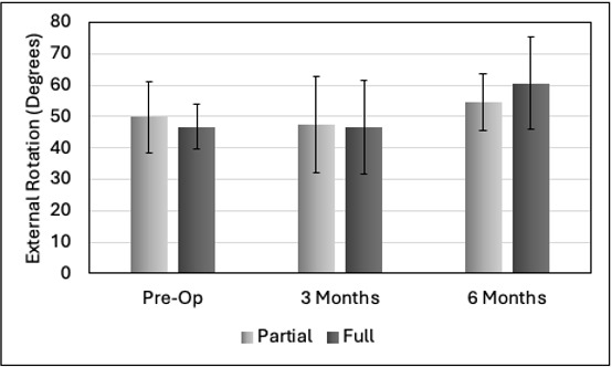
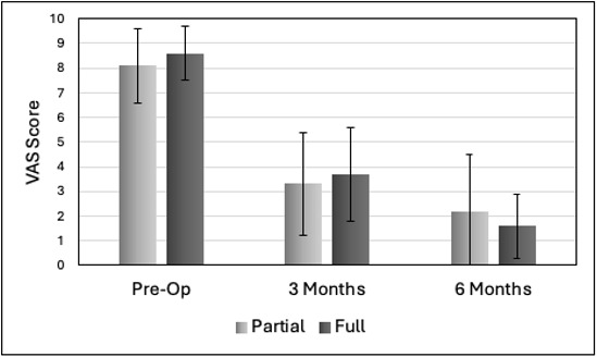
Flexion, external rotation, and VAS pain outcomes for the PTT and FTT groups are compared in Figures 9-11, respectively. The p values for the PTT and FTT comparisons of flexion, external rotation, and VAS pain at pre-op were 0.0675, 0.459, and 0.335; at 3 months were 0.548, 0.881, and 0.687; and at 6 months were 0.510, 0.211, and 0.429, respectively. As such, there were no significant differences among these values at a given time interval with respect to the type of tear. Figure 12 compares strength values for the two groups. The greatest improvement in strength for the PTT group occurred during the first 3 months and for the FTT group during the 3- to 6-month interval. At pre-op and 3-month follow-up, the strength was significantly less for the FTT group compared to the PTT group (p = 0.0019 and 0.0142, respectively). However, there were no significant strength differences between the two groups at 6 months (p = 0.537).
A graph of different colored bars AI-generated content may be incorrect.
Figure 9.Flexion vs. interval for partial thickness tear and full thickness tear populations. No significant differences between the PTT and FTT groups.
A graph of different levels of growth AI-generated content may be incorrect.
Figure 10.External rotation vs. interval for partial tear and full tear groups. No significant differences between the PTT and FTT groups.
A graph of different levels of growth AI-generated content may be incorrect.
Figure 11.VAS pain score for partial tear and full tear groups. No significant differences between the PTT and FTT groups.
A graph of strength and score AI-generated content may be incorrect.
Figure 12.Percent of shoulders grouped by partial and full thickness tears, strength score, and time interval. *P < 0.001. **P = 0.014

Complications

Four patients (13.8%, 4/29) developed adhesive capsulitis, including three with partial-thickness tears and one with a full-thickness tear - one at 2 weeks, two at 6 weeks, and one at 12 weeks. There was no significant difference between the 11.1% (1/9) and 15% ( 3/20) rates for the FTT and PTT groups, respectively (p = 1.000). All patients responded successfully to corticosteroid injection without requiring additional surgical intervention. One patient sustained a postoperative traumatic injury resulting in biceps tendon failure that required revision surgery; this complication was considered unrelated to the rotator cuff repair construct or implant. No implant-related complications, including infection, inflammatory reaction, fixation failure, implant migration, or foreign body response—were observed. Furthermore, no patients demonstrated clinical findings suggestive of recurrent rotator cuff failure during the follow-up period.

Discussion

This clinical study examined the short-term clinical exam-based outcomes of RCR surgery utilizing an HA-based patch. Key findings were that the overall patient population showed significant improvements in ROM, pain, and strength by 3-6 months. There were no significant differences in ROM and pain between the PTT and FTT groups at pre-op, 3 months, or 6 months, suggesting that there was no influence of the tear type on these outcomes. However, strength at pre-op and 3 months was significantly less in the FTT group than the PTT group but with no significant difference at 6 months. This suggests that 6 months was required for the strength of the FTT group to ‘catch up’ to that of the PTT group which may reflect the greater tendon disruption in the former with additional time for healing required to restore strength. While this is a reasonable interpretation of our subgroup analysis, the comparison was underpowered resulting in this being hypothesis generation. A systematic review and meta-analysis by Warren et al (Warren et al. 2024) of a bovine collagen patch used for RCR compared outcomes between PTT and FTT subgroups. Complication rates were similar between the two groups but the FTT group was associated with higher retear and revision rates than the PTT group while shoulder stiffness rates were higher in the PTT group. As such, their study provides a substantially more robust analysis than ours. Finally, Park et al (Park et al. 2004) treated 22 cases of PTT and 20 cases of FTT with traditional arthroscopic repair (no patch) that included subacromial decompression. No differences were found in functional recovery or reduction in pain between the two groups over 2-year follow-up with the most substantial improvements occurring during the first six months after surgery.

Adhesive capsulitis, or frozen shoulder, was the only post-operative complication in our study with an overall rate of occurrence of 13.8% (4/29), and 11.2% (1/9) and 15% (3/20) in the FTT and PTT groups, respectively (p = 1.000). This occurs in 5% to 32% of shoulder surgeries and can be primary with a contributive medical history, secondary to shoulder surgery, or idiopathic (Aim et al. 2022). The occurrence in our study was well within that reported in the literature and appeared to be unrelated to use of the patch. We found no clinical exam-based evidence of retear in our study population, however we cannot rule out asymptomatic tears (Longo et al. 2021). An important finding of this series was the absence of complications attributable to the HA-based implant itself such as inflammatory reaction, implant migration, fixation failure, or foreign body response. Although the study was not powered to evaluate uncommon adverse events, these early findings support the short-term safety profile of this implant and are consistent with the previously published pilot experience reported by Moore et al (Moore et al. 2026).

Like our study, Moore et al (Moore et al. 2026) was limited to 6-month follow-up and included PTT and FTT patients (unstratified) but had the advantage of multiple patient reported outcome measures (PROMs) and MRI at follow-up. They found significant improvement in the PROMs with MRI demonstrating tendon healing and patch integration by 6 months and no complications or reoperations. Using the same patch for RCR, we also found significant improvement in ROM, pain, and strength by 6 months with no complications attributable to the device and the only reoperation unrelated to the index RC procedure. Thus, there is overall congruence between Moore et al (Moore et al. 2026) and our study with respect to improvement in function and reduction in pain by 6-month follow-up. While it is reasonable to surmise that tendon healing and patch integration in our study were similar to Moore et al (Moore et al. 2026) via MRI, we cannot support this with direct evidence.

Among the RC patches currently available, the study patch is unique in that it is based on HA. HA can be chemically modified via esterification to create hydrogel scaffolds with the physio-chemical properties, stability, and half-life suitable for specific applications (Chang et al. 2024). As such flexible knitted structures hydrolyze, HA is released with an extended half-life (Milella et al. 2002). HA participates in wound healing and tissue regeneration through its action as a signaling molecule that binds to cell surface receptors (Chang et al. 2024; Chen and Abatangelo 1999; Oliva et al. 2021). Fetal wound healing is characterized by a lack of fibrous scarring with the HA concentration in fetal wounds remaining elevated longer than in adult wounds (weeks vs. days) (Chen and Abatangelo 1999; Longaker et al. 1991). Consequently, prolonged application of HA at the wound site in adults may modulate healing by making the local environment more fetal-like (Chen and Abatangelo 1999; Longaker et al. 1991). Several clinical studies have investigated periarticular injections of HA for rotator cuff tendinopathy with positive results (Oliva et al. 2021). The HA component of the Integrity implant has also been used to treat full- and partial-thickness chondral defects in the knee (Buda et al. 2019; Gobbi et al. 2017). As such, a strong theoretical foundation exists to support the use of an HA-based patch for RCR.

One strength of this study is that it adds to the limited body of previously reported clinical evidence for this unique HA-based RC patch. Another is that it compares outcomes between PTT and FTT subgroups which helps fill a notable void in the literature. There were, however, several limitations. First, this was a clinical exam-based study and no post-operative MRI outcomes were available. As such, tendon healing, integration of the patch, and the existence of asymptomatic retears were not directly assessed. Second, there were no study arms comparing the Integrity™ implant with RCR using either no patch or a different patch. Third, although most healing, pain improvement, and functional gain are known to occur during the first 6 months after RCR surgery, follow-up to at least one year would be required to better understand the relevance of earlier outcomes. Fourth, although diabetes mellitus and smoking status were recorded, other patient-specific biological factors such as thyroid disease, inflammatory arthropathy, and prior shoulder surgery were not consistently captured and therefore could not be incorporated into the present analysis. Future studies that are comparative, with MRI analysis, 1-2 year follow-up, and a more complete description of patient biological factors that could influence outcomes would enable a better understanding of the utility of this patch for rotator cuff repair.

Conclusions

In this retrospective case series, augmentation of rotator cuff repair with a hyaluronic acid-based implant was associated with significant improvements in pain, strength, and shoulder function during the first six postoperative months without implant-related complications. Comparable short-term outcomes were observed between partial- and full-thickness tear cohorts; however, these subgroup comparisons should be considered hypothesis-generating because of the limited sample size. Larger prospective comparative studies incorporating MRI assessment and longer follow-up are needed to define the role of this implant in rotator cuff repair.

Submitted: May 04, 2026 EDT

Accepted: August 30, 2026 EDT

References

Aim, F., R. Chevallier, B. Marion, S. Klouche, C. Bastard, and T. Bauer. 2022. “Psychological Risk Factors for the Occurrence of Frozen Shoulder after Rotator Cuff Repair.” Orthop Traumatol Surg Res 108 (2): 103212. https:/​/​doi.org/​10.1016/​j.otsr.2022.103212.
Google Scholar
Avanzi, P., L. D. Giudici, A. Capone, G. Cardoni, G. Lunardi, G. Foti, et al. 2019. “Prospective Randomized Controlled Trial for Patch Augmentation in Rotator Cuff Repair: 24-Month Outcomes.” J Shoulder Elbow Surg 28 (10): 1918–27. https:/​/​doi.org/​10.1016/​j.jse.2019.05.043.
Google Scholar
Barber, F. A., J. P. Burns, A. Deutsch, M. R. Labbe, and R. B. Litchfield. 2012. “A Prospective, Randomized Evaluation of Acellular Human Dermal Matrix Augmentation for Arthroscopic Rotator Cuff Repair.” Arthroscopy 28 (1): 8–15. https:/​/​doi.org/​10.1016/​j.arthro.2011.06.038.
Google Scholar
Bryant, D., R. Holtby, K. Willits, R. Litchfield, D. Drosdowech, A. Spouge, et al. 2016. “A Randomized Clinical Trial to Compare the Effectiveness of Rotator Cuff Repair with or without Augmentation Using Porcine Small Intestine Submucosa for Patients with Moderate to Large Rotator Cuff Tears: A Pilot Study.” J Shoulder Elbow Surg 25 (10): 1623–33. https:/​/​doi.org/​10.1016/​j.jse.2016.06.006.
Google Scholar
Buda, R., M. Baldassarri, L. Perazzo, D. Ghinelli, and G. Pagliazzi. 2019. “A Useful Combination for the Treatment of Patellofemoral Chondral Lesions: Realignment Procedure plus Mesenchymal Stem Cell-Retrospective Analysis and Clinical Results at 48 Months of Follow-Up.” Eur J Orthop Surg Traumatol 29 (2): 461–70. https:/​/​doi.org/​10.1007/​s00590-018-2310-z.
Google Scholar
Cai, Y. Z., C. Zhang, R. L. Jin, T. Shen, P. C. Gu, X. J. Lin, et al. 2018. “Arthroscopic Rotator Cuff Repair With Graft Augmentation of 3-Dimensional Biological Collagen for Moderate to Large Tears: A Randomized Controlled Study.” Am J Sports Med 46 (6): 1424–31. https:/​/​doi.org/​10.1177/​0363546518756978.
Google Scholar
Chalmers, P. N., and R. Z. Tashjian. 2020. “Patch Augmentation in Rotator Cuff Repair.” Curr Rev Musculoskelet Med 13 (5): 561–71. https:/​/​doi.org/​10.1007/​s12178-020-09658-4.
Google Scholar
Chang, W., L. Chen, and K. Chen. 2024. “The Bioengineering Application of Hyaluronic Acid in Tissue Regeneration and Repair.” Int J Biol Macromol 270 (Pt 2): 132454. https:/​/​doi.org/​10.1016/​j.ijbiomac.2024.132454.
Google Scholar
Chen, W. Y., and G. Abatangelo. 1999. “Functions of Hyaluronan in Wound Repair.” Wound Repair Regen 7 (2): 79–89. https:/​/​doi.org/​10.1046/​j.1524-475X.1999.00079.x.
Google Scholar
Cobb, T. E., R. A. C. Dimock, S. D. Memon, P. Consigliere, S. Ajami, M. Imam, et al. 2022. “Rotator Cuff Repair With Patch Augmentation: What Do We Know?” Arch Bone Jt Surg 10 (10): 833–46. https:/​/​doi.org/​10.22038/​ABJS.2022.61345.3012.
Google Scholar
Gobbi, A., C. Scotti, G. Karnatzikos, A. Mudhigere, M. Castro, and G. M. Peretti. 2017. “One-Step Surgery with Multipotent Stem Cells and Hyaluronan-Based Scaffold for the Treatment of Full-Thickness Chondral Defects of the Knee in Patients Older than 45 Years.” Knee Surg Sports Traumatol Arthrosc 25 (8): 2494–501. https:/​/​doi.org/​10.1007/​s00167-016-3984-6.
Google ScholarPubMed CentralPubMed
Iannotti, J. P., M. J. Codsi, Y. W. Kwon, K. Derwin, J. Ciccone, and J. J. Brems. 2006. “Porcine Small Intestine Submucosa Augmentation of Surgical Repair of Chronic Two-Tendon Rotator Cuff Tears. A Randomized, Controlled Trial.” J Bone Joint Surg Am 88 (6): 1238–44. https:/​/​doi.org/​10.2106/​00004623-200606000-00010.
Google Scholar
Kleyweg, R. P., F. G. van der Meche, and P. I. Schmitz. 1991. “Interobserver Agreement in the Assessment of Muscle Strength and Functional Abilities in Guillain-Barre Syndrome.” Muscle Nerve 14 (11): 1103–9. https:/​/​doi.org/​10.1002/​mus.880141111.
Google Scholar
Longaker, M. T., E. S. Chiu, N. S. Adzick, M. Stern, M. R. Harrison, and R. Stern. 1991. “Studies in Fetal Wound Healing. V. A Prolonged Presence of Hyaluronic Acid Characterizes Fetal Wound Fluid.” Ann Surg 213 (4): 292–96. https:/​/​doi.org/​10.1097/​00000658-199104000-00003.
Google ScholarPubMed CentralPubMed
Longo, U. G., A. Carnevale, I. Piergentili, A. Berton, V. Candela, E. Schena, et al. 2021. “Retear Rates after Rotator Cuff Surgery: A Systematic Review and Meta-Analysis.” BMC Musculoskelet Disord 22 (1): 749. https:/​/​doi.org/​10.1186/​s12891-021-04634-6.
Google ScholarPubMed CentralPubMed
Milella, E., E. Brescia, C. Massaro, P. A. Ramires, M. R. Miglietta, V. Fiori, et al. 2002. “Physico-Chemical Properties and Degradability of Non-Woven Hyaluronan Benzylic Esters as Tissue Engineering Scaffolds.” Biomaterials 23 (4): 1053–63. https:/​/​doi.org/​10.1016/​S0142-9612(01)00217-4.
Google Scholar
Moore, A., R. Patel, H. Cardona, V. Sochor, E. Cohen, and C. Baker. 2026. “MRI and Patient Reported Outcomes of a Hyaluronic Acid Based Rotator Cuff Augmentation Implant: A Pilot Case Series.” J Orthop Experience Innovation 7 (1): 10. https:/​/​doi.org/​10.60118/​001c.157512.
Google Scholar
Oliva, F., E. Marsilio, G. Asparago, A. Frizziero, A. C. Berardi, and N. Maffulli. 2021. “The Impact of Hyaluronic Acid on Tendon Physiology and Its Clinical Application in Tendinopathies.” Cells 10 (11). https:/​/​doi.org/​10.3390/​cells10113081.
Google ScholarPubMed CentralPubMed
Parikh, N., D. J. Martinez, I. Winer, L. Costa, D. Dua, and P. Trueman. 2021. “Direct and Indirect Economic Burden Associated with Rotator Cuff Tears and Repairs in the US.” Curr Med Res Opin 37 (7): 1199–211. https:/​/​doi.org/​10.1080/​03007995.2021.1918074.
Google Scholar
Park, J. Y., K. T. Chung, and M. J. Yoo. 2004. “A Serial Comparison of Arthroscopic Repairs for Partial- and Full-Thickness Rotator Cuff Tears.” Arthroscopy 20 (7): 705–11. https:/​/​doi.org/​10.1016/​S0749-8063(04)00598-5.
Google Scholar
Tashjian, R. Z. 2012. “Epidemiology, Natural History, and Indications for Treatment of Rotator Cuff Tears.” Clin Sports Med 31 (4): 589–604. https:/​/​doi.org/​10.1016/​j.csm.2012.07.001.
Google Scholar
Warren, J. R., E. R. Domingo-Johnson, A. A. Sorensen, A. L. Cheng, K. H. Latz, and A. Cil. 2024. “Bioinductive Patch as an Augmentation for Rotator Cuff Repair, a Systematic Review and Meta-Analysis.” J Shoulder Elbow Surg 33 (11): 2515–29. https:/​/​doi.org/​10.1016/​j.jse.2024.05.002.
Google Scholar
Yu, C., S. Feng, Y. Li, and J. Chen. 2023. “Application of Nondegradable Synthetic Materials for Tendon and Ligament Injury.” Macromol Biosci 23 (12): e2300259. https:/​/​doi.org/​10.1002/​mabi.202300259.
Google Scholar
Zhao, J., M. Luo, G. Liang, J. Pan, Y. Han, L. Zeng, et al. 2022. “What Factors Are Associated with Symptomatic Rotator Cuff Tears: A Meta-Analysis.” Clin Orthop Relat Res 480 (1): 96–105. https:/​/​doi.org/​10.1097/​CORR.0000000000001949.
Google Scholar

Attachments

Powered by Scholastica, the modern academic journal management system