
Introduction
Soft-tissue deficiency is a recurring problem in complex primary and revision total joint arthroplasty. Multiple operations, extensile exposures, scar, infection, tendon failure, and tissue loss can leave a mechanically sound reconstruction with an incomplete or tenuous soft-tissue envelope. The consequences may include persistent dead space, drainage, wound breakdown, exposure of synthetic material or prosthetic components, instability, and infection.
Extensor mechanism disruption after total knee arthroplasty illustrates this problem particularly well. Chronic or tissue-deficient disruption often requires allograft or synthetic mesh reconstruction, yet failure, infection, extensor lag, and reoperation remain substantial despite contemporary reconstruction strategies. Systematic reviews and multicenter series have reported broadly similar outcomes for synthetic mesh and allograft, with failure rates remaining clinically important at intermediate and longer-term follow-up (Bates and Springer 2015; Browne and Hanssen 2011; Abdel et al. 2018; Buller et al. 2020; Richardson et al. 2024; Anderson et al. 2023; Gencarelli et al. 2023; Shau et al. 2018).
Tapestry® is a resorbable, acellular scaffold composed of aligned type I bovine collagen and poly(D,L-lactide) (PDLLA). In the applications described here, the underlying tendon repair, mesh reconstruction, capsular repair, or other mechanical reconstruction is completed first; Tapestry is then used as an adjunct over residual areas in which native soft-tissue coverage remains attenuated. The clinical question is therefore not whether Tapestry can replace a reconstruction, but whether it can provide a useful biologic coverage layer when the surgeon has otherwise completed the reconstruction and still has inadequate tissue coverage.
Other commercially available bioinductive collagen scaffolds have been studied primarily in rotator cuff surgery. The most extensively studied porous type I bovine collagen implant has prospective multicenter and randomized trial data demonstrating increased tendon thickness and, in some settings, lower retear rates; however, these data address tendon healing rather than revision arthroplasty soft-tissue coverage. Published evidence for other commercially available bioinductive matrices remains limited, and Tapestry-specific clinical evidence is sparse outside the shoulder arthroplasty setting (Iban et al. 2026; Bushnell et al. 2021; Ruiz Ibán et al. 2024; Maghdouri-White et al. 2021; Nathani 2022). These studies provide biologic context for the present observations but should not be considered direct evidence for Tapestry use in arthroplasty.
The purpose of this report is to describe the use of Tapestry in difficult revision hip and knee arthroplasty cases and, more importantly, to share observations when selected treated areas were subsequently re-exposed. This report emphasizes practical indications, the appearance of the tissue at second look, and the limitations of these early observations.
Clinical Applications and Practical Technique
1. Complete the definitive reconstruction first. Implant revision, tendon repair, anchor fixation, mesh reconstruction, capsular approximation, and any required flap procedure should be performed according to the underlying pathology. Tapestry is not load-bearing and should not compensate for an inadequate mechanical repair.
2. Mobilize scar and maximize viable native-tissue coverage without excessive tension. When direct closure is possible, I favor native tissue closure. Tapestry is most useful when a limited residual area remains inadequately covered despite reasonable mobilization. A large defect, threatened skin envelope, exposed prosthesis, or defect requiring vascularized tissue should prompt consideration of plastic-surgery coverage or another reconstructive strategy.
3. After hemostasis and removal of loose or nonviable tissue, trim and orient Tapestry to cover the residual defect with broad contact and overlap onto viable tissue. Orient the blue lines along the anticipated direction of tensile stress. I avoid folding the implant or placing it under tension. The goal is a simple onlay that remains in contact with the underlying repair and adjacent viable tissue.
4. Secure the implant with interrupted sutures around the perimeter or at the corners. When needed, I add a central figure-of-eight suture to maintain apposition and limit tenting or fluid accumulation beneath the implant. Fixation is intended to hold the scaffold in place rather than add mechanical strength.
5. Postoperative mobility limitations are dictated by the underlying reconstruction. Range-of-motion restrictions, weight bearing, bracing, hip precautions, and progression of activity should follow the tendon, mesh, abductor, or capsular repair rather than the collagen implant.
Knee Applications
Extensor mechanism reconstruction with polypropylene mesh
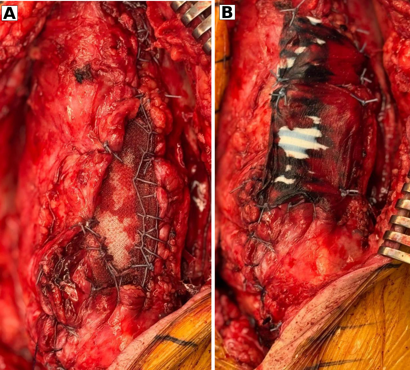
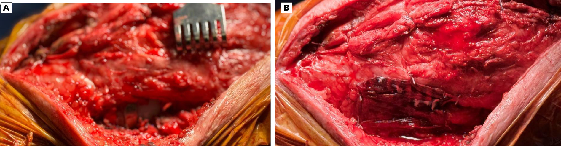
For catastrophic extensor mechanism deficiency, a Marlex mesh reconstruction (Browne and Hanssen 2011; Abdel et al. 2018; 2019) can be used, with the mesh serving as the load-bearing construct. Mobilizing the remaining quadriceps and retinacular tissues as much as possible and closing those tissues over the mesh is important. If the remaining soft tissue is attenuated or does not provide complete coverage, Tapestry can be used as an onlay over the reconstructed segment, extending onto viable tissue proximally and distally (Figure 1).
This distinction is important: Tapestry supplements the soft-tissue envelope; it is not part of the mechanical extensor mechanism reconstruction. Published Marlex literature supports the mechanical role of polypropylene mesh, but contemporary series demonstrate that failure and reoperation remain common, with 5-year survivorship free of revision of approximately 52% in one recent cohort (Buller et al. 2020; Richardson et al. 2024; Anderson et al. 2023; Gencarelli et al. 2023; Shau et al. 2018). No clinical comparative data show that adding a collagen scaffold improves mesh survivorship or extensor function.
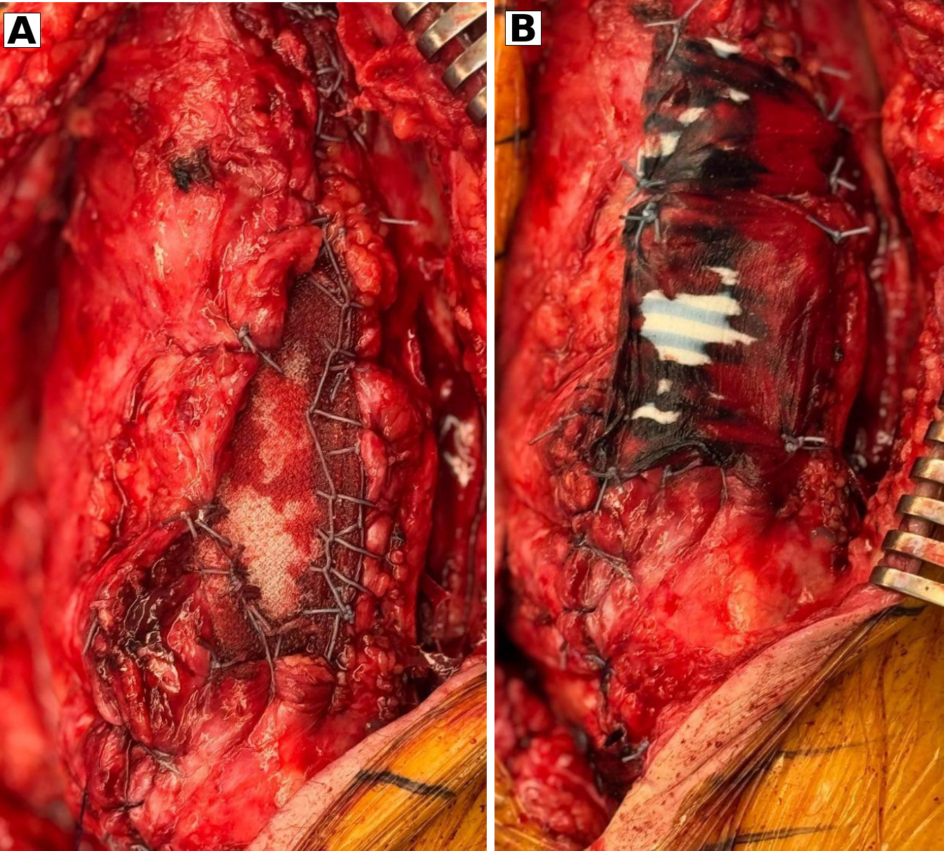
Two reoperations were informative but are not comparative evidence. In one earlier mesh reconstruction performed without Tapestry, erosive failure was followed by re-exposure of mesh that appeared poorly incorporated into the adjacent tendon. In a separate Tapestry-covered mesh reconstruction later explanted because of periprosthetic joint infection, the mesh and overlying tissue were densely adherent and difficult to separate. These observations raised the question that motivated this report: whether the additional collagen scaffold could contribute to a thicker, more substantial soft-tissue layer over an otherwise completed reconstruction.
Primary extensor mechanism repair
When a primary suture or anchor repair can restore tendon continuity without substantial segmental tissue loss, Tapestry can be used as an onlay without mesh. Complete and tension the tendon repair first, then secure the implant over the repaired quadriceps or patellar tendon with overlap onto viable tissue. Tapestry remains an adjunct to the completed repair, not the mechanical reconstruction itself.
Proximal tibial deep soft-tissue deficiency
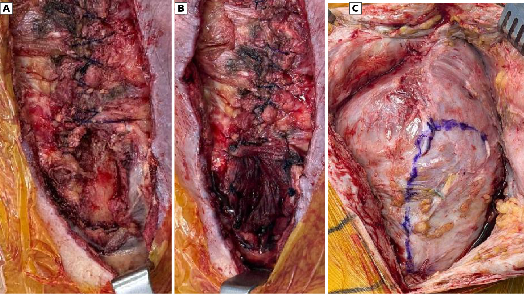
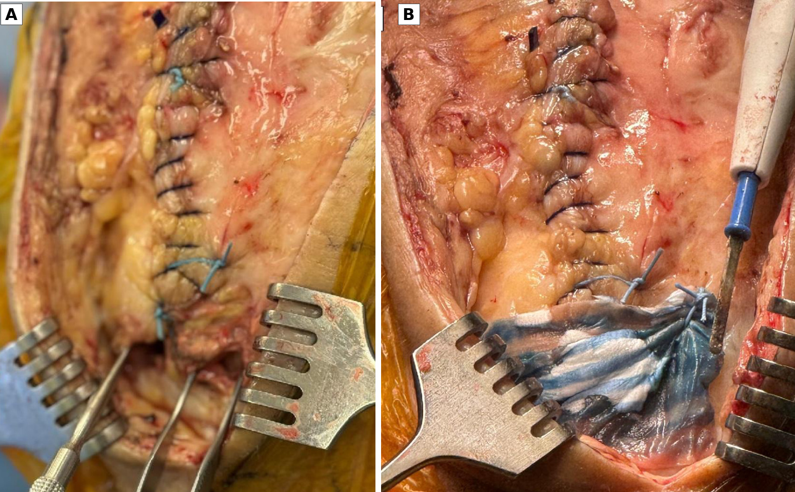
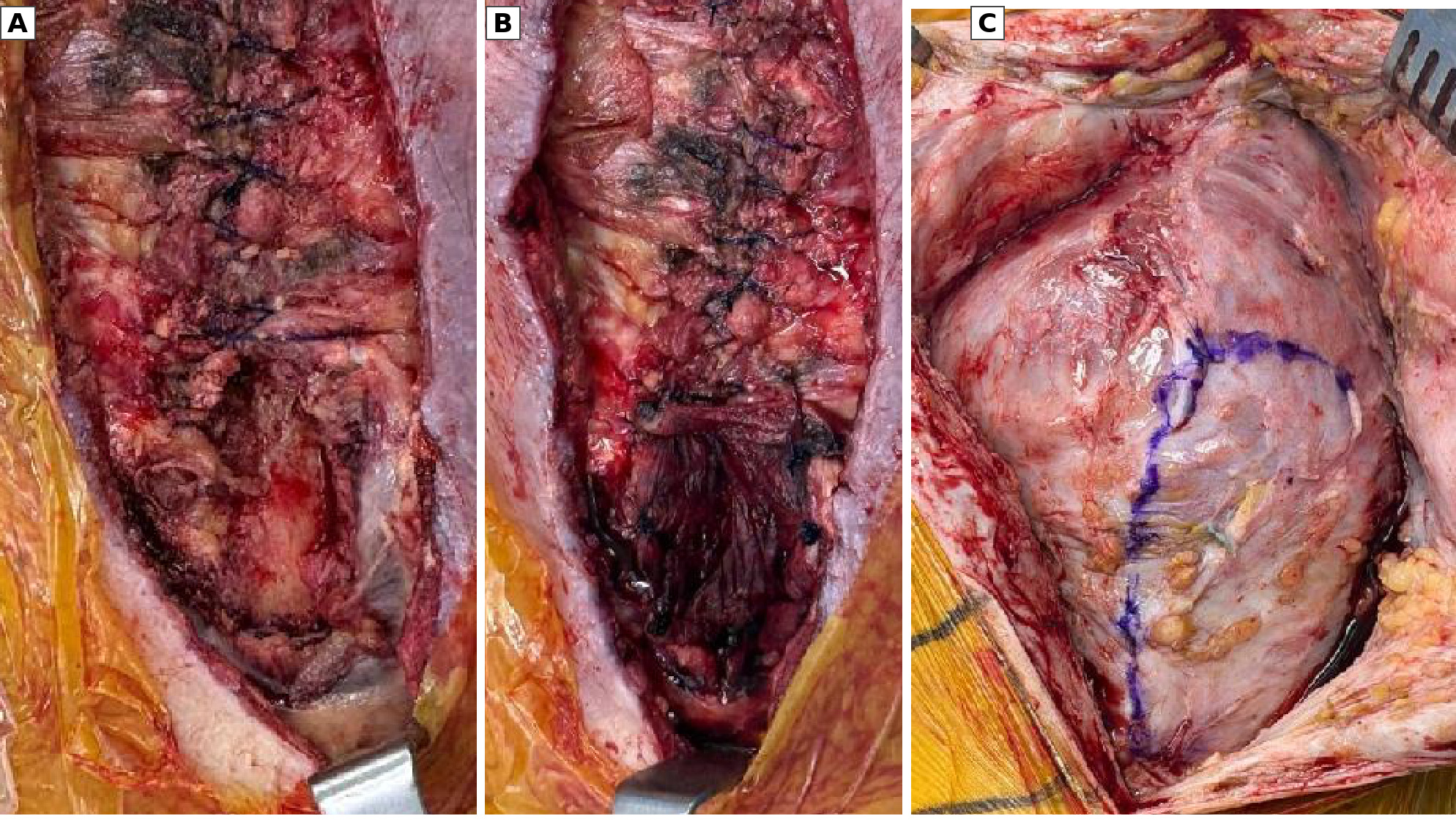
The proximal medial tibia can be vulnerable after repeated revision exposure or extensive debridement. When the capsule, retinaculum, or periosteal layer is attenuated, complete deep closure may not be possible, even with mobilization of surrounding tissue. For a limited residual defect with viable margins and an acceptable superficial envelope, Tapestry can be trimmed and oriented to span the deep deficiency and secured to the surrounding tissue (Figures 2 and 3).
Lateral retinacular/capsular deficiency
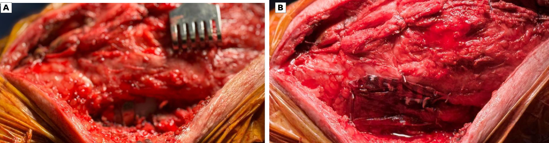
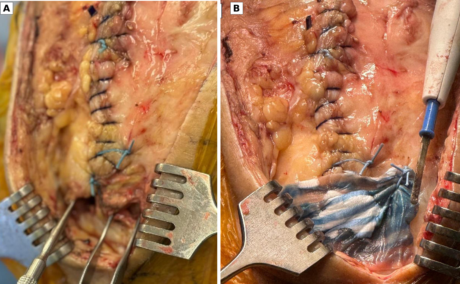
During revision total knee arthroplasty for patellar maltracking or instability, a lateral release may be necessary even when available tissue is compromised. The priority is a secure arthrotomy closure without sacrificing the medial repair to force a tenuous lateral closure. After reasonable lateral approximation, Tapestry can be placed over the residual retinacular or capsular gap as a coverage layer (Figure 4). In my experience, hematomas or effusions have appeared less severe in this setting when Tapestry was used.
Hip Applications
Abductor repair
A chronic gluteus medius or minimus tear may be encountered during primary or revision total hip arthroplasty. Complete the tendon repair to the greater trochanter using appropriate anchor or transosseous fixation and tensioning. Tapestry can then be placed as an onlay across the repair and adjacent viable tendon. This is a soft-tissue coverage and biologic-interface adjunct, not a substitute for secure tendon fixation.
Posterior capsular deficiency
In complex revision total hip arthroplasty through a posterior approach, scar and tissue loss may prevent complete capsular closure. First optimize component position and other determinants of stability. Attempt to mobilize, rotate, and approximate the remaining capsule as closely as possible, then secure Tapestry over the limited residual defect to supplement coverage. Postoperative precautions remain based on the overall stability construct.
Selected Second-Look Observations
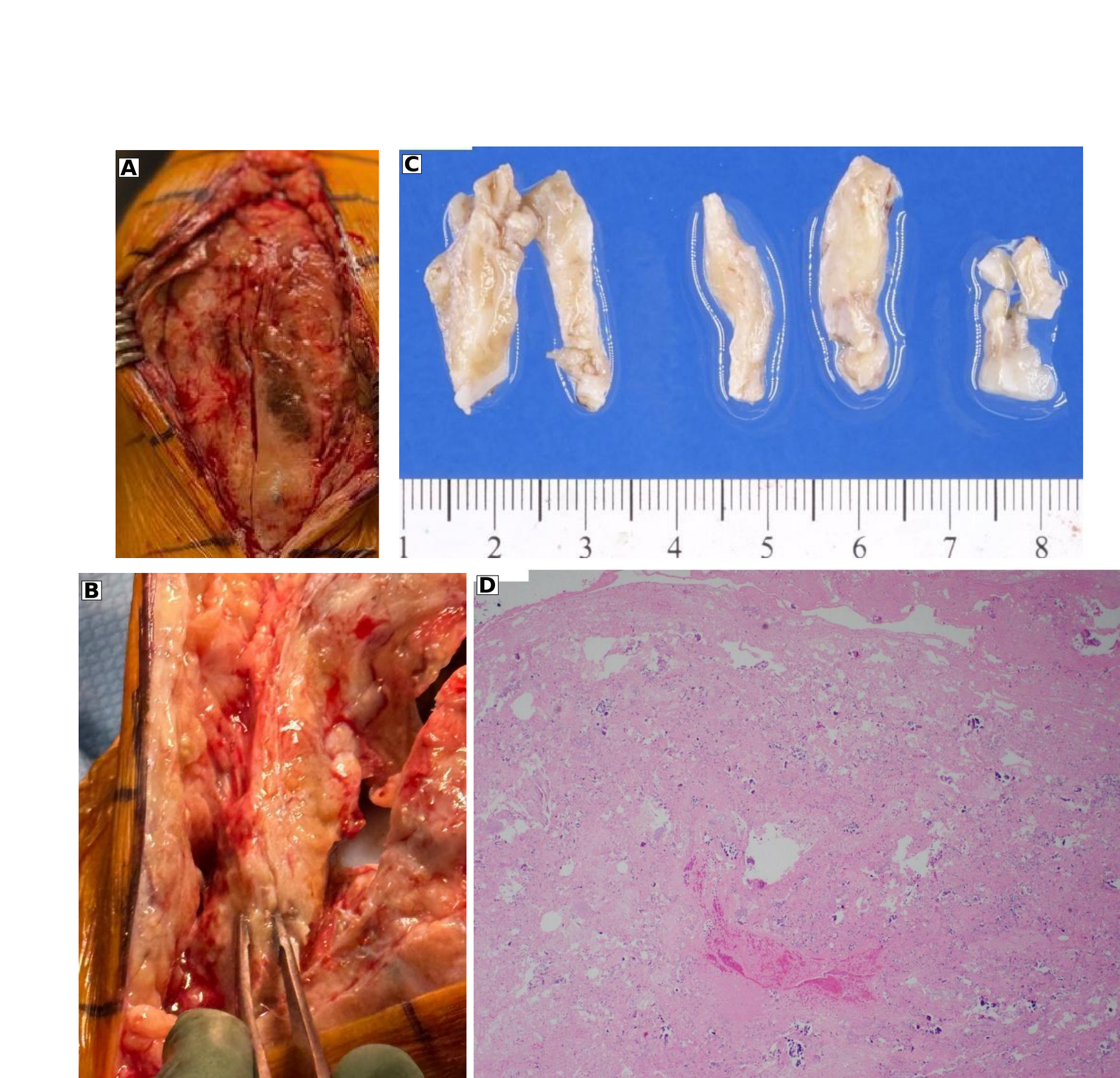
The most informative observations in this early experience arose from clinically indicated reoperations and planned second-stage procedures that permitted direct inspection of previously augmented sites. These procedures were not performed for research purposes and therefore represent selected observations rather than protocolized follow-up. At approximately 3 to 4 months after implantation, several treated regions demonstrated a macroscopically continuous soft-tissue layer over the prior area of deficiency. The treated region appeared subjectively thicker than adjacent native tissue, and in some cases a discrete boundary between the implanted scaffold and surrounding tissue could no longer be identified grossly. Because tissue thickness was not measured prospectively and the scaffold itself was not separately identified at reoperation, these findings are best described as gross tissue integration at the treated site rather than evidence of complete scaffold resorption or de novo collagen regeneration.
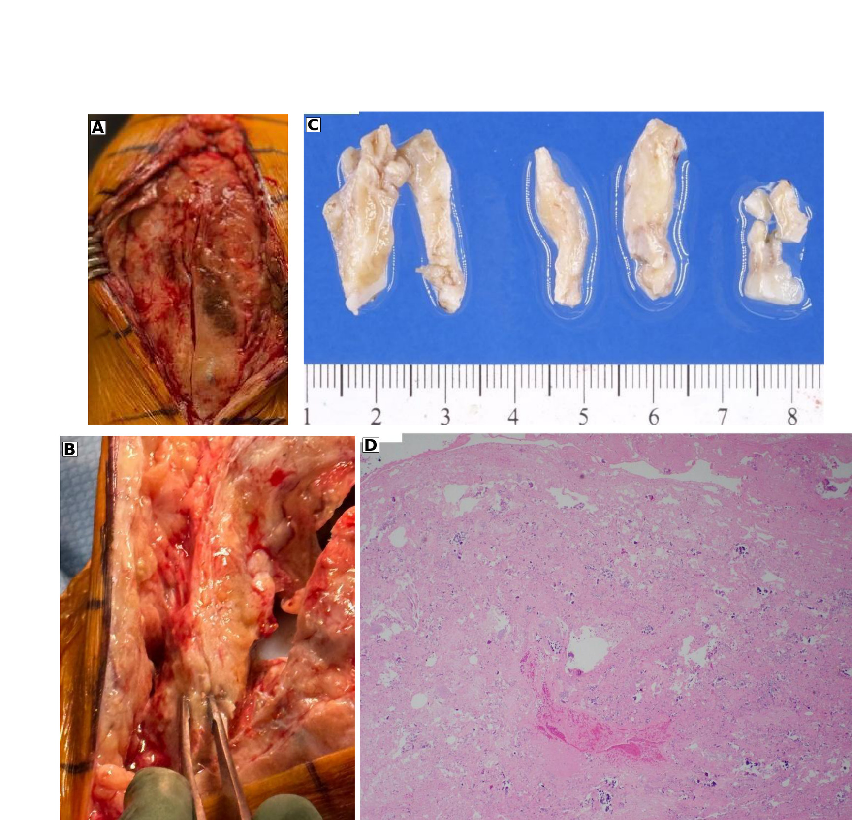
In one planned second-stage procedure, arthrotomy through the previously augmented region demonstrated a substantial, continuous tissue layer spanning the prior defect (Figure 5A-B). Tissue obtained from this region during the clinically indicated procedure was submitted for pathologic evaluation; the gross specimen and histologic appearance are shown in Figure 5C-D. Histologic examination demonstrated cellular connective tissue containing vascular channels. These findings support the presence of viable fibrovascular tissue at the treated site but cannot determine the relative contributions of newly formed tissue, native scar, residual scaffold, or the underlying repair, nor can histologic appearance alone establish the mechanical properties of the reconstructed tissue layer.
The observed tissue response is biologically plausible in the context of other resorbable collagen scaffolds. Human imaging and biopsy studies of a porous type I bovine collagen implant used in rotator cuff surgery have demonstrated increased tendon thickness, progressive host-cell and collagen integration, and tendon-like connective tissue at later follow-up (Bushnell et al. 2021; Bokor et al. 2016; Arnoczky et al. 2022). Preclinical evaluation of the Tapestry scaffold has likewise demonstrated collagenous connective-tissue ingrowth into and around the implant (US Food and Drug Administration 2020). These findings provide biologic context for the present observations but should not be extrapolated directly to revision arthroplasty, where the tissue environment, mechanical loading, infection risk, and indications are substantially different. The present findings therefore provide preliminary clinical evidence of gross tissue continuity and histologically viable connective tissue at selected Tapestry-treated sites, but they do not establish reproducibility, mechanical strength, or clinical efficacy.
Special Consideration: Staged Periprosthetic Joint Infection
I have also used Tapestry during selected first-stage procedures for periprosthetic joint infection when a deep soft-tissue defect remained after debridement, thorough irrigation, and spacer placement. This is an area in which I am deliberately cautious. I have not observed a soft-tissue failure attributable to Tapestry in these cases, and in staged infection cases in which I used the implant, infections subsequently cleared with the overall staged treatment. These observations do not establish that Tapestry reduces infection risk, improves eradication, or is safe in an infected field. Use in the setting of fungal infection, poor host grade, or inadequate skin coverage warrants particular caution. I therefore consider infection-related use an observation from early clinical experience, not a recommendation. The second-stage reimplantation procedures did not require additional Tapestry application because the previously augmented tissue provided adequate coverage at reoperation.
Discussion
Complex and revision arthroplasty often presents a difficult-to-quantify problem: the mechanical reconstruction may be well executed, but the surrounding biologic envelope remains thin, discontinuous, or difficult to close. This 57-case experience also provides practical feedback regarding when the implant may be useful: after definitive reconstruction and reasonable native-tissue approximation have been achieved, but the quality or completeness of the deep soft-tissue envelope remains concerning. In that role, the implant is simple to trim, conforms to irregular surfaces, and can be secured with limited sutures. The collagen matrix scaffold may be most intuitive over a repaired tendon or mesh reconstruction, but the same coverage concept has also been applied to selected periosteal, retinacular, capsular, and hip abductor deficiencies.
The most compelling information from this experience comes from the second looks: in selected reoperations at approximately 3 to 4 months, the treated regions appeared subjectively thicker and continuous with the surrounding tissue. One specimen underwent pathologic evaluation, providing an additional opportunity to examine what had formed at the implant site. Similar gross, imaging, and histologic observations have been reported with other bioinductive collagen scaffolds, particularly the most extensively studied porous type I bovine collagen implant, although those data are primarily from rotator cuff repair and cannot be directly extrapolated to revision arthroplasty (Bushnell et al. 2021; Bokor et al. 2016; Arnoczky et al. 2022). The most extensively studied commercially available bioinductive collagen implant has the strongest clinical evidence in this class, including prospective multicenter studies and randomized controlled trials in rotator cuff repair demonstrating increased tendon thickness and lower retear rates in selected populations (Bushnell et al. 2021; Ruiz Ibán et al. 2024; Bokor et al. 2016; Arnoczky et al. 2022). A 2025 systematic review concluded that clinical outcomes generally improve, although comparative evidence remains heterogeneous and higher-quality trials are still needed (Hurley et al. 2025). Recent reviews of commercially available bioinductive membranes similarly conclude that evidence for less-studied products remains limited (Iban et al. 2026). The present findings suggest that a substantial tissue layer may develop at Tapestry-treated sites in this setting, but they do not establish what that tissue contributes mechanically or clinically.
Limitations
This experience should not be interpreted as evidence of efficacy. Without a denominator by indication, standardized follow-up, or a comparison group, the absence of observed soft-tissue failures cannot establish superiority or equivalence to conventional closure or other biologic or reconstructive options, and the infection observations cannot establish infection safety. The value of this report is to describe a reproducible clinical problem, practical examples of how I addressed it, and the appearance of treated tissue at subsequent surgery.
A prospective study should now be feasible to record the indication, defect dimensions, implant size and orientation, fixation method, on-label or off-label use, wound complications, infection, reoperation, and indication-specific functional outcomes. In patients undergoing reoperation for an independent clinical indication, standardized photography and, when clinically appropriate, imaging or histologic analysis could further characterize the treated tissue.
Conclusion
In this early 57-procedure experience, Tapestry was used as a non-load-bearing onlay to supplement soft-tissue coverage after selected complex primary and revision hip and knee arthroplasty reconstructions. At subsequent operations approximately 3 to 4 months later, treated regions appeared subjectively thicker and continuous with surrounding tissue, with pathologic evaluation available in 1 specimen. No clinically apparent soft-tissue failures were observed at treated sites, although 1 postoperative infection occurred after an extensor mechanism reconstruction. These findings are encouraging and hypothesis-generating. The principal contribution of this report is to raise awareness of a practical adjunct for difficult soft-tissue coverage and to provide early clinical second-look information that can inform prospective evaluation.
Disclosures
The author has no conflicts of interest to disclose. No external funding or product support was received for this work.