
INTRODUCTION
In kinematically aligned (KA) total knee arthroplasty (TKA), the goal is to restore the limb and knee alignments to the pre-arthritic state, which in most patients precludes the need to release the collateral, retinacular, and posterior cruciate ligaments. KA ignores the locations of the center of the femoral head and center of the ankle because setting components to these landmarks alters limb alignment and joint-line obliquity, creating kinematic conflict among the ligaments during knee motion that can lead to stiffness, instability, poor clinical outcome scores, and shortened implant survival (Bae et al. 2024; Eckhoff et al. 2005; Franceschetti et al. 2025; MacDessi et al. 2021).
From a surgical perspective, the initial step in KA is to restore the pre-arthritic distal and posterior femoral joint lines. For the femoral condyle(s) on which articular cartilage is unworn, this is achieved by resecting articular cartilage and bone to a depth equal to the condyle of the femoral component. For the femoral condyle on which articular cartilage is worn, the cartilage is removed down to the level of subchondral bone and the resection thickness is decreased by 2 mm (Nam et al. 2014). Hence, articular cartilage is removed to the subchondral bone generally on the medial femoral condyle in varus knees and on the lateral femoral condyle in valgus knees. To perform these resections, a femoral referencing guide that directly contacts the surfaces is used.
The next step in KA involves restoring the pre-arthritic varus-valgus (V-V) angle and posterior tibial slope (PTS). Currently, KA is performed using manual instruments designed for mechanical alignment (MA), which include a proximal tibial cutting block attached to an extramedullary rod with a fixture that clamps just above the ankle. A single stylus in the cutting block is set on the lateral articular surface to set the depth of the resection. The surgeon adjusts the V-V angle and the PTS by changing the position of the extramedullary rod at the ankle. The PTS is fine-tuned until an angel wing, inserted in the medial saw slot, is parallel to the medial tibial articular surface. This setup has two limitations relevant to KA: visual setting of the V-V angle introduces variability, and the extramedullary fixture has limited adjustment range when the native PTS is shallow or steep, which varies by up to 17° across the population (Calek et al. 2022).
The study introduces a new dual-styli and angel-wing tibial resection guide designed to simplify tibial resection by registering the subchondral bone on both the medial and lateral tibia, thereby eliminating the need for the extramedullary rod and ankle reference. The primary objectives were to describe the design and perform a preliminary evaluation by reporting the frequency of tibial recuts and the use of thinner inserts, which indicate a more conservative resection thickness. A secondary objective was to evaluate the incidence of radiographic V-V angle and PTS baseplate outliers, defined as deviations greater than ±3° from preoperative values.
METHODS
An institutional review board approved the cross-sectional study using de-identified data (IRB # Pro00073629). Each patient met the Centers for Medicare & Medicaid Services guidelines for medical necessity for TKA, had Kellgren–Lawrence Grade III-IV osteoarthritis, and received treatment regardless of the severity of varus or valgus deformity or flexion contracture.
Between December 2024 and July 2025, two surgeons performed 204 caliper-verified KA TKAs with manual instrumentation and a KA-optimized implant system (GMK SpheriKA, Medacta International, Castel San Pietro, Switzerland, www.medacta.com, accessed on October 5, 2025) (Table 1). In every case in which the dual-styli and angel-wing tibial resection guide was available, it was used, indicating consecutive patient inclusion with 195 KA TKAs included in the study. Nine cases were excluded because the dual-styli and angel-wing guide was not available due to supply chain delays during the early study period; in these cases, conventional extramedullary tibial instrumentation was used. The KA-optimized implant system consists of a tibial insert with a flat lateral articular surface, medial ball-in-socket conformity, and posterior cruciate ligament (PCL) retention (Elorza et al. 2023); a femoral component featuring spherical condyles, a trochlear groove with a lateral wall opening to 20° valgus, and a flattened medial wall (Howell, Zabiba, Nedopil, et al. 2025; Howell, Zabiba, et al. 2026; Howell, Zabiba, Sadoghi, et al. 2025); and an anatomic baseplate that orients the anteroposterior axis of the insert parallel to the flexion-extension plane of the knee (Nedopil et al. 2021).
Description of the Tibial Resection Guide
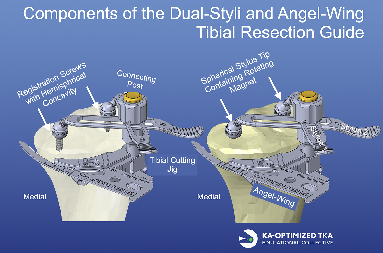
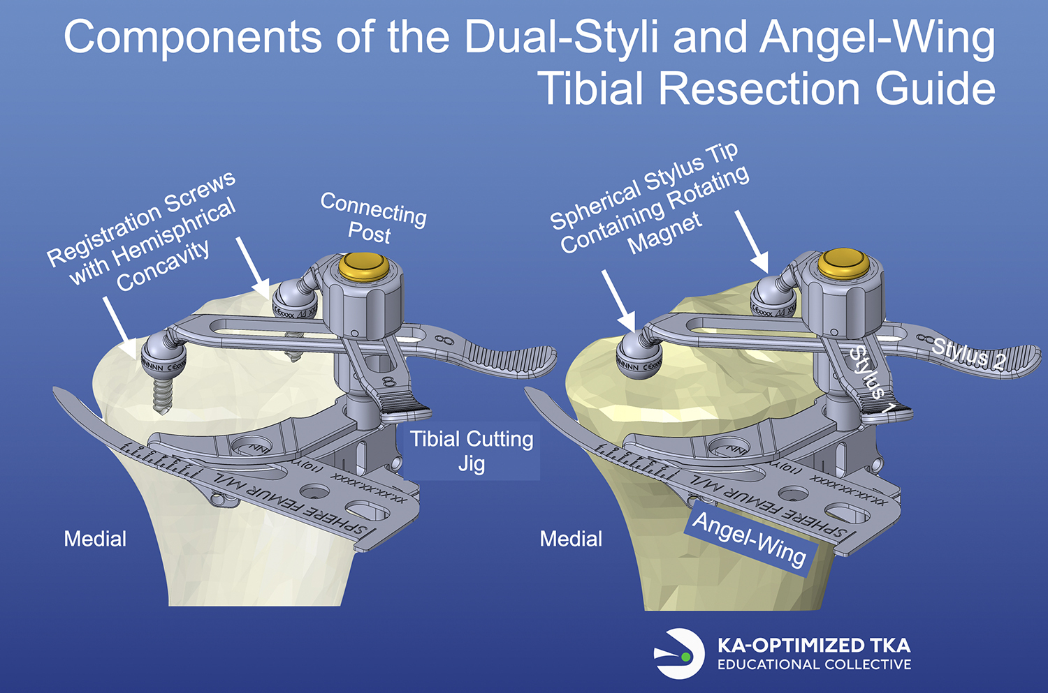
The components and operation of the new dual-styli and angel-wing tibial resection guide, including two tibial registration screws, two rotating and sliding styli, a connecting post, a tibial cutting jig, and an angel wing, are shown and described in Figure 1.
Steps for Using the Tibial Resection Guide
Using the tibial resection guide involved four key steps: exposing subchondral bone, placing registration screws, mounting and adjusting the tibial cutting jig, and pinning the jig in place. After knee exposure, the femur was resurfaced to the pre-arthritic state as previously described (Nedopil et al. 2018; Scott and Horton 2025). A caliper was used to measure the thickness of the medial and lateral distal and posterior femoral resections, which were verified as correct when they are within ± 0.5 mm of the condyle thickness of the femoral component, after compensating 1 mm for the saw blade’s kerf and 2 mm when cartilage is missing from wear.
The tibia was prepared by exposing the medial side 12 mm distal to the joint line and the entire tibial articular surface. With a 10-mm curette, an anteroposterior groove was created by removing cartilage down to subchondral bone in the center of the medial and lateral tibial compartments, with one edge of the groove at the base of the tibial spines, away from areas of tibial bone wear. In the center of each groove, a registration screw was drilled into the subchondral bone. The styli magnetically lock into the registration screws, and the length and rotation of the styli were set to rest the tibial cutting jig on the anterior surface of the tibia. The jig was secured by turning the tightening knob. The angel wing was inserted into the saw slot’s medial side, and the jig angulated until it was parallel to the medial tibial slope. The jig was fixed to the tibia with three pins: two drilled in parallel with one another and one drilled at an angle to the other two. These steps set the plane of the jig to restore the pre-arthritic V-V angle and PTS and to remove 8 mm of bone from the proximal tibia. To see a demonstration of the guide’s use intraoperatively, the interested reader should view Lesson 9 in the KA-optimized Core Curriculum (Howell, Hull, et al. 2026).
Balancing Step One: Verify the Varus-Valgus Orientation of the Tibial Resection is Correct or Requires a Recut
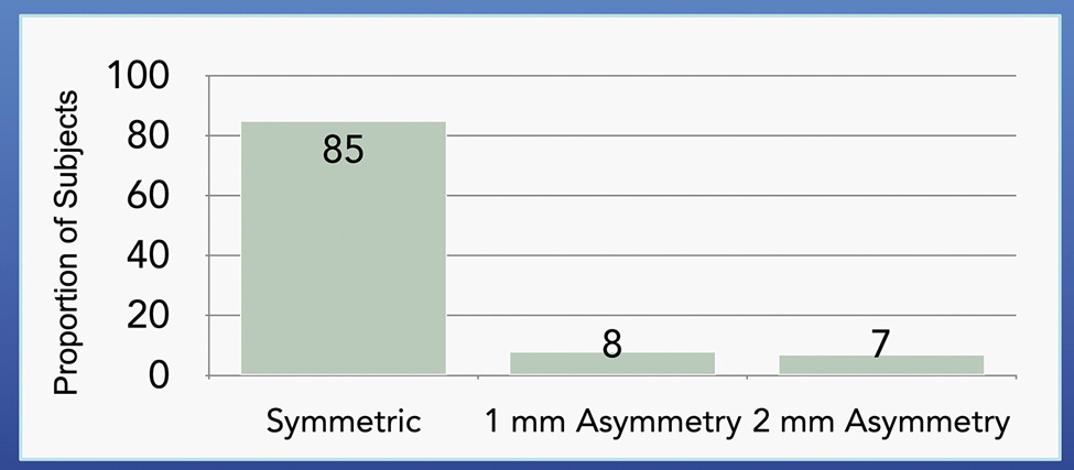
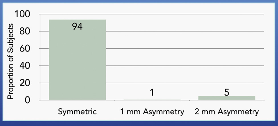
As a first preliminary verification check of balancing step one, a caliper was used to measure the thickness difference between the medial and lateral tibial resection at the base of the tibial spines (Howell, Hull, et al. 2026). A difference of 1 mm or less indicates that a spacer block will detect a symmetric extension space, while a difference of 2 mm or more indicates that the extension space likely will be asymmetric, and a tibial recut may be necessary. The second and definitive verification check was to determine whether the medial and lateral gaps are negligible and equal in extension with a spacer block during a varus-valgus stress. When the gap in one compartment exceeded 1 mm, a recut was performed by removing bone from the opposite tibial compartment using a skimmer saw cut or a recut guide to achieve a tight rectangular space.
Balancing Step Two: Verify the Posterior Slope Orientation of the Tibial Resection is Correct or Requires a Recut
As a first preliminary check of balancing step two, a caliper was used to measure the thickness difference between the anterior and posterior rims of the medial tibial resection The second verification was to view the medial side of the tibial resection, place a flat object like an osteotome on the anterior and posterior rim, and determine whether the slope of the osteotome was parallel to the tibial resection plane. When the osteotome was not parallel and the thickness difference exceeded 2 mm, a recut was performed using a recut guide by removing bone from the under-resected region.
Balancing Step Three: Use an Insert Goniometer to Choose the Optimal Insert Thickness Within ± 1mm
The final balancing step was to use an insert goniometer to select the optimal insert thickness. The optimal thickness is the one that restores full extension and provides the maximum tibial internal rotation without anterior lift-off of the insert at 90° of flexion. If insert lift-off was observed, then balancing step two above was repeated.
Radiographic Measurement Technique for Determining the Deviation in Baseplate V-V and Slope from Preoperative
All radiographs were obtained according to the Knee Society radiographic evaluation system and methodology (Meneghini et al. 2015). Long-leg alignment films were not used, as the measured variables are knee-level measurements. Pre- and post-operative radiographic analysis determined the deviation of the V-V angle and PTS of the tibial baseplate from the preoperative knee. One author (AZ) measured the posterior tibial slope (PTS: posterior slope > 0°) as the angle between a line along the posterior tibial cortex and a line parallel to the medial tibial plateau. The same author measured the anatomic medial proximal tibial angle (aMPTA; varus angle < 90°) (Horos 4.0.1, horosproject.com, last accessed 11/05/25) as the angle between the anatomic axis and the proximal tibial joint line. The author (AZ) was blinded to surgeon identity, recut status, and all clinical details at the time of measurement. Deviation in the V-V angle was defined as post-aMPTA minus pre-aMPTA; a positive value indicated greater baseplate valgus than preoperative. Deviation in PTS was defined as post-PTS minus pre-PTS; a positive value indicated a greater baseplate slope than preoperative. Deviations > 3° in magnitude were considered outliers. The ±3° threshold for defining outliers was selected based on its established use in prior arthroplasty literature as a clinically meaningful deviation from intended alignment (Bosco et al. 2025; Deckey et al. 2021). Intraobserver reliability (i.e. repeatability or precision) in each angle was determined by having one observer measure each angle in three trials on 20 patients selected randomly. The repeatability was the square root of the pooled variance. The precision in the deviation was calculated as: deviation precision = angle repeatability x square root (2(1-r)) where r is the correlation coefficient between the two angles used to determine the deviation. Because of the high correlations (r ≈ 0.9), the deviation precisions were limited to 0.2° for the V-V angle and 0.3° for the PTS.
Data Analysis
Statistical software (JMP Pro, version 18.0.1, Cary, NC, https://www.jmp.com, accessed on January 12, 2026) was used to determine the frequencies of V-V angle and PTS tibial recuts and the distribution of insert thicknesses. The mean and standard deviation (SD) are reported for the radiographic V-V angle and PTS angular differences from preoperative values. Upper (UCL) and lower (LCL) 95% confidence limits for proportions were computed using the Wilson score interval.
RESULTS
The 62 KA TKAs performed by the surgeon with 40 years of experience and the 133 KA TKAs performed by the surgeon with only 6 years of experience had similar distributions of age, sex classification, Kellgren-Lawrence osteoarthritic classification, insert thickness, and a similar mean changes in V-V angle and PTS of the baseplate relative to preoperative (Table 1).
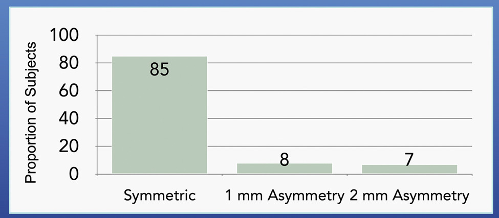
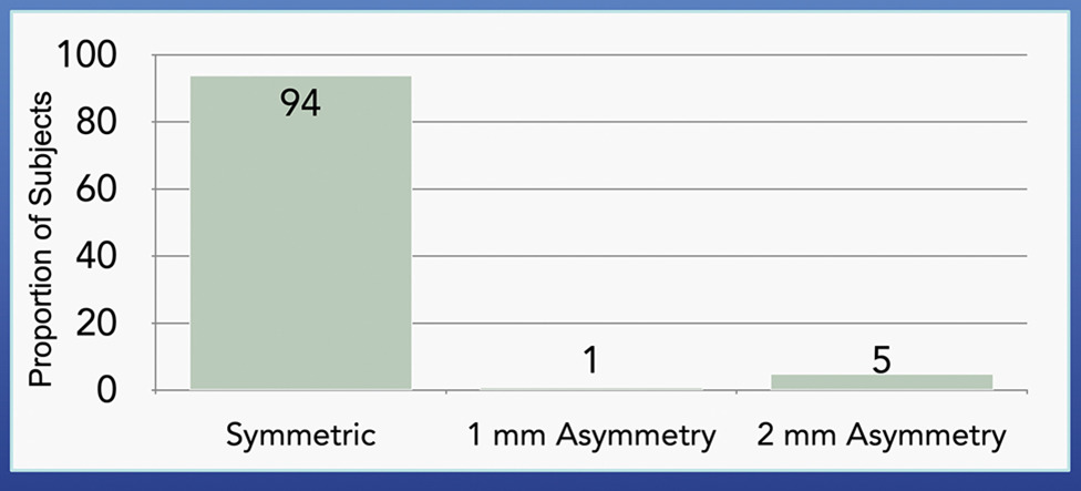
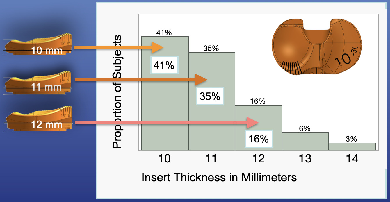
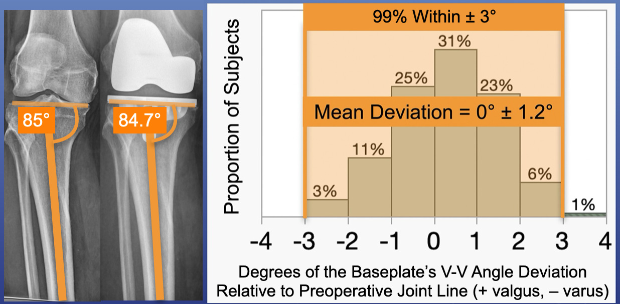
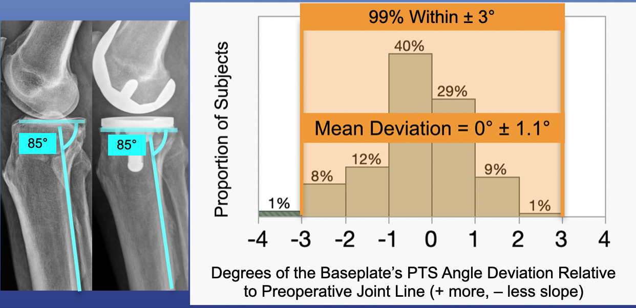
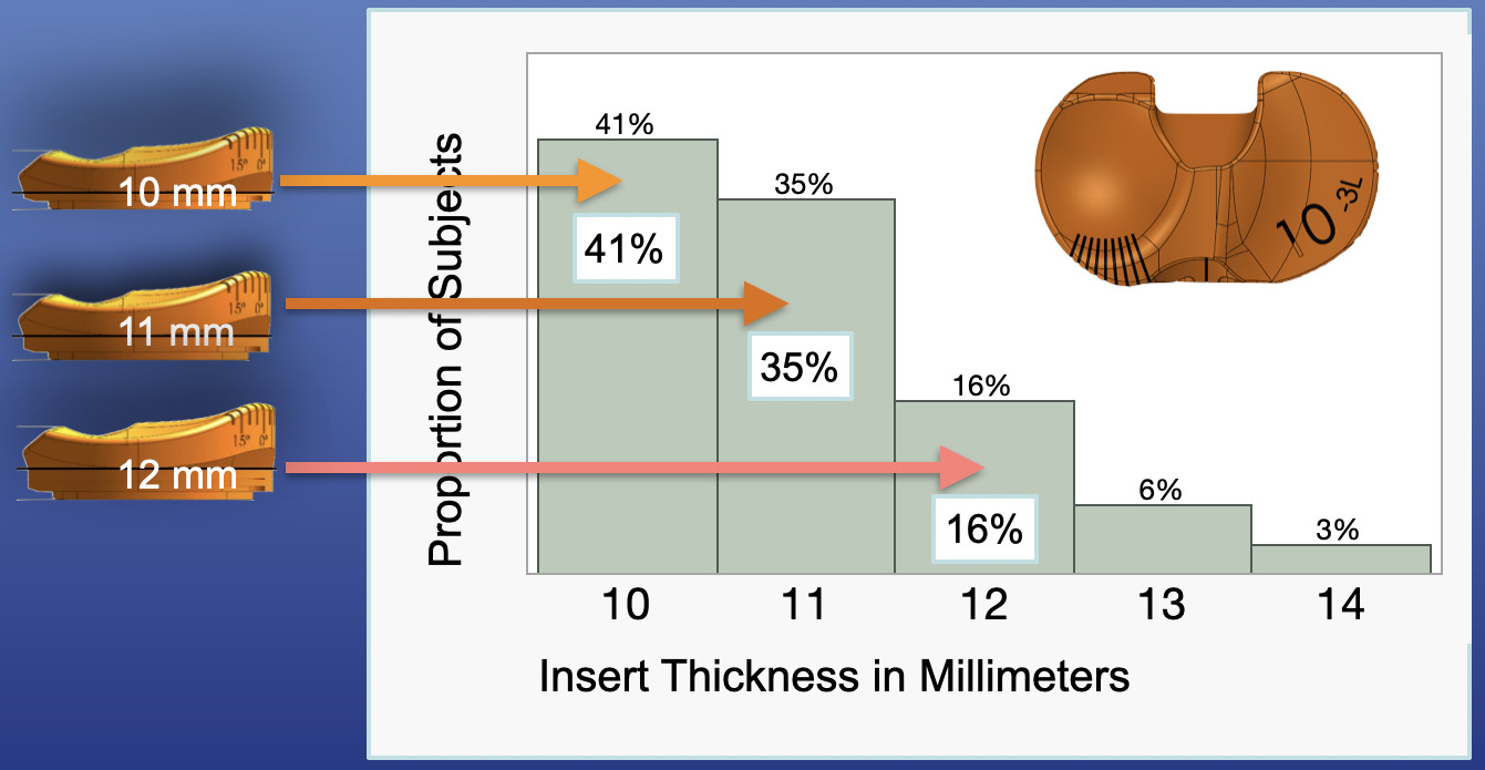
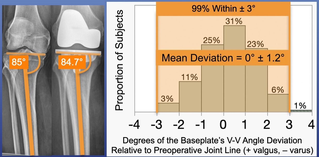
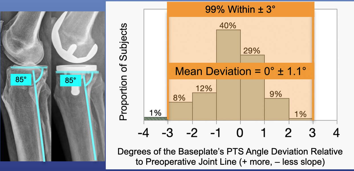
Of the 195 KA TKAs, 181 of 195 (93%, UCL/LCL: 96%/88%) and 185 of 195 (95%, UCL/LCL: 97%/91%) of the tibial resection planes were not recut in the V-V angle and PTS, respectively (Figures 2 and 3). In 177 of 195 (91%, UCL/LCL: 94%/86%) of cases, the insert thickness ranged from 10 to 12 mm and no insert exceeded 14 mm (Figure 4). Baseplate outlier alignments more than ±3° from preoperative occurred in V-V in one of 195 cases (i.e., < 1%) and in PTS in 1 of 195 cases (i.e., < 1%) (Figures 5 and 6). These two outliers occurred in two different patients. Neither outlier required revision surgery during the follow-up period.
DISCUSSION
In this early clinical evaluation, the most important findings are that the new dual-styli and angel-wing tibial resection guide overcame two limitations of conventional extramedullary tibial instruments (i.e. visual setting of the V-V angle and limited adjustment range), had a low frequency of V-V angle and PTS recuts, used thinner inserts indicating a conservative tibial resection thickness, and had few outlier alignments from preoperative.
This new dual-styli and angel-wing tibial resection guide is an evolution of commonly available extramedullary tibial resection instrumentation that clamps to the ankle and addresses two key limitations of this setup. Instead of visually setting the V-V angle based on a single stylus, which typically contacts the lateral articular surface, the two styli are connected to two registration screws drilled into the subchondral bone at the base of the lateral and medial tibial spines (Figure 1). By registering the screws drilled into the subchondral bone directly in both compartments, the uncertainty associated with visually aligning the V-V angle is eliminated. Also, by eliminating the extramedullary fixture, which interferes with the tibia and has inherent limitations of excursion when shallower or steeper tibial slope is required, the dual-styli and angel-wing resection guide extends the range of adjustment to restore the patient’s pre-arthritic PTS.
Although the dual-styli and angel-wing resection guide eliminated the visual alignment of the V-V angle, the PTS is still set by visual alignment. However, PTS restoration with an angel wing, placed either medially as done with the dual-styli angel-wing reference guide or on top of the medial tibial articular surface, is reasonably accurate (Franceschetti et al. 2025; Nedopil et al. 2017). Evidence of this accuracy is further confirmed by the results of the present study, showing that 95% of patients did not require a tibial recut. (Figure 4).
Since manual instruments are used to perform KA without relying on radiographic measurements by placing the femoral and tibial guides directly on the articular surfaces of the femur and tibia, the number of error sources are minimized. For the femur, the main source of error is the use of a fixed 2 mm adjustment for the thickness of the articular cartilage (Campi et al. 2025; Giurazza et al. 2025; Sandhar et al. 2026). For the tibia, the main sources of error associated with using the new dual-styli angel-wing resection guide for the V-V angle is the screw position choice, as well as whether there is bone wear under the screws. The main error source for the PTS is relying on visually aligning the angel wing parallel to the slope of the medial articular surface. However, errors from these sources had a minimal impact on tibial baseplate positioning, as the percentage of outliers exceeding 3° was < 1% for both the V-V angle (Figure 5) and the PTS (Figure 6).
Not only were the number of error sources minimized leading to a low frequency of V-V angle and PTS recuts, but also thinner inserts were used indicating conservative tibial resection thickness. Beyond indicating a conservative tibial resection, the narrow distribution of insert thicknesses (91% between 10–12 mm) also has practical implications for inventory management, particularly in ambulatory surgery centers where maintaining a broad range of insert sizes is logistically challenging.
Since this was a preliminary evaluation, several future studies for a more thorough evaluation are warranted. One is that results should be compared to those of control group where extramedullary instrumentation was used. In addition to the process-based measurements herein, this comparison should include operative efficiency and patient-reported outcomes. Also, a multicenter study is needed to determine inter-surgeon variability in recuts, insert thickness distribution, and alignment outliers. Next, the present study does not provide information about the learning curve, which is the number of cases a typical surgeon needs to perform to obtain the low recut rate and distribution of insert thicknesses that indicate a conservative tibial resection thickness.
CONCLUSION
Surgeons using the dual-styli and angel-wing tibial resection guide infrequently required recuts and infrequently used inserts thicker than 12 mm. The < 1% incidence of postoperative V-V angle and PTS baseplate outliers demonstrated reliable restoration of preoperative tibial alignment in this preliminary series.