Loading [Contrib]/a11y/accessibility-menu.js
Skip to main content
null
J Orthopaedic Experience & Innovation
  • Menu
  • Articles
    • Brief Report
    • Case Report
    • Data Paper
    • Editorial
    • Hand
    • Meeting Reports/Abstracts
    • Methods Article
    • Product Review
    • Research Article
    • Review Article
    • Review Articles
    • Systematic Review
    • All
  • For Authors
  • Editorial Board
  • About
  • Issues
  • Blog
  • "Open Mic" Topic Sessions
  • Advertisers
  • Recorded Content
  • CME
  • JOEI KOL Connect
  • search

RSS Feed

Enter the URL below into your favorite RSS reader.

https://journaloei.scholasticahq.com/feed
Research Article
Vol. 5, Issue 1, 2024June 09, 2024 EDT

Routine Chest X-Ray is Not Cost-Effective After Clavicle ORIF: A Cost Effectiveness Analysis with National Estimations

Samuel Rosas, MD, T. David Luo, MD, Amy P. Trammell, MD, Marcel G. Brown, MD, Matthew Gwilt, Jonathan C. Levy, MD, Holly T. Pilson, MD, Jason J. Halvorson, MD, Eben A. Carroll, Sharon N. Babcock, MD,
ClavicleOpen Reduction and Internal Fixation (ORIF)CostChest X-Ray (CXR)Radiographs
Copyright Logoccby-nc-nd-4.0 • https://doi.org/10.60118/001c.91276
J Orthopaedic Experience & Innovation
Rosas, Samuel, T. David Luo, Amy P. Trammell, Marcel G. Brown, Matthew Gwilt, Jonathan C. Levy, Holly T. Pilson, Jason J. Halvorson, Eben A. Carroll, and Sharon N. Babcock. 2024. “Routine Chest X-Ray Is Not Cost-Effective After Clavicle ORIF: A Cost Effectiveness Analysis with National Estimations.” Journal of Orthopaedic Experience & Innovation 5 (1). https:/​/​doi.org/​10.60118/​001c.91276.
Save article as...▾
Download all (6)
  • Click here : https://joeipub.com/learning
    Download
  • Figure 1. Two Way Dichotomous Model of patients undergoing Clavicle ORIF with and without PACU CXR with payoff for each of the reported strategies after Model Rollback:
    Download
  • Figure 2. Comparisons of each strategy using WTP Curves
    Download
  • Figure 3. Cost Effectiveness at Different Thresholds of WTP
    Download
  • Figure 4. Montecarlo Simulation Results
    Download
  • Figure 5. Incremental Cost-Effectiveness of the Two Models
    Download

Sorry, something went wrong. Please try again.

If this problem reoccurs, please contact Scholastica Support

Error message:

undefined

View more stats

Abstract

Introduction

Clavicular fractures can be managed surgically or via a non-operative approach. In those electing for surgical fixation, post-operative CXR may be routine practice in the post-anesthesia care unit (PACU) for a rare complication of pneumothorax (PTX), thus driving up the cost for managing these fractures. The value and associated cost of this routine CXR in PACU following open reduction internal fixation (ORIF) of clavicles has not been thoroughly assessed. In this study we investigate the cost effectiveness of obtaining CXR after operative treatment of clavicle fracture.

Materials and Methods

Study was designed as a healthcare two-way dichotomous model with decision trees built in where a CXR was either obtained or not. Literature review was performed to determine the cost estimates of CXR, the cost of ORIF, and associated hospital stay expenses. Annual volume of clavicle ORIF’s performed were derived from the PearlDiver database, queried for CPT-23515 (Clavicle ORIF). Estimation of CXR rates were performed from 3% to 98% of patients undergoing clavicle ORIF. Net monetary analysis with associated sensitivity analysis of 10,000 repetitions was performed.

Results

The strategy without CXR proved to be the most cost-effective strategy with a net monetary benefit (NMB) of $32,022.50. At $50,000 willingness to pay (WTP), the no CXR strategy was the preferred option. This strategy was found to be optimal 76% of the time followed by routine CXR 23% of the time and less than 1% being indifferent. The national annual costs of routine CXR after clavicle ORIFs was estimated to range from $7,100 to $349,860, which over 10 years represents anywhere from $81,540 to $2,663,640 in additional expenses.

Conclusion

The provided study demonstrates that routine CXR after clavicle ORIF is not cost effective. Due to the exceedingly low incidence of PTX as a post-operative complication in clavicle ORIF, we recommend that hospitals and surgeons refrain from ordering radiographs in post-operative patients, with the exception of those displaying concerning clinical signs for PTX or those with known pre-operative pulmonary injury. Adopting this strategy will limit unnecessary healthcare costs accrued by patients and may be the more clinically appropriate management.

Click here : https://joeipub.com/learning

Introduction

Clavicle fractures account for 2-10% of all fractures in adults, of which the majority occur in the middle third of the clavicle (Canadian Orthopaedic Trauma Society 2007a). Traditionally, these fractures have been treated nonoperatively (Canadian Orthopaedic Trauma Society 2007a). However, recent literature lacks a clear consensus regarding whether surgical or non-surgical management yields better outcomes in union rates and patient-perceived functional improvements (Canadian Orthopaedic Trauma Society 2007a; Heyworth et al. 2022; Axelrod et al. 2020). As a result, both options are routinely utilized in current practice. This lack of clarity in the approach to clavicle fractures has led to multiple studies in order to further delineate and compare the outcomes and cost-effectiveness of open reduction and internal fixation (ORIF) versus nonoperative management of these fractures (Canadian Orthopaedic Trauma Society 2007a; Heyworth et al. 2022; Axelrod et al. 2020; Nicholson et al. 2019; Liu et al. 2019).

Although rare, pneumothorax (PTX) is one of the most feared complications following surgical treatment of clavicle fractures. In one series of 1,350 patients who underwent ORIF of midshaft clavicle fractures, pneumothoraxes’ were identified in 16 patients (1.2%) (Leroux et al. 2014). Increased patient age and surgeries performed at an academic hospital were significantly associated with a greater odds ratio of PTX. Due to this feared complication, many surgeons routinely obtain chest x-rays (CXR) in the post-anesthesia care unit (PACU) following clavicle ORIF. Despite this increased utilization of CXR, the value of these x-rays has not been assessed. In the current healthcare climate where costs are greatly scrutinized, it is important to establish the utility and cost effectiveness of a diagnostic test.

The purpose of this study is to evaluate the cost-effectiveness of routine CXR after clavicle ORIF. Given the low incidence of PTX post-clavicle ORIF, we hypothesized that obtaining CXR would not be a cost-effective strategy.

Methods

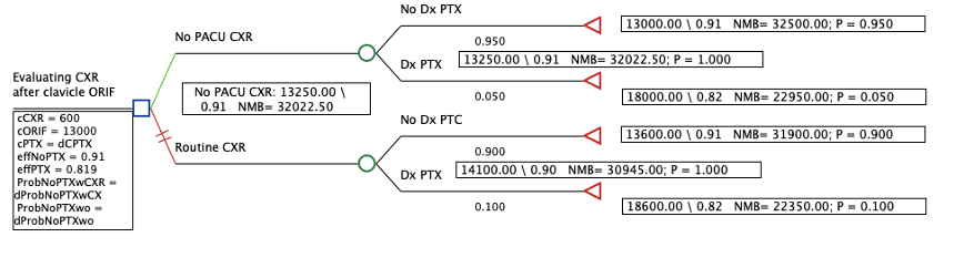
This study was designed as a healthcare two-way dichotomous model (Figure 1). Decision trees were built where a CXR was either obtained or not. The clinical scenario most closely mimicking the model studied herein, represents an outpatient clavicle ORIF in the non-acute trauma setting, in a non-polytrauma patient. This scenario was chosen since most clavicle fractures are treated non-emergently and as an outpatient surgery in the United States (Yang, Werner, and Gwathmey 2015; Schairer et al. 2017; Pang et al. 2017). Moreover, given that CXR does not typically change clinical practice beyond PTX, no other complications were included in this study (Leroux et al. 2014; Shubert et al. 2019a).

Figure 1
Figure 1.Two Way Dichotomous Model of patients undergoing Clavicle ORIF with and without PACU CXR with payoff for each of the reported strategies after Model Rollback:

Legend: Green is the preferred strategy. This supports the use of no PACU CXR after clavicle ORIF with a net monetary benefit of 31,545 based on 10,000 simulations.

Health States and Probabilities

The literature was queried to evaluate the incidence of PTX after clavicle ORIF. Provided the scarcity of data on PTX as an outcome of clavicle ORIF, our review was open to all types of articles ranging from case reports to systematic reviews and meta-analysis. Other data obtained from the literature included: cost estimates of CXR, cost of clavicle ORIF, length of stay for PTX treatment, rate of PTX treatment requiring more than observation, cost of PTX treatment and the health utility values of patients undergoing ORIF.

Literature reported incidence of PTX after clavicle ORIF demonstrates a rate of 1.2% (16 out of 1,350 patients). Of these 16 patients, 8 required chest tube insertion. In all instances the authors could not report whether the reported PTX occurred secondary to injury or due to surgical treatment which is significant given that an estimated 3% of patients with clavicle fractures also sustain a PTX regardless of surgical intervention (Leroux et al. 2014). Evaluation of the American Board of Orthopaedic Surgery part II data by Navarro et al. did not report any PTXs as a complication after ORIF for clavicle fractures undergoing surgical management in their evaluation of submitted cases (Navarro et al. 2016). A summary of the model parameters and their sources is outlined below (Table 1).

Table 1.Literature Review for Determination of Probabilities
Reported PTX after ORIF in the Literature
Event Probability References
PTX after ORIF 0 - 1.2% Leroux et al. JBJS 2014 (Leroux et al. 2014) (Leroux et al. 2014) (Shubert et al. 2019b), (Asadollahi et al. 2016), (Canadian Orthopaedic Trauma Society 2007b), (Chen et al. 2012),
PTX after ORIF 0% Shubert et al. JSES 2018 (Shubert et al. 2019b), (Asadollahi et al. 2016) (Chen et al. 2012),
PTX after ORIF 0%
ABOS Part 2 data. 0% Navarro et al. JSES 2016 (Navarro et al. 2016) (Navarro et al. 2016)
Single patient report after MIPO, LOS* 9 days Kim et al. Medicine 2019 (M. K. Kim et al. 2019) (M. K. Kim et al. 2019)
Single patient, PTX after closed fracture, LOS 5 days Kao and Goh. Hong Kong J of Emerg Med 2006 (Kao and Goh 2006) (Kao and Goh 2006)
Single patient, PTX after closed fracture delayed Gandham and Nagar. BMJ Case Rep 2013 (Gandham and Nagar 2013) (Gandham and Nagar 2013)
0% Shin et al. J Trauma and Acute Care Surg 2012 (Shin, Do, and Jang 2012) (Shin, Do, and Jang 2012)
0% Service et al. OTA Poster 2015 (Service B et al. 2015) (Service B et al. 2015)
0% McKnight et al. JSES 2016 (McKnight et al. 2016) (McKnight et al. 2016)

*LOS=Length of Stay

Given that most PTX are transitional states of disease of a worse health state for a defined period of time and not classified as chronic conditions, a discounted health utility state was calculated for those who sustained a PTX, which was 10% less of the utility of those 1 year out of a clavicle ORIF. Provided there is no reported health utility state value in the literature of patients who recovered from a PTX after clavicle ORIF, the 10% discounted health utility state is necessarily an estimated discounted value. Health utility states used were 0.91 one year after ORIF and 0.819 (0.91 – 10% = 0.819) one year after ORIF with PTX.

Costs and Cost Savings Estimation

The cost of ORIF, CXR and hospitalization were obtained from the literature (Table 2). For cost savings estimations the Pearldiver Database (Pearldiver Inc, Colorado Springs, Colorado) was queried for CPT-23515 (Clavicle ORIF) (Pang et al. 2017). Annual volume of procedures performed from 2010 to 2018 were obtained from the PearlDiver database using the CPT code above. The cost of developing a PTX was calculated based on average daily inpatient hospital stay costs from length of stay reported in the literature for this complication (Table 2).

Table 2.Input Data for Model Creation
Input Data for Model Creation
Price Reference
Cost of CXR $300 -⁠ $594 (Chui et al. 2018; Louie et al. 2015)
Cost of ORIF $13,000 (Liu et al. 2019; Walton et al. 2015)
Cost of PTX $5,000 * (Beattie et al. 2020)
PTX LOS 1.6 - 4.38 (Zhan and Miller 2003; M. Kim and Moore 2020; Williams and Penn 2021; Brown et al. 2020)

* Assumption Length of Stay (LOS) 1 or 2

To estimate potential cost savings of not obtaining CXRs, we elected to evaluate different rates of obtaining CXR after clavicle ORIF based on the annual number of clavicle ORIFs. Estimation of CXR rates were performed from 3% to 98% of patients undergoing clavicle ORIF and cost savings calculated as: Cost of CXR x n undergoing CXR = annual savings. Lastly, number needed to treat (NNT) was also evaluated regarding costs. The following formulas were used to calculate NNT.

Absolute Risk Reduction = (Control event rate) − (Experimental event rate)

NNT = 1/ARR.

Cost estimates were not adjusted for inflation during estimation of potential cost savings provided the short time frame and because costs would increase similarly between the different rates of ORIFs undergoing CXR used in our analysis. An estimation of cost savings was performed after an optimal strategy was decided based on the Montecarlo simulations (see below). A Montecarlo simulation is the preferred method of assessing the probability of different outcomes when random variables exist. Uncertainty is addressed by running the simulation numerous times to yield estimates that accurately approximate real-life circumstances.

Data analysis and Deterministic Sensitivity Analysis

The probabilistic sensitivity analysis with 2 arm decision tree and Montecarlo simulations were designed to evaluate the cost effectiveness of our clinical question. Given the lack of retrospective or prospective data on this subject, we elected to perform a deterministic sensitivity analysis that, represents multiple iterations (10,000) and computations of the probabilities (Table 1). The probability distribution of no PTX without CXR in PACU was estimated using a Beta distribution with a mean of 90% and standard deviation (SD) of 5% while the probability distribution of no PTX with CXR in PACU was based on a gamma distribution with mean of 95% and SD of 2%. These probabilities and their mean occurrence rates were chosen based on the sporadic occurrence of the outcomes of interest. Furthermore, the high SDs are reflective of the rarity of these events as well. The health utility values were used as an effectiveness marker. The commonly referenced threshold of $50,000 willingness to pay was used for this cost effectiveness analysis (Nwachukwu and Bozic 2015).

For estimation of the optimal clinical strategy to determine whether CXR in PACU was cost effective, the net monetary benefit (NMB) was chosen as the outcome of interest. Net monetary benefit (NMB) represents the most cost-effective strategy considering the costs and health utility outcomes.

The TreeAge pro Healthcare version 2021 R1.0 software (Williamstown, Massachusetts) was used for the modeling and simulation and all statistical analysis. Microsoft Excel (V16.4, Redmond, Washington) was used to compute cost savings. Data obtained from the PearlDiver Server was extracted from the Mariner dataset which is held within a HIPAA compliant database that de-identifies patients; therefore, an IRB review was not indicated.

Results

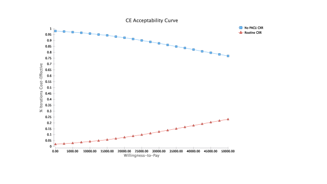
Montecarlo simulations proved ORIF of the clavicle without CXR in the PACU to be the most cost-effective option with a NMB of $32,022.50 (Figure 1). Payoffs with each of the reported strategies after model rollback are further demonstrated (Figure 1). Moreover, at the chosen willingness to pay (WTP) threshold of $50,000, the no CXR strategy was the dominating option. Comparisons of the strategies can be seen with the WTP curve (Figure 2).

Figure 2
Figure 2.Comparisons of each strategy using WTP Curves

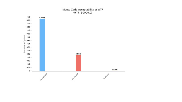
The results of the Montecarlo simulations with cost effectiveness at different thresholds of WTP based on chosen strategy are shown below (Figure 3). As WTP increases, obtaining CXR becomes cost effective following ORIF. The no CXR strategy was found to be optimal 76% of the time at WTP of $50,000 followed by routine CXR 23% of the time and less than 1% being indifferent (Figure 4).

Figure 3
Figure 3.Cost Effectiveness at Different Thresholds of WTP
Figure 4
Figure 4.Montecarlo Simulation Results

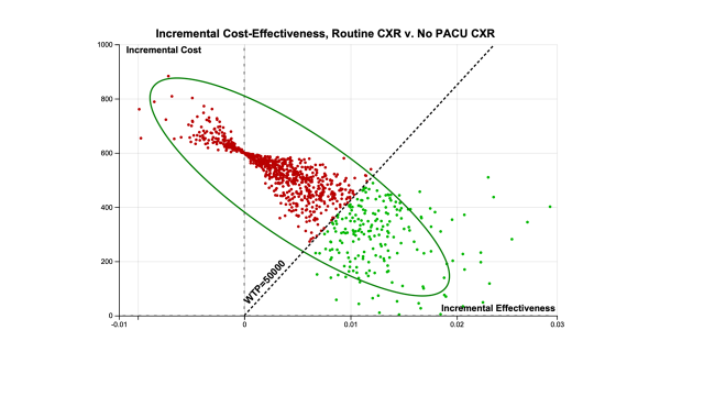
The incremental cost effectiveness comparison of the two strategies is depicted below (Figure 5). The repetitive simulation of the events demonstrates the low incremental effectiveness that is obtained from routine CXR after ORIF (red region), and the green region represents the more cost-effective strategy of no PACU CXR.

Figure 5
Figure 5.Incremental Cost-Effectiveness of the Two Models

Estimation of Possible Cost Savings

From 2010 to 2018 there were a total of 9,060 clavicle ORIFs identified within the PearlDiver Database. The mean annual number of ORIFs was 1,006 (SD 143.4). These procedures represented $38,369,304 in reimbursements.

The national annual cost of routine CXR after clavicle ORIF was estimated to range from $7,100 to $349,860 (3% vs 98% undergoing CXR, respectively), which over 10 years represents anywhere from $81,540 to $2,663,640. If no CXR had been obtained for all the ORIFs performed from 2010 to 2018, $2,718,000 would have been saved. As described by Leroux et al. 1.2% were found to have a PTX based on CXR, this would incur a cost of $32,616 for 108.7 ORIFs (Leroux et al. 2014) . If CXR was able to identify all PTX after clavicle bringing the missed incidence to 0, then 83 CXR would be the NNT, representing a cost of $24,900 (83 x $300.0).

Discussion

Clavicle fractures are a common occurrence in the United States. While the literature remains divided on whether surgical fixation or non-operative management is the preferred treatment strategy, high-level studies have shown the possible benefit of higher union rates with clavicle ORIF (Canadian Orthopaedic Trauma Society 2007a). As such, the rate of clavicle ORIF remains around a thousand cases performed annually. One of the more feared complications of clavicle ORIF is postoperative PTX. To avoid a missed PTX, many surgeons cautiously order CXR in the PACU after clavicle ORIF. The value and associated cost of this routine CXR in PACU following ORIF of clavicles has not been reviewed.

The model without CXR proved to be the most cost-effective strategy. Net monetary benefit (NMB) is a method of expressing cost effectiveness that considers both the willingness to pay threshold and health benefits (QALYs) for a given intervention. By converting QALYs into monetary units, the cost of each treatment strategy can then be deducted. With a NMB of $32,022.50 and at a WTP of $50,000, the no CXR strategy was the preferred option (Figure 1). 76% of the time, this strategy was found to be optimal followed by routine CXR 23% of the time and less than 1% being indifferent (Figure 4). The national annual costs of routine CXR after clavicle ORIFs were estimated to range from $7,100 to $349,860, which over 10 years could have represented anywhere from $81,540 to $2,663,640 based on annual percent imaged.

Despite the presented cost-effective findings after forgoing post-operative CXR, surgeons may be reluctant to adopt this practice and should continue to use their clinical judgement in regard to the need for postoperative CXR. Risk factors for PTX such as infraclavicular blocks, or worsening emphysema, are well documented in the literature and can help guide surgeons in determining which patients would most benefit from a CXR in the PACU following clavicular ORIF (Gauss et al. 2014). Other indications such as pleuritic chest pain or changes in vitals in the operating room or PACU, could also be used as an indication for postoperative CXR. Acknowledging these very real and legitimate reasons to obtain a postoperative CXR for clavicle ORIF, physicians should continue to use clinical judgement in the PACU regarding the need for a CXR.

In addition to the risk factors noted to increase incidence of PTX following clavicle ORIF mentioned above, several other considerations are worth discussing. First, is the predicted sensitivity of CXR in identifying PTX. Recent studies have found that CXR had a lower pooled sensitivity of detecting PTX, while chest ultrasound (CUS) demonstrated a significantly improved pooled sensitivity in comparison (Ebrahimi et al. 2014; Alrajab et al. 2013). This presents an alternative more cost-effective strategy than obtaining post-operative radiographs to help guide surgeons in patients with signs concerning for PTX (Alrajab et al. 2013). As mentioned previously, monitoring of intraoperative vital signs can assist in raising suspicion for PTX. Changes in airway pressures or hemodynamic stability could suggest PTX, in which intraoperative fluoroscopy has successfully been used to diagnose PTX (Heyba, Rashad, and Al-Fadhli 2020). While these alternative methods have been shown to successfully detect PTX, their true cost-effectiveness and practical utility in clavicle post-ORIF is harder to infer.

Our study is not without limitations. The data used for this analysis is based on a retrospective review of the literature and point estimates. It is likely that the individual studies referenced held different protocols in determining the incidence of post-operative complications of pneumothorax. This includes limitations in identifying whether pneumothorax was secondary to the initial presenting injury or due to ORIF, and identification of delayed presentations of PTX beyond the post-operative hospital monitoring period. There is also a possibility that PTX is underreported due to potential inaccuracies in diagnosis. Despite this, we anticipate minimal variation in the reported incidence and expect no significant impact on our reported outcomes of cost effectiveness strategies.

Further, limitations exist in regard the need for estimations of procedural cost and health utility discounts. Regional and institutional costs may vary for imaging studies and operative procedures outside of our reported ranges. A discounted health utility state was assigned for those who sustained PTX at 10% less of the utility of those one year out of clavicle ORIF. Given the paucity of literature referencing utility states of individuals recovering from PTX after clavicle ORIF, this estimation may over or undervalue the true discounted utility rate. Moreover, the cost estimates of not obtaining CXR and developing a PTX leading to severe long-term morbidity or mortality have not been included in our model given the rarity of these events. As such, our data warrants prospective studies to validate our findings. Nonetheless, our study offers annual and 10-year approximations of cost savings across several decision strategies, with clear evidence that the no CXR option yields the most financial sense. By referencing numerous smaller, single-institution studies in our literature review for cost and outcomes data, our results are substantiated and allow hospitals and surgeons to best allocate resources and eliminate expenditures to patients.

The distribution of the health states of patients who receive postoperative CXR after clavicle ORIF is great, spanning from young active adults to elderly patients with varying degrees of comorbidities, associated injuries and inciting traumatic events. Future studies should investigate patient associated injuries and comorbidities that would place a patient at higher risk for PTX after clavicle ORIF with the goal of elucidating whether postoperative CXR is appropriate. Nonetheless, our results demonstrate that for the majority of patients undergoing clavicle ORIF, routine PACU CXR is not warranted.

Conclusion

Routine CXR following clavicle ORIF may be an example of defensive medicine, where fear of potential pneumothorax and a poor patient outcome causes reflexive ordering of post-operative radiographs. We have found that this practice is neither cost-effective, nor in most cases, clinically necessary. Elimination of PACU CXR represents an opportunity for cost savings at the institutional level as well as limiting excessive expense on behalf of the patient.

We recommend that surgeons use their best clinical judgment in deciding whether to order CXR in high-risk patients or those symptomatic post-procedure, as certain instances do warrant investigation. For the vast majority of asymptomatic patients, we believe following the no CXR strategy is the more appropriate clinical and fiscal option.

Submitted: June 08, 2023 EDT

Accepted: December 17, 2023 EDT

References

Alrajab, Saadah, Asser M. Youssef, Nuri I. Akkus, and Gloria Caldito. 2013. “Pleural Ultrasonography versus Chest Radiography for the Diagnosis of Pneumothorax: Review of the Literature and Meta-Analysis.” Critical Care 17 (5): R208. https:/​/​doi.org/​10.1186/​cc13016.
Google ScholarPubMed CentralPubMed
Asadollahi, Saeed, Raphael C. Hau, Richard S. Page, Martin Richardson, and Elton R. Edwards. 2016. “Complications Associated with Operative Fixation of Acute Midshaft Clavicle Fractures.” Injury 47 (6): 1248–52. https:/​/​doi.org/​10.1016/​j.injury.2016.02.005.
Google Scholar
Axelrod, Daniel E., Seper Ekhtiari, Anthony Bozzo, Mohit Bhandari, and Herman Johal. 2020. “What Is the Best Evidence for Management of Displaced Midshaft Clavicle Fractures? A Systematic Review and Network Meta-Analysis of 22 Randomized Controlled Trials.” Clinical Orthopaedics & Related Research 478 (2): 392–402. https:/​/​doi.org/​10.1097/​corr.0000000000000986.
Google ScholarPubMed CentralPubMed
Beattie, Genna, Caitlin M. Cohan, Kathryn Chomsky-Higgins, Annie Tang, Lara Senekjian, and Gregory P. Victorino. 2020. “Is a Chest Radiograph after Thoracostomy Tube Removal Necessary? A Cost-Effective Analysis.” Injury 51 (11): 2493–99. https:/​/​doi.org/​10.1016/​j.injury.2020.07.055.
Google Scholar
Brown, Simon G.A., Emma L. Ball, Kyle Perrin, Stephen E. Asha, Irene Braithwaite, Diana Egerton-Warburton, Peter G. Jones, et al. 2020. “Conservative versus Interventional Treatment for Spontaneous Pneumothorax.” New England Journal of Medicine 382 (5): 405–15. https:/​/​doi.org/​10.1056/​nejmoa1910775.
Google Scholar
Canadian Orthopaedic Trauma Society. 2007a. “Nonoperative Treatment Compared with Plate Fixation of Displaced Midshaft Clavicular Fractures.” The Journal of Bone & Joint Surgery 89 (1): 1–10. https:/​/​doi.org/​10.2106/​jbjs.f.00020.
Google Scholar
———. 2007b. “Nonoperative Treatment Compared with Plate Fixation of Displaced Midshaft Clavicular Fractures.” The Journal of Bone & Joint Surgery 89 (1): 1–10. https:/​/​doi.org/​10.2106/​jbjs.f.00020.
Google Scholar
Chen, Yun-Feng, Hai-Feng Wei, Chi Zhang, Bing-Fang Zeng, Chang-Qing Zhang, Jian-Feng Xue, Xue-Tao Xie, and Ye Lu. 2012. “Retrospective Comparison of Titanium Elastic Nail (TEN) and Reconstruction Plate Repair of Displaced Midshaft Clavicular Fractures.” Journal of Shoulder and Elbow Surgery 21 (4): 495–501. https:/​/​doi.org/​10.1016/​j.jse.2011.03.007.
Google Scholar
Chui, Jason, Rasha Saeed, Luke Jakobowski, Wanyu Wang, Basem Eldeyasty, Fang Zhu, LeeAnne Fochesato, Ronit Lavi, and Daniel Bainbridge. 2018. “Is Routine Chest X-Ray After Ultrasound-Guided Central Venous Catheter Insertion Choosing Wisely?: A Population-Based Retrospective Study of 6,875 Patients.” Chest 154 (1): 148–56. https:/​/​doi.org/​10.1016/​j.chest.2018.02.017.
Google Scholar
Ebrahimi, A., M. Yousefifard, H. Mohammad Kazemi, H.R. Rasouli, H. Asady, A. Moghadas Jafari, and M. Hosseini. 2014. “Diagnostic Accuracy of Chest Ultrasonography versus Chest Radiography for Identification of Pneumothorax: A Systematic Review and Meta-Analysis.” Tanaffos 13 (4): 29–40.
Google Scholar
Gandham, S., and A. Nagar. 2013. “Delayed Pneumothorax Following an Isolated Clavicle Injury.” BMJ Case Reports 2013 (feb20 1): bcr1120115168–bcr1120115168. https:/​/​doi.org/​10.1136/​bcr-11-2011-5168.
Google ScholarPubMed CentralPubMed
Gauss, A., I. Tugtekin, M. Georgieff, A. Dinse-Lambracht, D. Keipke, and G. Gorsewski. 2014. “Incidence of Clinically Symptomatic Pneumothorax in Ultrasound-Guided Infraclavicular and Supraclavicular Brachial Plexus Block.” Anaesthesia 69 (4): 327–36. https:/​/​doi.org/​10.1111/​anae.12586.
Google Scholar
Heyba, Mohammed, Areej Rashad, and Abdul-Aziz Al-Fadhli. 2020. “Detection and Management of Intraoperative Pneumothorax during Laparoscopic Cholecystectomy.” Case Reports in Anesthesiology 2020 (April):1–7. https:/​/​doi.org/​10.1155/​2020/​9273903.
Google ScholarPubMed CentralPubMed
Heyworth, Benton E., Andrew T. Pennock, Ying Li, Elizabeth S. Liotta, Brittany Dragonetti, David Williams, Henry B. Ellis, et al. 2022. “Two-Year Functional Outcomes of Operative vs Nonoperative Treatment of Completely Displaced Midshaft Clavicle Fractures in Adolescents: Results From the Prospective Multicenter FACTS Study Group.” The American Journal of Sports Medicine 50 (11): 3045–55. https:/​/​doi.org/​10.1177/​03635465221114420.
Google Scholar
Kao, YH, and SH Goh. 2006. “Isolated Clavicle Fracture with Secondary Pneumothorax: A Case Report.” Hong Kong Journal of Emergency Medicine 13 (2): 113–15. https:/​/​doi.org/​10.1177/​102490790601300204.
Google Scholar
Kim, Mi Kyeong, Hyun-Jung Lee, Ann Hee You, and Hee Yong Kang. 2019. “Pneumothorax after Minimally Invasive Plate Osteosynthesis for Midshaft Clavicle Fracture: A Case Report.” Medicine (Baltimore) 98 (33): e16836. https:/​/​doi.org/​10.1097/​md.0000000000016836.
Google ScholarPubMed CentralPubMed
Kim, Michelle, and James E. Moore. 2020. “Chest Trauma: Current Recommendations for Rib Fractures, Pneumothorax, and Other Injuries.” Current Anesthesiology Reports 10 (1): 61–68. https:/​/​doi.org/​10.1007/​s40140-020-00374-w.
Google ScholarPubMed CentralPubMed
Leroux, Timothy, David Wasserstein, Patrick Henry, Amir Khoshbin, Tim Dwyer, Darrell Ogilvie-Harris, Nizar Mahomed, and Christian Veillette. 2014. “Rate of and Risk Factors for Reoperations After Open Reduction and Internal Fixation of Midshaft Clavicle Fractures: A Population-Based Study in Ontario, Canada.” Journal of Bone and Joint Surgery 96 (13): 1119–25. https:/​/​doi.org/​10.2106/​jbjs.m.00607.
Google Scholar
Liu, Jane, Karan Srivastava, Travis Washington, Joseph Hoegler, S. Trent Guthrie, William Hakeos, and Vasilios Moutzouros. 2019. “Cost-Effectiveness of Operative Versus Nonoperative Treatment of Displaced Midshaft Clavicle Fractures: A Decision Analysis.” Journal of Bone and Joint Surgery 101 (1): 35–47. https:/​/​doi.org/​10.2106/​jbjs.17.00786.
Google Scholar
Louie, Raphael J., Jennifer E. Tonneson, Minda Gowarty, Philip P. Goodney, Richard J. Jr. Barth, and Kari M. Rosenkranz. 2015. “Complete Blood Counts, Liver Function Tests, and Chest x-Rays as Routine Screening in Early-Stage Breast Cancer: Value Added or Just Cost?” Breast Cancer Research and Treatment 154 (1): 99–103. https:/​/​doi.org/​10.1007/​s10549-015-3593-y.
Google Scholar
McKnight, Braden, Nathanael Heckmann, J. Ryan Hill, William C. Pannell, Amir Mostofi, Reza Omid, and George F. “Rick” III Hatch. 2016. “Surgical Management of Midshaft Clavicle Nonunions Is Associated with a Higher Rate of Short-Term Complications Compared with Acute Fractures.” Journal of Shoulder and Elbow Surgery 25 (9): 1412–17. https:/​/​doi.org/​10.1016/​j.jse.2016.01.028.
Google Scholar
Navarro, Ronald A., Jonathan D. Gelber, John J. Harrast, John G. III Seiler, Kent R. Jackson, and Ivan A. Garcia. 2016. “Frequency and Complications after Operative Fixation of Clavicular Fractures.” Journal of Shoulder and Elbow Surgery 25 (5): e125–29. https:/​/​doi.org/​10.1016/​j.jse.2015.11.065.
Google Scholar
Nicholson, J. A., N. Clement, E. Goudie, and C. M. Robinson. 2019. “Routine Fixation of Displaced Midshaft Clavicle Fractures Is Not Cost-Effective: A Cost Analysis from a Randomized Controlled Trial.” The Bone & Joint Journal 101-B (8): 995–1001. https:/​/​doi.org/​10.1302/​0301-620x.101b8.bjj-2018-1253.r2.
Google Scholar
Nwachukwu, Benedict U., and Kevin J. Bozic. 2015. “Updating Cost Effectiveness Analyses in Orthopedic Surgery: Resilience of the $50,000 per QALY Threshold.” The Journal of Arthroplasty 30 (7): 1118–20. https:/​/​doi.org/​10.1016/​j.arth.2015.02.017.
Google Scholar
Pang, Eric Quan, Steven Zhang, Alex H.S. Harris, and Robin N. Kamal. 2017. “Treatment Trends in Older Adults With Midshaft Clavicle Fractures.” The Journal of Hand Surgery 42 (11): 875–82. https:/​/​doi.org/​10.1016/​j.jhsa.2017.06.099.
Google Scholar
Schairer, William W., Benedict U. Nwachukwu, Russell F. Warren, David M. Dines, and Lawrence V. Gulotta. 2017. “Operative Fixation for Clavicle Fractures—Socioeconomic Differences Persist Despite Overall Population Increases in Utilization.” Journal of Orthopaedic Trauma 31 (6): e167–72. https:/​/​doi.org/​10.1097/​bot.0000000000000820.
Google Scholar
Service B, HG, R Palmer, J Langford, G Haidukewych, and K Koval. 2015. “Is a Postoperative Chest Radiograph Necessary After Open Reduction and Internal Fixation of a Clavicle Fracture in a Trauma Population?” In OTA, edited by OTA, 509.
Google Scholar
Shin, Sang-Jin, Nam-Hoon Do, and Kee-Young Jang. 2012. “Risk Factors for Postoperative Complications of Displaced Clavicular Midshaft Fractures.” Journal of Trauma and Acute Care Surgery 72 (4): 1046–50. https:/​/​doi.org/​10.1097/​ta.0b013e31823efe8a.
Google Scholar
Shubert, Daniel J., Kevin H. Shepet, Abigail F. Kerns, and Michelle A. Bramer. 2019a. “Postoperative Chest Radiograph after Open Reduction Internal Fixation of Clavicle Fractures: A Necessary Practice?” Journal of Shoulder and Elbow Surgery 28 (5): e131–36. https:/​/​doi.org/​10.1016/​j.jse.2018.09.016.
Google Scholar
———. 2019b. “Postoperative Chest Radiograph after Open Reduction Internal Fixation of Clavicle Fractures: A Necessary Practice?” Journal of Shoulder and Elbow Surgery 28 (5): e131–36. https:/​/​doi.org/​10.1016/​j.jse.2018.09.016.
Google Scholar
Walton, Blaine, Karim Meijer, Keith Melancon, and Michael Hartman. 2015. “A Cost Analysis of Internal Fixation versus Nonoperative Treatment in Adult Midshaft Clavicle Fractures Using Multiple Randomized Controlled Trials.” Journal of Orthopaedic Trauma 29 (4): 173–80. https:/​/​doi.org/​10.1097/​bot.0000000000000225.
Google Scholar
Williams, Owen David, and Michael Penn. 2021. “Can Patients with Traumatic Pneumothorax Be Managed without Insertion of an Intercostal Drain?” Trauma 23 (1): 74–79. https:/​/​doi.org/​10.1177/​1460408620946261.
Google Scholar
Yang, Scott, Brian C. Werner, and Frank W. Gwathmey. 2015. “Treatment Trends in Adolescent Clavicle Fractures.” Journal of Pediatric Orthopaedics 35 (3): 229–33. https:/​/​doi.org/​10.1097/​bpo.0000000000000258.
Google Scholar
Zhan, Chunliu, and M.R. Miller. 2003. “Excess Length of Stay, Charges, and Mortality Attributable to Medical Injuries during Hospitalization.” JAMA 290 (14): 1868–74. https:/​/​doi.org/​10.1001/​jama.290.14.1868.
Google Scholar

This website uses cookies

We use cookies to enhance your experience and support COUNTER Metrics for transparent reporting of readership statistics. Cookie data is not sold to third parties or used for marketing purposes.

Powered by Scholastica, the modern academic journal management system