Loading [Contrib]/a11y/accessibility-menu.js
Skip to main content
null
J Orthopaedic Experience & Innovation
  • Menu
  • Articles
    • Brief Report
    • Case Report
    • Data Paper
    • Editorial
    • Hand
    • Meeting Reports/Abstracts
    • Methods Article
    • Product Review
    • Research Article
    • Review Article
    • Review Articles
    • Systematic Review
    • All
  • For Authors
  • Editorial Board
  • About
  • Issues
  • Blog
  • "Open Mic" Topic Sessions
  • Advertisers
  • Recorded Content
  • CME
  • JOEI KOL Connect
  • search

RSS Feed

Enter the URL below into your favorite RSS reader.

https://journaloei.scholasticahq.com/feed
Research Article
Vol. 4, Issue 2, 2023December 07, 2023 EDT

Registration of Knee Articulating Surface Utilizing a Portable Handheld Laser Scanner: A Cadaveric Study

Morteza Meftah, MD, Akram A. Habibi, MD, Gordon Goodchild,
Laser scannertotal knee arthroplastycadaveric studybone topography
Copyright Logoccby-nc-nd-4.0 • https://doi.org/10.60118/001c.87400
J Orthopaedic Experience & Innovation
Meftah, Morteza, Akram A. Habibi, and Gordon Goodchild. 2023. “Registration of Knee Articulating Surface Utilizing a Portable Handheld Laser Scanner: A Cadaveric Study.” Journal of Orthopaedic Experience & Innovation 4 (2). https:/​/​doi.org/​10.60118/​001c.87400.
Save article as...▾
Download all (4)
  • Click here to start your pathway to continuing education!
    Download
  • Figure 1. Registration of a cadaveric knee utilizing the handheld laser scanner.
    Download
  • Figure 2. Auto-registration tool for femoral and tibial surfaces of one cadaver knee. A) handheld scanner, B) MRI scan with point cloud, C) handheld and MRI scans overlaid, D) handheld scan aligned with MRI scan.
    Download
  • Figure 3. Registration RMS error for 6 cadaver knees where n is the number of individuals that scanned a specimen.
    Download

Sorry, something went wrong. Please try again.

If this problem reoccurs, please contact Scholastica Support

Error message:

undefined

View more stats

Abstract

INTRODUCTION

Image-based navigation systems for total knee arthroplasty (TKA) require intra-operative registration of anatomical landmarks to the pre-operative images. Unfortunately, this process is long, tedious, and involves multiple steps with an optical tracking probe to bony identify landmarks. We hypothesized that use of a handheld laser scanner can achieve an accurate and fast registration of the cartilage surface.

METHODS

Magnetic resonance imaging (MRI) scans were obtained for six cadavers and images were segmented to produce 3-dimenstional (3D) models of the articulating surface of the tibia and femur using commercial software (MIMICS, Materialise). A portable, hand-held laser scanner (E4D Technologies) was used to generate surface models of the same tibia and femur. An iterative closest point (ICP) algorithm was then used to align the handheld scans to the MRI scans. The accuracy of the registration fit was determined by the Root Mean Square (RMS) difference between the two surfaces.

RESULTS

There were a total of 72 scans. All 6 cadaveric knees were successfully registered to their MRI images with <1mm mean RMS error. The mean time for successful registration was 54 +/- 24 seconds. The inter- and intra-observer reliability were 0.92 and 0.87 respectively.

CONCLUSION

A handheld laser scanner can produce fast and accurate registration of the tibia and femur during a TKA. Registration was accurate within less than 1mm with multiple operators of different training. Further studies should assess in vivo accuracy of scanned bone topography and implant positioning compared to other technologies.

Click here to start your pathway to continuing education!

Introduction

The alignment of total knee arthroplasty (TKA) implants plays a significant role in implant survivorship and functionality (Sharkey et al. 2014; Sikorski 2008; Fang, Ritter, and Davis 2009; Liu et al. 2016). Malpositioning can lead to instability, pain, decreased range of motion, and implant loosening (Kumar and Dorr 1997; Hofmann et al. 2003; Sharkey et al. 2014; Bellemans et al. 2002). Computer-assisted technology was designed to improve the accuracy and reproducibility of implant positioning with higher precision of bony cuts and improved assessment of soft tissue tension. The first computer-assisted TKA navigation was performed in 1997 which ushered in a new method of performing knee arthroplasty (Leitner et al. 1997; Delp et al. 1998).

The adoption of computer-assisted surgery (CAS) for total knee navigation has been steadily increasing. The Australian Orthopaedic Association National Joint Replacement Registry reported that CAS TKA have risen from 2.4% to 32% since 2003 and 2019 respectively (“Australian Orthopaedic Association National Joint Replacement Registry (AOANJRR). Hip, Knee & Shoulder Arthroplasty: 2020 Annual Report” 2020). Friederich et al. conducted a survey among 389 Swiss orthopaedic surgeons from 2006-2007 and found that 25.7% utilized a CAS when performing TKA in at least 75% of cases (Friederich and Verdonk 2008). Conversely, the adoption of navigated TKA in the United States has increased at a much slower rate when compared to other countries. Antonios et al. examined the Nationwide Inpatient Sample and found that from 2005 to 2014, the use of navigation in TKA procedures had increased from 1.2% to 6.3% in the United States (Antonios et al. 2019).

Navigation has been shown to improve overall implant positioning compared to conventional TKA techniques (Haaker et al. 2005; Anderson, Buehler, and Markel 2005; Chin et al. 2005). Although a majority of current navigation and robotic platforms are image-less, there are disadvantages when compared to image-based platforms.

Operative times with CAS are increased by 23% when compared to conventional techniques with one study comparing 108 patients finding that a mean of eleven minutes was required to initially setup and register the navigation (Dutton et al. 2008; Bauwens et al. 2007). This increased time was in-part due to a pointer probe which was used to register the bone morphology for the navigation system.

This increase in operative time is mainly related to the registration phase, in both image-based and image-less systems. The image-based systems are more accurate for detection of anatomical landmarks pre-operatively based on surface topography (bony or cartilage). They also provide the opportunity for surgeons to “pre-plan” the implant positioning prior to surgery to save time intra-operatively according to the overall alignment, thickness of bony resection, and implant sizing (Bae and Song 2011). The downside is that an MRI or CT scan is required, which adds cost or risk of radiation exposure.

In image-less systems, since there are no prior advanced images available, the surgeon is required to either “paint” the articulating surface with a pointer probe to obtain a more accurate joint surface and a complete anatomical surface, or manually choose the most important anatomical landmarks, which may not be consistently accurate (Jenny and Boeri 2004; Robinson et al. 2006). Manual use of pointer probes introduce user error and variability when recording bone morphology. Stulberg et al. demonstrated inaccuracies when registering a navigation system with a handheld pointer and demonstrated up to 12o of variability in the coronal plane and attributed the variability to knee morphology, software inaccuracy, and surgeon technique (Stulberg, Loan, and Sarin 2002).

A new technology utilizing laser scanning offers a quick and accurate method of acquiring bone morphology and does not rely on costly preoperative advanced imaging or additional time to paint the joint surface during surgery (Joshi and Rowe 2017). Our study sought to examine the utility of an intraoperative handheld laser scanner to acquire anatomical landmarks of the tibio-femoral joint. We hypothesize that the use of a laser scanner will allow for efficient registration and provide an accurate model of the tibio-femoral joint in a few seconds when compared to preoperative magnetic resonance imaging (MRI).

Methods

Specimen preparation and MRI imaging

This study involved cadaveric specimens which did not require institutional review board approval. The study included six fresh frozen cadaver knee joint specimens with native tibiofemoral joints. The specimens were initially imaged with a high-resolution MRI knee scan using a dedicated coil. This was combined with a low-resolution series of the lower lib with imaging of the Ankle, Knee and Hip. The images were segmented to produce 3-dimentional (3D) model of the articulating surface of the tibia and femur of each specimen using commercially available software (MIMICS, Materialise).

Surgical procedure and Laser Scan

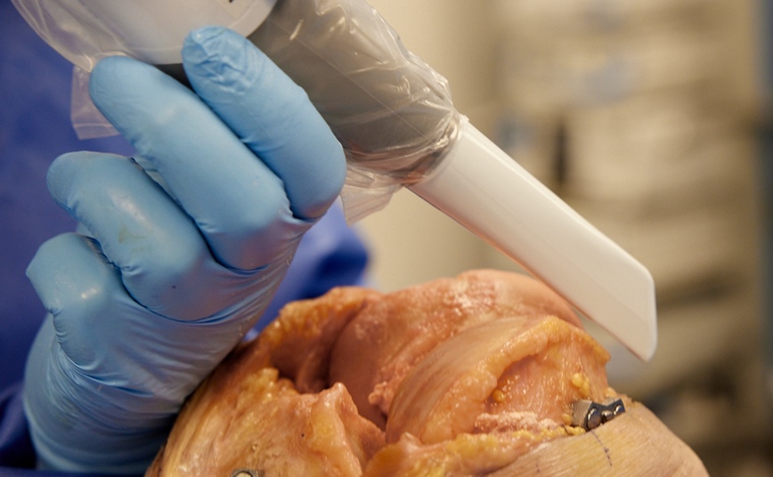
Each specimen was placed in the supine position and secured to the table proximally. A leg holder was positioned to hold the knee at 90o of flexion. A standard medial parapatellar incision was carried out on each specimen. Soft tissue dissection was completed to allow for exposure of the tibia and femur. Care was taken to ensure that the articular surface was not damaged during the preparation. Once adequate exposure of the tibia and femur was achieved, a portable handheld laser scanner (E4D Technologies, Dallas, TX) was used to scan the surface topology of the cartilage (Figure 1). The femur and tibia were scanned separately. Each specimen was scanned 3 times by four different operators to calculate inter- and intra-observer reliability. Operators included a senior orthopedic surgeon and a fellow specialized in arthroplasty, as well as 2 engineers experienced in navigation technologies. Time for each scan was recorded.

Figure 1
Figure 1.Registration of a cadaveric knee utilizing the handheld laser scanner.

Laser Scan Registration Validation

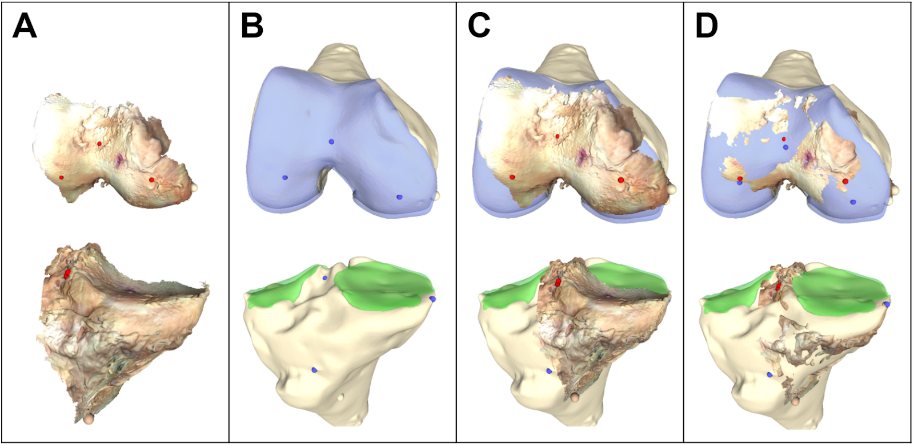
After the completion of laser scanning, the 3D models were registered to the segmented MRI scans that were completed prior to the procedure. An Iterative Closest Point (ICP) algorithm was used to align the handheld laser scans to the MRI scans within the registration application (Caira Surgical, New York, NY) (Figure 2). The accuracy of the registration fit was determined by the Root Mean Square (RMS) difference between the two surfaces in the region of interest. The ICP algorithm attempted to identify the rigid transformation that best fits a cloud of points with a model (Du et al. 2017). The algorithm uses a least-squares method to minimize the sum of the squared differences between both sets of points. The algorithm was preset to use 85% of the point cloud for matching to allow for surface variation and outliers.

Figure 2
Figure 2.Auto-registration tool for femoral and tibial surfaces of one cadaver knee. A) handheld scanner, B) MRI scan with point cloud, C) handheld and MRI scans overlaid, D) handheld scan aligned with MRI scan.

Results

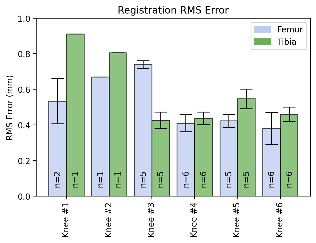
A total of 72 scans were completed by four different operators on the 6 cadaveric specimens. All scans were successfully registered and validated with the use of the ICP algorithm on the pre-procedure MRI. Each scan had an RMS error of <1mm for both the femoral and tibial scans (Figure 3). The mean time for successful registration with the handheld laser scanner was 54 ± 24 seconds. The inter- and intra-observer reliability were 0.92 and 0.82, respectively.

Figure 3
Figure 3.Registration RMS error for 6 cadaver knees where n is the number of individuals that scanned a specimen.

Discussion

The main goal of this study was to assess the utility of a handheld laser scanner for intraoperative registration of the tibial and femoral articular topography. This goal was achieved by comparing the 3D model formed by the laser scanner to models segmented from MRI scans collected prior to dissection of the cadaveric specimens. The results from our study demonstrated that surfaces of the tibia and femur could successfully be scanned using a handheld laser. When comparing the 3D models generated from the handheld laser scanner to the segmented MRI scans, the model had an average of less than 1 mm difference in RMS when compared to the scan. This was similarly seen by Chan et al. who utilized a light scanner to record bone topography and had an RMS error of 1.0 – 1.2mm in their in vitro study (Chan et al. 2016). However, the study utilized CT scan for segmented models to compare with the 3D scans, negating the cartilage. The current study was able to use MRI models, allowing for visualization of the cartilage.

Initial cost, user variability, need for specialized training, and increased operative time have hindered the adoption of CAS when performing a total knee arthroplasty in the United States (Jones and Jerabek 2018; Dutton et al. 2008; Bauwens et al. 2007). The use of a handheld laser scanner is able to overcome some of these limitations seen in other navigation systems. The laser scanner used in this study was able to achieve a highly accurate and reproducible registration of the joint surface topography with a mean time of 54 seconds. This is in contrast to the 5 to 11 minutes that may be necessary for probe-based navigation systems demonstrated in a study by Dutton et al (Dutton et al. 2008). Moreover, these results are based on a single surgeon performing 108 TKA which demonstrates the amount of time needed to register probe-based navigation systems even with experienced surgeons. Multiple operators in our study were able to accurately scan the tibio-femoral bone topography with minimal training and experience. Other navigation systems can require up to ten procedures for surgeons to become comfortable enough to acquire reproducible registration (Stulberg, Loan, and Sarin 2002). Similarly, robotics within other fields such as spine surgery may benefit from utilizing a laser scanner to reduce the registration time. Kostrzewski et al. utilized cadaveric models to demonstrate that registration and verification of takes approximately 7 minutes and 54 seconds to complete for cervical spine surgery (Kostrzewski et al. 2012).

The accuracy and reproducibility of measurements in a navigation system are crucial to ensuring appropriate positioning of TKA implants. Previous authors have discussed that deviations in coronal alignment of greater than 3° lead to increased risks of failure of the implants in a TKA (Berend et al. 2004; Ritter et al. 1994). The accuracy offered by laser scanners for TKA could help to reduce the risk of implant malpositioning. Winemaker et al. demonstrated that patients undergoing TKA with large preoperative alignment deformities averaging 17.1o had asymmetric flexion or extension gaps ≥3 mm when compared with patients with less preoperative deformity (Winemaker 2002). This asymmetry placed patients at increased risk of instability. The laser scanning technology assessed in this study is able to produce 3D models with a less than 1mm RMS when compared to MRI models. This accuracy may provide improvements in gap balancing especially in patients with larger deformities.

As this technology continues to improve, the authors believe that it will become integrated with the current navigation and robotics technology utilized in arthroplasty today. Rather than relying or CT scans or manual probing for knee surface topography for robotics, we hope to see this technology used to capture knee topography in real time during the procedure. Moreover, as technology continues to advance, we envision the ability to determine rotation utilizing a fiducial for augments 6 degrees of freedom tracking (Meftah 2022) based on bony anatomy similar to manual probing used in current navigation technology. The information gathered from the laser scanner will be combined with other information including standing alignment film radiographs to provide surgeons with the necessary information to accurately make their bony cuts and appropriate position implants.

There are several limitations to this study. Similar to other cadaveric studies, a limited number of specimens were tested in our study, however we had a very high inter- and intra-observer reliability. Lack of soft tissue bleeding was an additional limitation to this study, although scanning femur or tibia within 30 seconds can reduce the risk of blood interfering with registration. Additionally, preservation methods utilized for the cadavers may affect the MRI model, even though the cadavers were fresh-frozen.

In conclusion, the results of this cadaveric study demonstrated that a handheld laser scanner is a feasible method for registering the tibio-femoral joint. The technology is able to provide accurate, reproducible bone topography in a fraction of the time of other registration methods. These results demonstrate the utility of this technology when performing navigated TKA. Future studies should focus on assessing the technology through in vivo testing and the accuracy of implant positioning when compared to other navigation modalities. In addition, future studies should examine the integration of the laser registration with radar tracking technology to orient the scan with the surgical coordinate system.

Submitted: June 10, 2023 EDT

Accepted: August 17, 2023 EDT

References

Anderson, Kevin C., Knute C. Buehler, and David C. Markel. 2005. “Computer Assisted Navigation in Total Knee Arthroplasty: Comparison With Conventional Methods’.” The Journal of Arthroplasty 20 (October): 132–38.
Google Scholar
Antonios, Joseph K., Shane Korber, Lakshmanan Sivasundaram, Cory Mayfield, Hyunwoo Paco Kang, Daniel A. Oakes, and Nathanael D. Heckmann. 2019. “Trends in Computer Navigation and Robotic Assistance for Total Knee Arthroplasty in the United States: An Analysis of Patient and Hospital Factors.” Arthroplasty Today 5 (1): 88–95. https:/​/​doi.org/​10.1016/​j.artd.2019.01.002.
Google ScholarPubMed CentralPubMed
“Australian Orthopaedic Association National Joint Replacement Registry (AOANJRR). Hip, Knee & Shoulder Arthroplasty: 2020 Annual Report.” 2020. AOA. 2020. https:/​/​aoanjrr.sahmri.com/​documents/​10180/​689619/​Hip%2C+Knee+%26+Shoulder+Arthroplasty+New/​6a07a3b8-8767-06cf-9069-d165dc9baca7.
Bae, Dae Kyung, and Sang Jun Song. 2011. “Computer Assisted Navigation in Knee Arthroplasty.” Clinics in Orthopedic Surgery 3 (4): 259. https:/​/​doi.org/​10.4055/​cios.2011.3.4.259.
Google ScholarPubMed CentralPubMed
Bauwens, Kai, Gerrit Matthes, Michael Wich, Florian Gebhard, Beate Hanson, Axel Ekkernkamp, and Dirk Stengel. 2007. “Navigated Total Knee Replacement. A Meta-Analysis.” The Journal of Bone & Joint Surgery 89 (2): 261–69. https:/​/​doi.org/​10.2106/​jbjs.f.00601.
Google Scholar
Bellemans, J., S. Banks, J. Victor, H. Vandenneucker, and A. Moemans. 2002. “Fluoroscopic Analysis of the Kinematics of Deep Flexion in Total Knee Arthroplasty. . Influence of Posterior Condylar Offset.” The Journal of Bone and Joint Surgery. British Volume 84-B (1): 50–53. https:/​/​doi.org/​10.1302/​0301-620x.84b1.0840050.
Google Scholar
Berend, Michael E, Merrill A Ritter, John B Meding, Philip M Faris, E Michael Keating, Ryan Redelman, Gregory W Faris, and Kenneth E Davis. 2004. “Tibial Component Failure Mechanisms in Total Knee Arthroplasty.” Clinical Orthopaedics & Related Research 428 (428): 26–34. https:/​/​doi.org/​10.1097/​01.blo.0000148578.22729.0e.
Google Scholar
Chan, Brandon, Jason Auyeung, John F. Rudan, Randy E. Ellis, and Manuela Kunz. 2016. “Intraoperative Application of Hand-Held Structured Light Scanning: A Feasibility Study.” International Journal of Computer Assisted Radiology and Surgery 11 (6): 1101–8. https:/​/​doi.org/​10.1007/​S11548-016-1381-8/​FIGURES/​5.
Google ScholarPubMed
Chin, Pak Lin, Kuang Ying Yang, Seng Jin Yeo, and Ngai Nung Lo. 2005. “Randomized Control Trial Comparing Radiographic Total Knee Arthroplasty Implant Placement Using Computer Navigation Versus Conventional Technique.” The Journal of Arthroplasty 20 (5): 618–26. https:/​/​doi.org/​10.1016/​j.arth.2005.04.004.
Google Scholar
Delp, Scott L., David S. Stulberg, Brian Davies, Frederic Picard, and Francois Leitner. 1998. “Computer Assisted Knee Replacement.” Clinical Orthopaedics and Related Research 354 (354): 49–56. https:/​/​doi.org/​10.1097/​00003086-199809000-00007.
Google Scholar
Du, Shaoyi, Yiting Xu, Teng Wan, Huaizhong Hu, Sirui Zhang, Guanglin Xu, and Xuetao Zhang. 2017. “Robust Iterative Closest Point Algorithm Based on Global Reference Point for Rotation Invariant Registration.” Plos One 12 (11): e0188039. https:/​/​doi.org/​10.1371/​journal.pone.0188039.
Google ScholarPubMed CentralPubMed
Dutton, Andrew Quoc, Seng-Jin Yeo, Kuang-Ying Yang, Ngai-Nung Lo, Kui-Un Chia, and Hwei-Chi Chong. 2008. “Computer-Assisted Minimally Invasive Total Knee Arthroplasty Compared with Standard Total Knee Arthroplasty: A Prospective, Randomized Study.” The Journal of Bone and Joint Surgery-American Volume 90 (1): 2–9. https:/​/​doi.org/​10.2106/​jbjs.f.01148.
Google Scholar
Fang, David M., Merrill A. Ritter, and Kenneth E. Davis. 2009. “Coronal Alignment in Total Knee Arthroplasty: Just How Important Is It?” The Journal of Arthroplasty 24 (6): 39–43. https:/​/​doi.org/​10.1016/​j.arth.2009.04.034.
Google Scholar
Friederich, N., and R. Verdonk. 2008. “The Use of Computer-Assisted Orthopedic Surgery for Total Knee Replacement in Daily Practice: A Survey among ESSKA/SGO-SSO Members.” Knee Surgery, Sports Traumatology, Arthroscopy 16 (6): 536–43. https:/​/​doi.org/​10.1007/​S00167-008-0518-X/​FIGURES/​6.
Google ScholarPubMed
Haaker, Rolf G, Martin Stockheim, Michael Kamp, Gunnar Proff, Johannes Breitenfelder, and Andreas Ottersbach. 2005. “Computer-Assisted Navigation Increases Precision of Component Placement in Total Knee Arthroplasty.” Clinical Orthopaedics and Related Research, no. 433 (April), 152–59. https:/​/​doi.org/​10.1097/​01.blo.0000150564.31880.c4.
Google Scholar
Hofmann, S., J. Romero, E. Roth-Schiffl, and T. Albrecht. 2003. “[Rotational Malalignment of the Components May Cause Chronic Pain or Early Failure in Total Knee Arthroplasty].” Der Orthopäde 32 (6): 469–76. https:/​/​doi.org/​10.1007/​s00132-003-0503-5.
Google Scholar
Jenny, Jean–Yves, and Cyril Boeri. 2004. “Low Reproducibility of the Intra–Operative Measurement of the Transepicondylar Axis during Total Knee Replacement.” Acta Orthopaedica Scandinavica 75 (1): 74–77. https:/​/​doi.org/​10.1080/​00016470410001708150.
Google Scholar
Jones, Christopher W., and Seth A. Jerabek. 2018. “Current Role of Computer Navigation in Total Knee Arthroplasty.” The Journal of Arthroplasty 33 (7): 1989–93. https:/​/​doi.org/​10.1016/​j.arth.2018.01.027.
Google Scholar
Joshi, Shailesh V., and Philip J. Rowe. 2017. “A Novel Approach for Intra-Operative Shape Acquisition of the Tibio-Femoral Joints Using 3D Laser Scanning in Computer Assisted Orthopaedic Surgery.” The International Journal of Medical Robotics and Computer Assisted Surgery 14 (1): e1855. https:/​/​doi.org/​10.1002/​rcs.1855.
Google Scholar
Kostrzewski, S., J. M. Duff, C. Baur, and M. Olszewski. 2012. “Robotic System for Cervical Spine Surgery.” The International Journal of Medical Robotics and Computer Assisted Surgery 8 (2): 184–90. https:/​/​doi.org/​10.1002/​rcs.446.
Google Scholar
Kumar, P. J., and L. D. Dorr. 1997. “Severe Malalignment and Soft-Tissue Imbalance in Total Knee Arthroplasty.” The American Journal of Knee Surgery 10 (1): 36–41. https:/​/​europepmc.org/​article/​med/​9051176.
Google ScholarPubMed
Leitner, F., F. Picard, R. Minfelde, H. J. Schulz, P. Cinquin, and D. Saragaglia. 1997. “Computer-Assisted Knee Surgical Total Replacement.” Lecture Notes in Computer Science (Including Subseries Lecture Notes in Artificial Intelligence and Lecture Notes in Bioinformatics) 1205:629–38. https:/​/​doi.org/​10.1007/​BFB0029288/​COVER.
Google Scholar
Liu, Hai xiao, Ping Shang, Xiao Zhou Ying, and Yu Zhang. 2016. “Shorter Survival Rate in Varus-Aligned Knees after Total Knee Arthroplasty.” Knee Surgery, Sports Traumatology, Arthroscopy 24 (8): 2663–71. https:/​/​doi.org/​10.1007/​S00167-015-3781-7/​FIGURES/​7.
Google ScholarPubMed
Meftah, Morteza. 2022. Surgical System with Intra-operative 3D Scanners and Surgical Methods Using the Same. 20220241047, issued April 13, 2022. https:/​/​patentimages.storage.googleapis.com/​5e/​33/​ba/​94b65af953dea2/​US20220241047A1.pdf.
Ritter, M. A., P. M. Faris, E. M. Keating, and J. B. Meding. 1994. “Postoperative Alignment of Total Knee Replacement Its Effect on Survival.” Clinical Orthopaedics and Related Research 299 (299): 153–56. https:/​/​doi.org/​10.1097/​00003086-199402000-00021.
Google Scholar
Robinson, Mitchel, Donald G Eckhoff, Karl D Reinig, Michelle M Bagur, and Joel M Bach. 2006. “Variability of Landmark Identification in Total Knee Arthroplasty.” Clinical Orthopaedics &amp; Related Research 442 (January):57–62. https:/​/​doi.org/​10.1097/​01.blo.0000197081.72341.4b.
Google Scholar
Sharkey, Peter F., Paul M. Lichstein, Chao Shen, Anthony T. Tokarski, and Javad Parvizi. 2014. “Why Are Total Knee Arthroplasties Failing Today—Has Anything Changed After 10 Years?” The Journal of Arthroplasty 29 (9): 1774–78. https:/​/​doi.org/​10.1016/​j.arth.2013.07.024.
Google Scholar
Sikorski, J. M. 2008. “Alignment in Total Knee Replacement.” Journal of Bone and Joint Surgery - Series B 90 (9): 1121–27. https:/​/​doi.org/​10.1302/​0301-620X.90B9.20793/​ASSET/​IMAGES/​LARGE/​F5.MEDIUM.JPEG.
Google ScholarPubMed
Stulberg, S. David, Peter Loan, and Vineet Sarin. 2002. “Computer-Assisted Navigation in Total Knee Replacement: Results of an Initial Experience in Thirty-Five Patients.” The Journal of Bone and Joint Surgery-American Volume 84 (SUPPL. 2): 90–98. https:/​/​doi.org/​10.2106/​00004623-200200002-00011.
Google Scholar
Winemaker, M.J. 2002. “Perfect Balance in Total Knee Arthroplasty: The Elusive Compromise.” The Journal of Arthroplasty 17 (1): 2–10. https:/​/​doi.org/​10.1054/​arth.2002.29321.
Google Scholar

This website uses cookies

We use cookies to enhance your experience and support COUNTER Metrics for transparent reporting of readership statistics. Cookie data is not sold to third parties or used for marketing purposes.

Powered by Scholastica, the modern academic journal management system