Loading [Contrib]/a11y/accessibility-menu.js
Skip to main content
null
J Orthopaedic Experience & Innovation
  • Menu
  • Articles
    • Brief Report
    • Case Report
    • Data Paper
    • Editorial
    • Hand
    • Meeting Reports/Abstracts
    • Methods Article
    • Product Review
    • Research Article
    • Review Article
    • Review Articles
    • Systematic Review
    • All
  • For Authors
  • Editorial Board
  • About
  • Issues
  • Blog
  • "Open Mic" Topic Sessions
  • Advertisers
  • Recorded Content
  • CME
  • JOEI KOL Connect
  • search

RSS Feed

Enter the URL below into your favorite RSS reader.

https://journaloei.scholasticahq.com/feed
Case Report
Vol. 4, Issue 2, 2023November 19, 2023 EDT

High Grade Osteosarcoma at the Site of a Ceramic on Cross-Linked Polyethylene Cementless Hip Replacement: A Case Report and Review of the Literature

Lindsay C. Scally, Adam S. Levin, MD, Boby K. Manu, Frank R. DiMaio, MD, MBA,
High Grade OsteosarcomaTotal Hip ArthroplastyCeramic ArticulationsCase ReportSarcomaTHA
Copyright Logoccby-nc-nd-4.0 • https://doi.org/10.60118/001c.82075
J Orthopaedic Experience & Innovation
Scally, Lindsay C., Adam S. Levin, Boby K. Manu, and Frank R. DiMaio. 2023. “High Grade Osteosarcoma at the Site of a Ceramic on Cross-Linked Polyethylene Cementless Hip Replacement: A Case Report and Review of the Literature.” Journal of Orthopaedic Experience & Innovation 4 (2). https:/​/​doi.org/​10.60118/​001c.82075.
Save article as...▾
Download all (7)
  • Click here to learn more about Mako Total Knee 2.0
    Download
  • Click here to learn more about The Orthopaedic Implant Company
    Download
  • Figure 1. Pre-operative AP Radiograph (a) and AP Radiograph 1-year post-operative (b) confirming the success of the THA.
    Download
  • Figure 2. AP Pelvis Radiograph 3.2 Years post-operatively (a) and CT scan of Gluteus Medius Muscular Infiltration (b) which were unremarkable for acute etiology of the patient’s pain.
    Download
  • Figure 3. High grade sarcoma with numerous osteoclast type giant cells (a). Atypical, polygonal osteoblast production of lace-like osteoid (b). Necrotic tumor with numerous osteoclast type giant cells (c). Arrow illustrating atypical mitosis (d).
    Download
  • Click here to learn more about Zilretta
    Download
  • Click here to learn more about ConMed
    Download

Sorry, something went wrong. Please try again.

If this problem reoccurs, please contact Scholastica Support

Error message:

undefined

View more stats

Abstract

The following case report describes a 50-year-old male with a history of post-traumatic arthritis of the right hip after a previous open reduction of a traumatic hip dislocation as a young adult. He underwent an uncomplicated ceramic on highly cross-linked polyethylene primary total hip arthroplasty (THA), with unremarkable intra-operative pathology at that time. Three years postoperatively, the patient developed acute onset of hip and groin pain and associated radiographic findings which proved to be related to a high-grade osteosarcoma within the soft tissue juxtaposed to the ceramic on highly cross-linked polyethylene THA. While metal-associated primary bone sarcomas have been previously reported, we are unaware of any prior description of an osteosarcoma in the setting of a ceramic on highly cross-linked polyethylene articulation. This current report represents the first case of a primary osteosarcoma associated with this type of hip arthroplasty articulation.

Introduction

Total hip arthroplasty (THA) is a commonly performed procedure in the current practice of Orthopaedic Surgery, with over 400,000 primary and revision THAs performed yearly (Levett et al. 2023). This common procedure is not without its risks however, as the development of local malignancy associated with THA has become increasingly recognized in the literature. Although osteosarcomagenesis is rare, questions regarding patient risk and its association with implant articulations continue. Recent literature has been published implicating Metal on Metal (MoM) articulation debris as a source of local inflammatory disease (Bozic et al. 2012), as well as increasing suspicion that prosthetic joint implant articulations may provoke a biological reaction that leads to the development of malignant tumors (Gillespie et al. 1988). The question remains whether there are particular types of implants that increase the likelihood of developing such malignancies. Until now research seems to have pinned the development of bone sarcomas to MoM or metal-on-poly articulations, while ceramic articulations have rarely been implicated. In January 2023 the first report of osteosarcoma around a Ceramic-on-Ceramic (CoC) THA was published (Levett et al. 2023), and here we report the first case report in the literature describing a high-grade osteosarcoma developing at the site of a ceramic on UHMWPE cementless total hip arthroplasty three years after undergoing index arthroplasty procedure. Osteosarcomas are extremely rare, comprising only 4% of all diagnosed sarcomas, with known risk factors including radiation, chronic infections, or exposure to chemicals in drinking water such as Fluoride or Radium (Burningham et al. 2012). This report opens the argument that the literature may have been wrong in their association of osteosarcomagenesis with metal, as a patient with no other risk factors developed such a malignancy, and that we may have to allocate more resources into investigating ceramic articulations as well.

Click here to learn more about Mako Total Knee 2.0

Case History

The patient was a 50-year-old male who had initially sustained a dislocation of the right hip after a motor vehicle accident as a young adult. He ultimately developed post-traumatic arthritis of the right hip following an open reduction of the traumatic hip dislocation. The patient’s only pertinent past medical history included deep vein thrombosis and thoracotomy secondary to lung abscess.

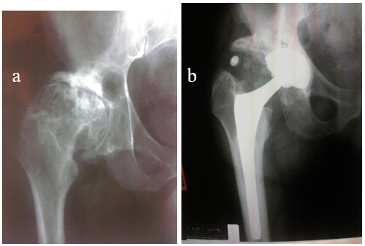
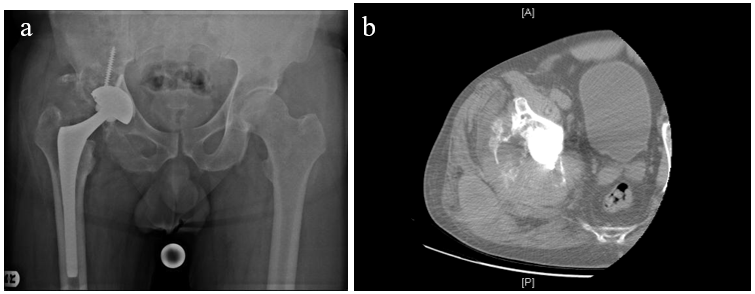
He underwent a total hip arthroplasty in January of 2008 for the post-traumatic arthritis, without postoperative complications. Radiographs at that time demonstrated no concerning findings (Fig. 1), and the patient recovered uneventfully from that procedure. At the time of the hip arthroplasty, no unexpected intraoperative findings were noted, and despite the prior trauma history the patient did not undergo perioperative hip radiotherapy for heterotopic ossification prophylaxis. The index pathology found no evidence of malignancy, noting “degenerative joint disease with a large subarticular pseudocyst and extensive fibro cartilaginous surface proliferation.” Three years postoperatively, the patient presented back to the primary arthroplasty surgeon, complaining of 1 week of ipsilateral hip and groin pain. On physical exam the patient had an antalgic gait but had painless PROM of the operative hip. Radiographs were without radiographic signs of loosening, nor aggressive features (Fig. 2).

Click here to learn more about The Orthopaedic Implant Company
Figure 1
Figure 1.Pre-operative AP Radiograph (a) and AP Radiograph 1-year post-operative (b) confirming the success of the THA.
Figure 2
Figure 2.AP Pelvis Radiograph 3.2 Years post-operatively (a) and CT scan of Gluteus Medius Muscular Infiltration (b) which were unremarkable for acute etiology of the patient’s pain.

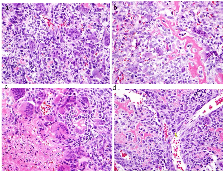
Three months later, the patient presented to our senior author with increasing pain in the right hip. At that time, in March 2011, the patient was admitted to the hospital and underwent a workup in suspicion of a periprosthetic joint infection. However, preliminary results were inconclusive for infection, and the decision was made to proceed with arthrotomy for tissue sampling. At the time of surgery, copious sanguineous fluid was yielded. Cultures and soft tissue samples for pathology were obtained. All final cultures were reported as negative; however, initial frozen sections revealed high grade osteosarcoma in all soft tissue samples (Fig. 3).

The patient was found to have developed a high-grade soft tissue osteosarcoma juxtaposed to the site of ceramic on cross linked polyethylene THA with metastatic lung disease, and was treated with palliative care, and subsequently expired.

Figure 3
Figure 3.High grade sarcoma with numerous osteoclast type giant cells (a). Atypical, polygonal osteoblast production of lace-like osteoid (b). Necrotic tumor with numerous osteoclast type giant cells (c). Arrow illustrating atypical mitosis (d).
Click here to learn more about Zilretta

Discussion

In a review of the available English language orthopaedic literature, there are no prior reports of osteosarcoma associated with alumina ceramic on UHMWPE articulations. Over the last 30 years, there have been approximately 40 number of cases associating in 8 series of bone and/or soft tissue tumors around the site of THA (Levett et al. 2023; Gillespie et al. 1988; Keel et al. 2001; Schuh et al. 2004; Lucas et al. 2001; Min et al. 2008; Rana et al. 2001; Syed et al. 2002; Stephensen et al. 1999; Visuri, Pulkkinen, and Paavolainen 2006; Memoli et al. 1986; Paavolainen et al. 1999). In humans, complications resulting from a THA vary from infection to the development of periprosthetic tumors. In our review, it is suggested that factors related to the surgery provide the trigger to tumorigenesis, though a definitive reason has not been determined. More likely, the presence of a foreign body is the predisposition. Such tumors may likely occur as a result from the wear and debris of implants, and can present as benign pseudotumors or could be associated with sarcoma (Levett et al. 2023). Serum levels of heavy metal ions in MoM articulations have been extensively reviewed, and osteosarcoma associated with CoC have recently been reported. The literature suggests that over time metallic implants will eventually corrode, and it is arguable that products of corrosion may be important in tumorigenesis (Bozic et al. 2012). This would seem less likely in ceramic articulations, where only the stem taper may be implicated. Although there are assumed etiologies of the development of periprosthetic masses and pseudotumors, the metastasis of such sarcomas is where the question remains. The majority of case reports of osteosarcoma following THA have been in canines, rather than humans. Interestingly, the wide time range to development of osteosarcoma in both canines and humans has been similar with intervals reported between 5 months and 19.5 years (Bozic et al. 2012; Gillespie et al. 1988; Burningham et al. 2012; Keel et al. 2001; Schuh et al. 2004; Langkamer et al. 1997; Goodfellow 1992; Solomon and Sekel 1992; Prasad et al. 2002; Dunn et al. 2012; Murphy, Parker, and Woodard 1997). Oncogenesis around the prosthesis has been revealed in animal experiments (Rana et al. 2001). According to large cohorts, however, the incidence of malignant tumors in THA patients is lower or similar to that in the general population (Syed et al. 2002). The causes of osteosarcomas can range from pubertal growth spurts, recessive DNA mutations, hereditary cancers, environmental/chemical exposures, and prior high dose radiation treatments (Burningham et al. 2012); none of which were relevant with our patient. In addition, the patient’s preliminary labs and imaging ruled out any preexisting malignancy or infection at the site of his THA, suggesting a potential association with his ceramic on UHMWPE cementless THA and the development of his osteosarcoma.

Click here to learn more about ConMed

Summary

We report a patient with hip pain three years post THA, diagnosed with high-grade osteosarcoma at the site of the arthroplasty with metastatic lung disease, causing his eventual expiration. While the Orthopaedic implant may be associated in some individuals with local malignancy, the potential risk at this time remains theoretical, and causality has not yet been established in humans. It is undeniable that the benefits of THA far outweigh the risks of possible malignancy, while the nature of tumor development remains obscure (Prasad et al. 2002; Apley 1989). Moreover, the possibility remains that the development of a malignancy may be coincidental. Therefore, we conclude that it is prudent to report all documented cases to help shed light on this exceedingly rare but devastating situation.


Acknowledgement

The author(s) confirm that informed consent has been obtained from the involved patient(s) or if appropriate from the parent, guardian, power of attorney of the involved patient(s); and, they have given approval for this information to be published in this case report (series).

Submitted: April 26, 2023 EDT

Accepted: June 23, 2023 EDT

References

Apley, AG. 1989. “Malignancy and Joint Replacement: The Tip of an Iceberg?” The Journal of Bone and Joint Surgery. British Volume 71-B (1): 1–1. https:/​/​doi.org/​10.1302/​0301-620x.71b1.2914975.
Google Scholar
Bozic, Kevin J., James Browne, Chris J. Dangles, Paul A. Manner, Adolph J. Yates Jr., Kristy L. Weber, Kevin M. Boyer, et al. 2012. “Modern Metal-on-Metal Hip Implants.” Journal of the American Academy of Orthopaedic Surgeons 20 (6): 402–6. https:/​/​doi.org/​10.5435/​jaaos-20-06-402.
Google Scholar
Burningham, Zachary, Mia Hashibe, Logan Spector, and Joshua D Schiffman. 2012. “The Epidemiology of Sarcoma.” Clinical Sarcoma Research 2 (14). https:/​/​doi.org/​10.1186/​2045-3329-2-14.
Google ScholarPubMed CentralPubMed
Dunn, A.L., E.A. Buffa, D.M. Hanshaw, and M. Farrell. 2012. “Osteosarcoma at the Site of Titanium Orthopaedic Implants in a Dog.” Australian Veterinary Journal 90 (1–2): 39–43. https:/​/​doi.org/​10.1111/​j.1751-0813.2011.00866.x.
Google Scholar
Gillespie, WJ, CM Frampton, RJ Henderson, and PM Ryan. 1988. “The Incidence of Cancer Following Total Hip Replacement.” The Journal of Bone & Joint Surgery. British Volume 70-B (4): 539–42. https:/​/​doi.org/​10.1302/​0301-620x.70b4.3403594.
Google Scholar
Goodfellow, J. 1992. “Malignancy and Joint Replacement.” The Journal of Bone and Joint Surgery. British Volume 74-B (5): 645–645. https:/​/​doi.org/​10.1302/​0301-620x.74b5.1527107.
Google Scholar
Keel, Suzanne B., Kenneth A. Jaffe, G. Petur Nielsen, and Andrew E. Rosenberg. 2001. “Orthopaedic Implant-Related Sarcoma: A Study of Twelve Cases.” Modern Pathology 14 (10): 969–77. https:/​/​doi.org/​10.1038/​modpathol.3880420.
Google Scholar
Langkamer, V.G., C.P. Case, C. Collins, I. Watt, J. Dixon, A.J. Kemp, and R.M. Atkins. 1997. “Tumors around Implants.” The Journal of Arthroplasty 12 (7): 812–18. https:/​/​doi.org/​10.1016/​s0883-5403(97)90013-9.
Google Scholar
Levett, Jordan J., Robert E. Turcotte, Sungmi Jung, John Antoniou, and Olga L. Huk. 2023. “Osteosarcoma Around a Ceramic-on-Ceramic Total Hip Arthroplasty.” Arthroplasty Today 19 (February):101094. https:/​/​doi.org/​10.1016/​j.artd.2022.101094.
Google ScholarPubMed CentralPubMed
Lucas, D. R., P. R. Miller, M. P. Mott, J. L. Kronick, and K. K. Unni. 2001. “Arthroplasty-Associated Malignant Fibrous Histiocytoma: Two Case Reports.” Histopathology 39 (6): 620–28. https:/​/​doi.org/​10.1046/​j.1365-2559.2001.01253.x.
Google Scholar
Memoli, Vincent A., Robert M. Urban, Joseph Alroy, and Jorge O. Galante. 1986. “Malignant Neoplasms Associated with Orthopedic Implant Materials in Rats.” Journal of Orthopaedic Research 4 (3): 346–55. https:/​/​doi.org/​10.1002/​jor.1100040311.
Google Scholar
Min, Woo-Kie, Shin-Yoon Kim, Chang-Wug Oh, Sung-Jung Kim, Tae-In Park, and Kyung-Hoi Koo. 2008. “Malignant Fibrous Histiocytoma Arising in the Area of Total Hip Replacement.” Joint Bone Spine 75 (3): 319–21. https:/​/​doi.org/​10.1016/​j.jbspin.2007.05.007.
Google Scholar
Murphy, S. T., R. B. Parker, and J. C. Woodard. 1997. “Osteosarcoma Following Total Hip Arthroplasty in a Dog.” Journal of Small Animal Practice 38 (6): 263–67. https:/​/​doi.org/​10.1111/​j.1748-5827.1997.tb03365.x.
Google Scholar
Paavolainen, Pekka, Eero Pukkala, Pekka Pulkkinen, and Tuomo Visuri. 1999. “Cancer Incidence in Finnish Hip Replacement Patients from 1980 to 1995.” The Journal of Arthroplasty 14 (3): 272–80. https:/​/​doi.org/​10.1016/​s0883-5403(99)90051-7.
Google Scholar
Prasad, P.S.V., J.B. Latham, J.K. Tucker, and R.Y. Ball. 2002. “Disseminated Osteosarcoma Arising in the Pelvis after Total Hip Arthroplasty.” The Journal of Arthroplasty 17 (3): 373–78. https:/​/​doi.org/​10.1054/​arth.2002.30290.
Google Scholar
Rana, B., S. Shetty, P. Grigoris, and R. Reid. 2001. “Sarcoma Arising Adjacent to a Total Hip Arthroplasty.” Scottish Medical Journal 46 (1): 17–19. https:/​/​doi.org/​10.1177/​003693300104600108.
Google Scholar
Schuh, Alexander, Gunther Zeiler, Ulrich Holzwarth, and Thomas Aigner. 2004. “Malignant Fibrous Histiocytoma at the Site of a Total Hip Arthroplasty.” Clinical Orthopaedics and Related Research 425 (August):218–22. https:/​/​doi.org/​10.1097/​00003086-200408000-00031.
Google Scholar
Solomon, Michael I., and Ronald Sekel. 1992. “Total Hip Arthroplasty Complicated by a Malignant Fibrous Histiocytoma.” The Journal of Arthroplasty 7 (4): 549–50. https:/​/​doi.org/​10.1016/​s0883-5403(06)80077-x.
Google Scholar
Stephensen, S. L., G. Schwarz Lausten, H. S. Thomsen, and B. Bjerregaard. 1999. “Liposarcoma in Association with Total Hip Replacement.” International Orthopaedics 23 (3): 187–89. https:/​/​doi.org/​10.1007/​s002640050344.
Google ScholarPubMed CentralPubMed
Syed, A.A., M. Agarwal, G. Fenelon, and M. Toner. 2002. “Osseous Malignant Non-Hodgkin’s B-Cell Lymphoma Associated with Total Hip Replacement.” Leukemia & Lymphoma 43 (11): 2213–16. https:/​/​doi.org/​10.1080/​1042819021000016140.
Google Scholar
Visuri, Tuomo, Pekka Pulkkinen, and Pekka Paavolainen. 2006. “Malignant Tumors at the Site of Total Hip Prosthesis. Analytic Review of 46 Cases.” The Journal of Arthroplasty 21 (3): 311–23. https:/​/​doi.org/​10.1016/​j.arth.2005.03.046.
Google Scholar

This website uses cookies

We use cookies to enhance your experience and support COUNTER Metrics for transparent reporting of readership statistics. Cookie data is not sold to third parties or used for marketing purposes.

Powered by Scholastica, the modern academic journal management system