Loading [Contrib]/a11y/accessibility-menu.js
Skip to main content
null
J Orthopaedic Experience & Innovation
  • Menu
  • Articles
    • Brief Report
    • Case Report
    • Data Paper
    • Editorial
    • Hand
    • Meeting Reports/Abstracts
    • Methods Article
    • Product Review
    • Research Article
    • Review Article
    • Review Articles
    • Systematic Review
    • All
  • For Authors
  • Editorial Board
  • About
  • Issues
  • Blog
  • "Open Mic" Topic Sessions
  • Advertisers
  • Recorded Content
  • CME
  • JOEI KOL Connect
  • search

RSS Feed

Enter the URL below into your favorite RSS reader.

http://localhost:22663/feed
Case Report
Vol. 4, Issue 2, 2023October 28, 2023 EDT

Utilization of 1.25mm wire and locking plate fixation for a comminuted low transverse distal humerus fracture, a case report of a 78-year-old woman.

Michael R McDermott, Naem A Mufarreh, DO, Brian A Handal, Sean L Gao, DO, H. Brent Bamberger, DO,
Distal humerusSkinny WireLow Transverse FractureORIFTechniqueTips and TricksImplanted WireOsteoporosis
Copyright Logoccby-nc-nd-4.0 • https://doi.org/10.60118/001c.82037
J Orthopaedic Experience & Innovation
McDermott, Michael R, Naem A Mufarreh, Brian A Handal, Sean L Gao, and H. Brent Bamberger. 2023. “Utilization of 1.25mm Wire and Locking Plate Fixation for a Comminuted Low Transverse Distal Humerus Fracture, a Case Report of a 78-Year-Old Woman.” Journal of Orthopaedic Experience & Innovation 4 (2). https:/​/​doi.org/​10.60118/​001c.82037.
Save article as...▾
Download all (8)
  • Click here to learn more about The Orthopaedic Implant Company
    Download
  • Figure 1. A-B: AP and lateral x-rays of an elbow demonstrating a low transverse extra-articular distal humerus fracture, AO/OTA.A.3.
    Download
  • Click here to learn more about Pacira
    Download
  • Figure 2. Interoperative C-arm radiographs showing initial wire placement (A) and final placement after the wires were cut and secured with a posteriorlateral plate (B). C & D are interoperative images highlighting how the cut wires were secured beneath the posteriorlateral plate.
    Download
  • Figure 3. Elbow range of motion measurements at seven weeks post skinny wire ORIF, represents as percentage of range of motion of uninjured arm.
    Download
  • Figure 4. A-C: AP, Lateral and radial head views of a right elbow demonstrating fracture healing, five months post skinny wire ORIF
    Download
  • Click here to learn more about ConMed
    Download
  • Click here to learn more about MotionSense
    Download

Sorry, something went wrong. Please try again.

If this problem reoccurs, please contact Scholastica Support

Error message:

undefined

View more stats

Abstract

Introduction

Low transverse distal humerus fractures can be challenging to treat in elderly populations—especially when there is severe comminution or underlying bone demineralization. Current operative interventions include total elbow arthroplasty or open reduction internal fixation (ORIF) with traditional plate and screw fixation. Herein, we present a unique case utilizing Kirschner wires (K-wires) and locking plate fixation as an alternative approach to managing low transverse distal humerus fractures.

Case

A 78-year-old right-hand dominant retired female presented with a displaced, comminuted, low transverse distal humerus fracture secondary to a fall from standing. Fixation of this fracture was achieved through a slight modification of the skinny K-wire and locking plate technique described by Olson et al. in 2021.

Methods

A triceps-sparing approach to the distal humerus was conducted through a direct posterior incision. The low transverse extra-articular fracture was identified and anatomically reduced. A posterior medial distal humerus plate was secured to reduce the medial column with 1.25x150mm K-wires subsequently placed through the trochlea. The K-wires were then bent as they exited the lateral side and cut short in the metaphysis, approximately 90° to the shaft of the humerus. After which, a posterolateral distal humerus plate was used to secure the lateral epicondyle and secures the K-wires beneath it.

Results

Seven weeks after the procedure, the patient had regained 81% elbow arc of motion, with a complete return of grip strength, pronation, and supination. At this time, radiographs demonstrated satisfactory bone healing, and the patient surpassed all therapy goals.

Conclusion

Our case demonstrates the modified use of the 1.25mm K-wire and locking plate technique as a satisfactory treatment option for comminuted low transverse distal humerus fracture in a 78-year-old female who sustained a ground-level fall. Ultimately resulting in the rapid return of function by postoperative week seven.

Introduction

Extra-articular distal humerus fractures are uncommon and account for 3-6% of all fractures (Ekholm et al. 2006; Epps and Grant 1991; Buhr and Cooke 1959). Adequate reduction often requires operative intervention consisting of open reduction and internal fixation (ORIF) or total elbow arthroplasty (TEA); however, despite proper fixation, patient satisfaction is often lacking (Ekholm et al. 2006; Epps and Grant 1991; Buhr and Cooke 1959; Obremskey et al. 2003; Lauder and Richard 2020; Aitken and Rorabeck 1986; Rose et al. 1982; Anglen 2005; Jupiter and Mehne 1992). Furthermore, the operative intervention of these fractures is time-sensitive in an effort to restore alignment and range of motion (ROM) of the shoulder and elbow (Self et al. 1995; Waddell, Hatch, and Richards 1988; O’Driscoll 2005). This is imperative as several factors can increase the morbidity associated with distal humerus fractures, including the degree of comminution and bone demineralization. This becomes ever more pressing as we note the substantial increase in fragility fractures within the past 50 years—as well as the subsequent morbidity associated with these injuries (Palvanen et al. 1998, 2003; Kannus et al. 2002; Curtis et al. 2017) despite the plethora of available treatment options (Curtis et al. 2017; Giangregorio et al. 2006; Kanis et al. 2014). Thus, as patients continue to have suboptimal results with medical treatment, we must develop new approaches to combat these fractures surgically. Herein, we present a unique fixation method to aid orthopaedic surgeons in addressing these fractures. Our case explicitly highlights the use of 1.25x150mm (skinny) K-wires and dual plating to operatively manage a comminuted, displaced, low transverse distal humerus fracture in a 78-year-old female.

Click here to learn more about The Orthopaedic Implant Company

Case

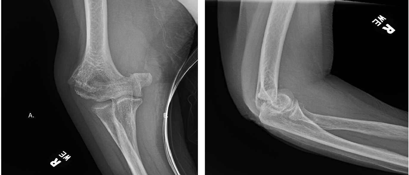
A 78-year-old, right-hand dominant female presented to the emergency department after a ground-level fall with right elbow pain and decreased range of motion. Her pertinent medical history was remarkable for hypertension, intentional tremor treated with deep brain stimulator placement, osteopenia, previous ORIF of bilateral distal radius fractures, as well as alcohol and tobacco use. Physical examination revealed significant lateral right elbow swelling, pain at the elbow with wrist rotation, and limited range of motion secondary to guarding. The right upper extremity was neurovascularly intact throughout all motor and sensory distributions. Radiographic examination revealed a comminuted extra-articular distal humerus (Figure 1). Given the extent of the comminution and bone demineralization, there was concern that traditional ORIF with plate and screw fixation would not sufficiently capture the comminuted fragments. Therefore, utilizing a slight modification of the technique established by Olson et al (Olson and Dyer 2021)., dual locking plates were used in conjunction with skinny K-wires to achieve fractures fixation.

Figure 1
Figure 1.A-B: AP and lateral x-rays of an elbow demonstrating a low transverse extra-articular distal humerus fracture, AO/OTA.A.3.

Surgical description

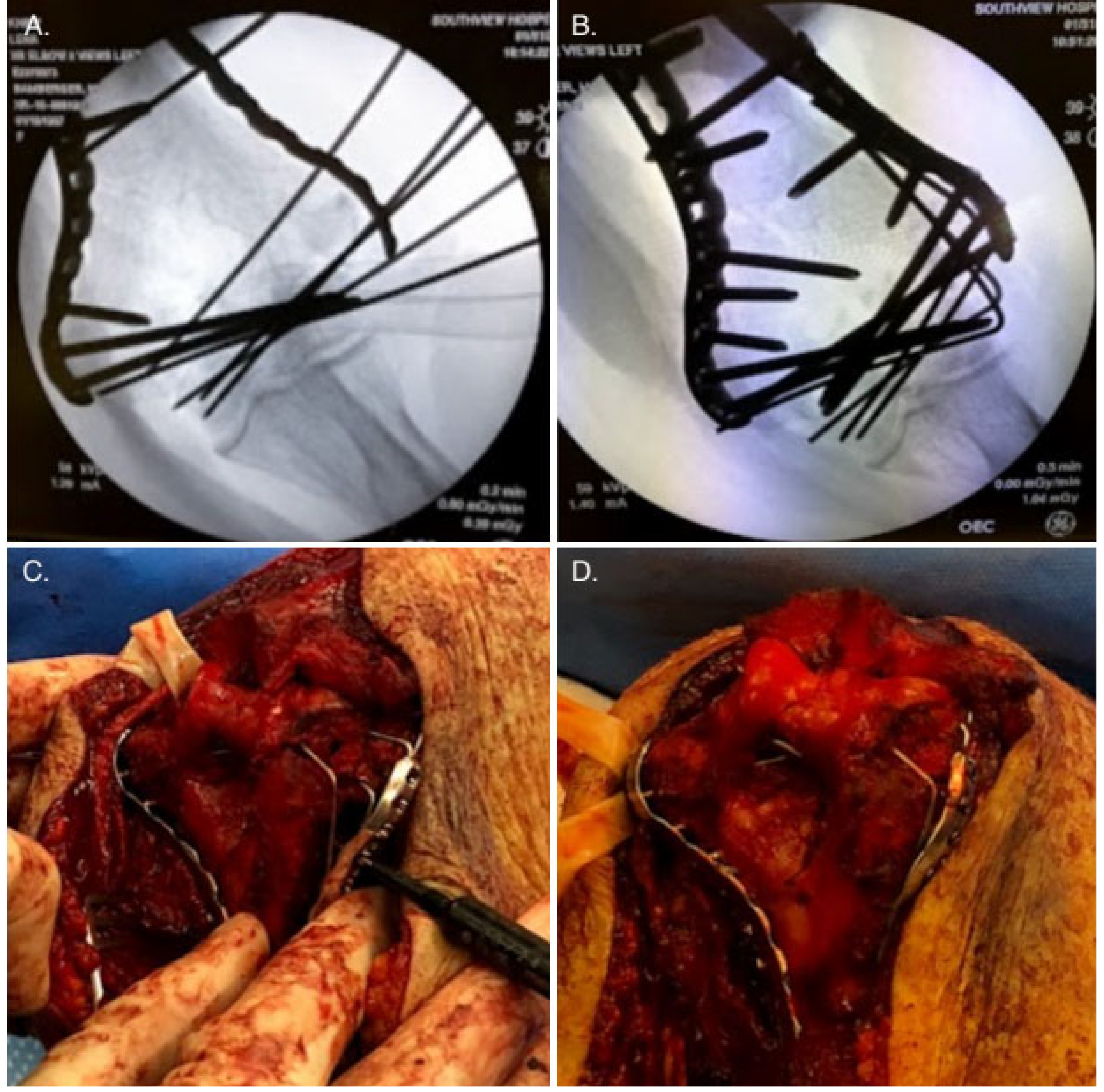
The patient was placed in the supine position on the operating table with the operative extremity flexed 90 degrees and placed across her chest. Utilizing a posterior incision, a triceps-sparing approach to the distal humerus was taken, creating a medial and lateral window. The fracture was identified as a comminuted low transverse fracture just distal to the olecranon fossa. The fracture was then manually reduced. Once anatomic alignment was achieved, provisional 1.25x150mm K-wires were placed to attach the disarticulate piece to the metaphyseal bone. A posteromedial distal humerus plate was secured with locking screws yielding a satisfactory reduction of the medial column. Skinny K-wires were subsequently placed in a lateral to medial fashion through the trochlea under direct fluoroscopy, thus securing the comminuted fragments. The wires were then bent along the lateral side of the contour toward the shaft and cut short in the metaphyseal bone lesion. The wires were secured in the same technique as Olson, with the subtle change of shortening the wires prior to administration of the plate, intraoperative images shown in figure 2. A posterolateral distal humerus plate was applied to the lateral epicondyle, securing the 1.25-mm k-wires beneath. The lateral plate was subsequently secured with locking screws. Intraoperative range of motion was assessed, and final C-arm radiographs revealed a satisfactory reduction of the articular fragment to the shaft with restoration of the underlying articular surface. Upon completion of the procedure, a soft dressing and ace wrap was applied to the operative extremity.

Click here to learn more about Pacira
Figure 2
Figure 2.Interoperative C-arm radiographs showing initial wire placement (A) and final placement after the wires were cut and secured with a posteriorlateral plate (B). C & D are interoperative images highlighting how the cut wires were secured beneath the posteriorlateral plate.

Results

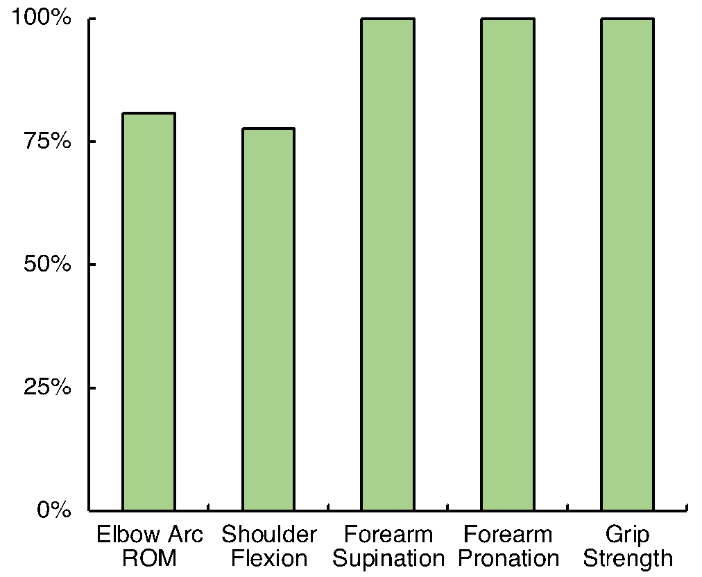
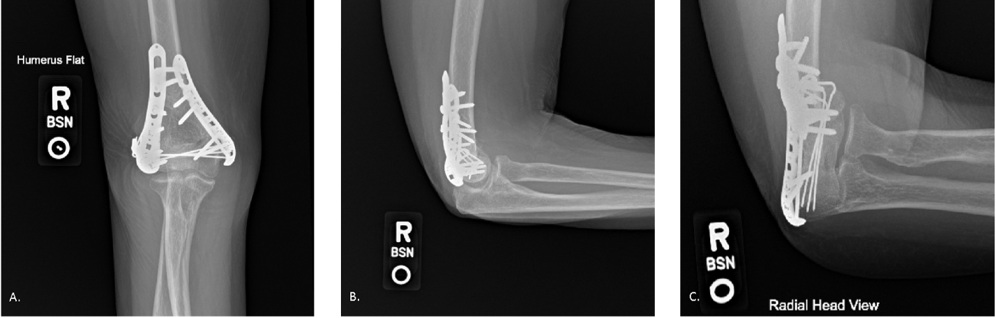
The patient began physical therapy six days after operative fixation, focusing on passive range of motion. Utilizing the contralateral upper extremity as a baseline: pain, range of motion, edema, and grip strength measures were trended (Figure 3). The patient self-reported her ability to perform activities of daily living (ADL) on a weekly basis. By postoperative week four the patient demonstrated substantial improvement in all tested categories, with graduation from therapy at postoperative week seven. At this point, she reported a 91% reduction in pain as well as the ability to use her right upper extremity for a majority of ADLs. Her grip strength had returned to baseline with a substantial reduction in edema of the affected extremity. Her total elbow range of motion was 81% of her non-injured extremity (117º elbow arc of motion). Additionally, the patient tolerated therapy and displayed successful bone healing on serial radiographs without the need for hardware removal or re-operation. Complete fracture healing was achieved at postoperative month five, as evidenced by radiographic examination (Figure 4).

Figure 3
Figure 3.Elbow range of motion measurements at seven weeks post skinny wire ORIF, represents as percentage of range of motion of uninjured arm.
Figure 4
Figure 4.A-C: AP, Lateral and radial head views of a right elbow demonstrating fracture healing, five months post skinny wire ORIF
Click here to learn more about ConMed

Discussion

Several surgical options are currently employed to treat low transverse distal humerus fractures. Yet, there are still many challenges to ORIF of said fractures, including complex fixation, post-operative stiffness, and fracture nonunion (Epps and Grant 1991; Buhr and Cooke 1959; Obremskey et al. 2003; Anglen 2005; Jupiter and Mehne 1992; Tejwani et al. 2009; Prasarn et al. 2011; Rajput et al. 2016). The incorporation of implanted K-wires for a distal humerus fracture was first suggested by Kamrani et al. in 2012 when they reported on 19 patients who underwent “pin and plate fixation” for AO 13. A.1, and C.1-C.3 fractures (Kamrani et al. 2011). Following that report, Abdoli et al. reported on 14 patients, adapting the “pin and plate fixation” technique for distal humerus articular fractures (Abdoli, Farhoud, and Kamrani 2020). Both Kamrani’s and Abdoli’s studies demonstrated successful healing and early mobilization of the elbow. However, they reported significant complications, with a reoperation rate of 42.1% (8/19) and 50% (7/14) for Kamrani and Abdoli, respectively. In both Kamrani’s and Abdoli’s technique, the wires were bent on the lateral side and secured to the bone by a 3 or 4 hole plate, leaving a majority of the wires uncovered by the plate. Both Kamrani’s and Abdoli’s high reoperation rate stemmed from prominent wires, skin irritation, wire migration into the joint, and symptomatic hardware from their olecranon osteotomy fixation. Despite this, both studies illustrated a substantial return of elbow function with a total mean arc motion total of 99° for Kamrani and 97° for Abdoli in cohorts with an average age of 46 and 36, respectively (Kamrani et al. 2011; Abdoli, Farhoud, and Kamrani 2020).

In 2021, Olson et al. first described their skinny wire and locking plate fixation technique in 30 patients with comminuted intra-articular distal humerus fractures. Olson reported excellent functional results in the cohort with an average age of 59. 26% of their patients had known osteoporosis, and most of the fractures resulted from a ground level fall. In their technique, the fractures were anatomically reduced, and multiple skinny wires were placed to secure the fragments. These wires were then bent as they exited laterally, molded to the shaft and a secured beneath a lateral plate that covered a majority of the wire. For plate selection, they used only a lateral plate in 10% (3/30), bicolumnar plating in 81% (24/30), and bicolumnar + posteriolateral plating in 9% (2/30) of cases. Olson’s skinny K-wire and locking plate fixation technique showed 100% fracture union and average arc elbow motion of 102°. Their technique only resulted in a 35% (11/30) reoperation rate, with 8 having symptomatic hardware leading to its removal (Olson and Dyer 2021; Dryer 2020).

The case presented herein was designed to confirm Olson’s skinny K-wire and locking plate fixation technique, with a few key modifications in an attempt to reduce the reoperation rate. The triceps-sparing approach eliminated the complications that Olson, Abdoli, and Kamrani faced related to their olecranon osteotomy. To reduce hardware prominence, posterolateral and posteromedial plates were selected rather than direct lateral and medial plates. In the procedure, the order of plate and wire placements was also adjusted. After achieving anatomic reduction, a posteromedial plate was used to secure the medial column. This provided some stability of the fracture while skinny wires were implanted, ultimately reducing the total number of wires needed to secure the comminuted fragments. As Olson described, the wires exited laterally were bent to the contour of the distal humerus, however, in this case, the wires were shortened before the posterolateral plate was used to secure them to the lateral column. While these are slight modifications, reducing the number of wires, shortening the length of the wires outside of bone, and plate selection were all done in an effort reduce the overall hardware burden for the patient. For this patient, there was no skin irritation, no concern for pin migration, and no reported symptomatic hardware. Radiographic evidence of fracture healing coincided with the 78-year- old patient’s timely reduction in pain and quick recovery of ROM. Olson, Kamrani, and Abdoli reported follow-ups at a year or longer demonstrating elbow arcs of 102°, 99°, and 97° respectively, while this case demonstrates an arc of 117° after just seven weeks of recovery (Olson and Dyer 2021; Kamrani et al. 2011; Abdoli, Farhoud, and Kamrani 2020).

While this report demonstrates a successful case for this patient, it does not provide the opportunity to statistically compare the outcomes due to an overall low power with an n-value of one. A greater number of cases would be required to adequately assess the quality of outcomes from this technique. Given this, we plan to follow with a case series to further establish this modification of the skinny K-wire and dual-locking plate fixation method as a viable treatment option for distal humerus fractures.

Click here to learn more about MotionSense

Conclusion

The skinny k-wire and dual locking plate fixation technique was designed by Olson et al. to provide a structurally sound option for treating significant comminution distal humerus fractures. This study demonstrates a slight modification of this technique that yielded an expedited recovery time, significant improvement in pain, restoration of range of motion, and preserved overall function. Successfully treating a 78-year-old woman with a low transverse distal humerus fracture with minimal minimizing postoperative complications such as the need for reoperation or salvage procedures.

Thus, we find skinny K-wire and dual plate fixation to be a viable treatment option for distal humerus fractures complicated by comminution and bone demineralization. While a larger patient cohort is needed to fully access this procedure—we believe it is necessary to share this slightly modified technique in the interim with the orthopaedic community—to aid in treating patients with complex, comminuted, and/or demineralized distal humerus fractures.


This article will be in press until the end of 2023.

Submitted: February 11, 2023 EDT

Accepted: June 23, 2023 EDT

References

Abdoli, Abbas, Amir Reza Farhoud, and Reza Shahryar Kamrani. 2020. “Treatment of Distal Humerus Articular Fracture with Pin-and-Plate Technique.” The Journal of Hand Surgery (Asian-Pacific Volume) 25 (03): 332–39. https:/​/​doi.org/​10.1142/​s242483552050037x.
Google Scholar
Aitken, G. K., and C. H. Rorabeck. 1986. “Distal Humeral Fractures in the Adult.” Clinical Orthopaedics and Related Research 207 (June):191–97. https:/​/​doi.org/​10.1097/​00003086-198606000-00034.
Google Scholar
Anglen, Jeffrey. 2005. “Distal Humerus Fractures.” Journal of the American Academy of Orthopaedic Surgeons 13 (5): 291–97. https:/​/​doi.org/​10.5435/​00124635-200509000-00001.
Google Scholar
Buhr, A.J., and A.M. Cooke. 1959. “Fracture Patterns.” The Lancet 273 (7072): 531–36. https:/​/​doi.org/​10.1016/​s0140-6736(59)92306-2.
Google Scholar
Curtis, Elizabeth M, Rebecca J Moon, Nicholas C Harvey, and Cyrus Cooper. 2017. “The Impact of Fragility Fracture and Approaches to Osteoporosis Risk Assessment Worldwide.” Bone 104 (November):29–38. https:/​/​doi.org/​10.1016/​j.bone.2017.01.024.
Google ScholarPubMed CentralPubMed
Dryer, G. 2020. “2020 CA Orthopedic Association Annual Meeting; Orthobullets.” Skinny Wires For Comminuted Distal Humerus Fractures - George S.M. Dyer, MD, FACS Lecture. 2020. https:/​/​www.youtube.com/​watch?app=desktop&v=WeLik4sc7ww.
Ekholm, R., J. Adami, J. Tidermark, K. Hansson, H. Törnkvist, and S. Ponzer. 2006. “Fractures of the Shaft of the Humerus. An Epidemiological Study of 401 Fractures.” The Journal of Bone and Joint Surgery. British Volume 88-B (11): 1469–73. https:/​/​doi.org/​10.1302/​0301-620x.88b11.17634.
Google Scholar
Epps, C.H., and R.E. Grant. 1991. “Fractures of the Shaft of the Humerus.” In Rockwood and Green’s Fractures in Adults, edited by Rockwood Jr OA, Green DR, and Buchoiz RW, 3rd ed., 1:843–69. Philadelphia: JB Lippincott.
Google Scholar
Giangregorio, L., A. Papaioannou, A. Cranney, N. Zytaruk, and J.D. Adachi. 2006. “Fragility Fractures and the Osteoporosis Care Gap: An International Phenomenon.” Seminars in Arthritis and Rheumatism 35 (5): 293–305. https:/​/​doi.org/​10.1016/​j.semarthrit.2005.11.001.
Google Scholar
Jupiter, Jesse B, and David K Mehne. 1992. “Fractures of the Distal Humerus.” Orthopedics 15 (7): 825–33. https:/​/​doi.org/​10.3928/​0147-7447-19920701-07.
Google Scholar
Kamrani, Reza Shariar, Saeed Reza Mehrpour, Mohamad Reza Aghamirsalim, Reza Sorbi, Ramin Zargar Bashi, and Alper Kaya. 2011. “Pin and Plate Fixation in Complex Distal Humerus Fractures: Surgical Technique and Results.” International Orthopaedics 36 (4): 839–44. https:/​/​doi.org/​10.1007/​s00264-011-1343-2.
Google ScholarPubMed CentralPubMed
Kanis, John A, Axel Svedbom, Nicholas Harvey, and Eugene V McCloskey. 2014. “The Osteoporosis Treatment Gap.” Journal of Bone and Mineral Research 29 (9): 1926–28. https:/​/​doi.org/​10.1002/​jbmr.2301.
Google Scholar
Kannus, Pekka, Seppo Niemi, Jari Parkkari, Mika Palvanen, Ari Heinonen, Harri Sievänen, Teppo Järvinen, Karim Khan, and Markku Järvinen. 2002. “Why Is the Age-Standardized Incidence of Low-Trauma Fractures Rising in Many Elderly Populations?” Journal of Bone and Mineral Research 17 (8): 1363–67. https:/​/​doi.org/​10.1359/​jbmr.2002.17.8.1363.
Google Scholar
Lauder, Alexander, and Marc J. Richard. 2020. “Management of Distal Humerus Fractures.” European Journal of Orthopaedic Surgery & Traumatology 30 (5): 745–62. https:/​/​doi.org/​10.1007/​s00590-020-02626-1.
Google Scholar
Obremskey, William T., Mohit Bhandari, Douglas R. Dirschl, and Emil Shemitsch. 2003. “Internal Fixation versus Arthroplasty of Comminuted Fractures of the Distal Humerus.” Journal of Orthopaedic Trauma 17 (6): 463–65. https:/​/​doi.org/​10.1097/​00005131-200307000-00014.
Google Scholar
O’Driscoll, Shawn W. 2005. “Optimizing Stability in Distal Humeral Fracture Fixation.” Journal of Shoulder and Elbow Surgery 14 (1): S186–94. https:/​/​doi.org/​10.1016/​j.jse.2004.09.033.
Google Scholar
Olson, Jeffrey J., and George S.M. Dyer. 2021. “Skinny Wire and Locking Plate Fixation for Comminuted Intra-Articular Distal Humerus Fractures: A Technical Trick and Case Series.” JSES Reviews, Reports, and Techniques 1 (1): 34–40. https:/​/​doi.org/​10.1016/​j.xrrt.2020.11.007.
Google ScholarPubMed CentralPubMed
Palvanen, Mika, P. Kannus, S. Niemi, and J. Parkkari. 1998. “Secular Trends in the Osteoporotic Fractures of the Distal Humerus in Elderly Women.” European Journal of Epidemiology 14 (2): 159–64. https:/​/​doi.org/​10.1023/​a:1007496318884.
Google Scholar
Palvanen, Mika, Seppo Niemi, Jari Parkkari, and Pekka Kannus. 2003. “Osteoporotic Fractures of the Distal Humerus in Elderly Women.” Annals of Internal Medicine 139 (3): W-61. https:/​/​doi.org/​10.7326/​0003-4819-139-3-200308050-00021-w2.
Google Scholar
Prasarn, Mark L, Jaimo Ahn, Omesh Paul, Elizabeth M. Morris, Stephen P. Kalandiak, David L. Helfet, and Dean G. Lorich. 2011. “Dual Plating for Fractures of the Distal Third of the Humeral Shaft.” Journal of Orthopaedic Trauma 25 (1): 57–63. https:/​/​doi.org/​10.1097/​bot.0b013e3181df96a7.
Google Scholar
Rajput, Ashok, Yatinder Kharbanda, Yashwant Singh Tanwar, Vishal Srivastava, Vikas Birla, and Ramsagar Pandit. 2016. “Retrospective Analysis of Extra-Articular Distal Humerus Shaft Fractures Treated with the Use of Pre-Contoured Lateral Column Metaphyseal LCP by Triceps-Sparing Posterolateral Approach.” Strategies in Trauma and Limb Reconstruction 12 (1): 1–9. https:/​/​doi.org/​10.1007/​s11751-016-0270-6.
Google ScholarPubMed CentralPubMed
Rose, Steven H., L. Joseph Melton III, Bernard F. Morrey, Duane M. Ilstrup, and B. Lawrence Riggs. 1982. “Epidemiologic Features of Humeral Fractures.” Clinical Orthopaedics and Related Research 168 (August):24–30. https:/​/​doi.org/​10.1097/​00003086-198208000-00003.
Google Scholar
Self, James, Steven F. Viegas, William L. Buford Jr., and Rita M. Patterson. 1995. “A Comparison of Double-Plate Fixation Methods for Complex Distal Humerus Fractures.” Journal of Shoulder and Elbow Surgery 4 (1): 10–16. https:/​/​doi.org/​10.1016/​s1058-2746(10)80002-3.
Google Scholar
Tejwani, Nirmal C., Anjali Murthy, Jason Park, Toni M. McLaurin, Kenneth A. Egol, and Fred J. Kummer. 2009. “Fixation of Extra-Articular Distal Humerus Fractures Using One Locking Plate versus Two Reconstruction Plates: A Laboratory Study.” Journal of Trauma: Injury, Infection & Critical Care 66 (3): 795–99. https:/​/​doi.org/​10.1097/​ta.0b013e318181e53c.
Google Scholar
Waddell, James P., Jeremy Hatch, and Robin Richards. 1988. “Supracondylar Fractures of the Humerus—Results of Surgical Treatment.” The Journal of Trauma: Injury, Infection, and Critical Care 28 (12): 1615–21. https:/​/​doi.org/​10.1097/​00005373-198812000-00001.
Google Scholar

This website uses cookies

We use cookies to enhance your experience and support COUNTER Metrics for transparent reporting of readership statistics. Cookie data is not sold to third parties or used for marketing purposes.

Powered by Scholastica, the modern academic journal management system