Loading [Contrib]/a11y/accessibility-menu.js
Skip to main content
null
J Orthopaedic Experience & Innovation
  • Menu
  • Articles
    • Brief Report
    • Case Report
    • Data Paper
    • Editorial
    • Hand
    • Meeting Reports/Abstracts
    • Methods Article
    • Product Review
    • Research Article
    • Review Article
    • Review Articles
    • Systematic Review
    • All
  • For Authors
  • Editorial Board
  • About
  • Issues
  • Blog
  • "Open Mic" Topic Sessions
  • Advertisers
  • Recorded Content
  • CME
  • JOEI KOL Connect
  • search

RSS Feed

Enter the URL below into your favorite RSS reader.

https://journaloei.scholasticahq.com/feed
Case Report
Vol. 4, Issue 1, 2023April 05, 2023 EDT

New reconstructive option in chronic distal biceps ruptures. A case report.

Miguel Tovar-Bazaga Sr., Maria Pérez-Cuesta Llaneras Sr., Alejandro Badia Sr., MD, FACS, FRCSI,
Distal bicepsChronic ruptureAcellular dermal matrix
Copyright Logoccby-nc-nd-4.0 • https://doi.org/10.60118/001c.72645
J Orthopaedic Experience & Innovation
Tovar-Bazaga, Miguel, Maria Pérez-Cuesta Llaneras, and Alejandro Badia. 2023. “New Reconstructive Option in Chronic Distal Biceps Ruptures. A Case Report.” Journal of Orthopaedic Experience & Innovation 4 (1). https:/​/​doi.org/​10.60118/​001c.72645.
Save article as...▾
Download all (13)
  • Click here to learn about Pacira
    Download
  • Click here to learn more about Insignia Hip Stem
    Download
  • Figure 1. MRI showing tendon rupture and retraction of distal biceps tendon.
    Download
  • Figure 2. Frayed and scarred distal biceps tendon.
    Download
  • Figure 3. Preparation of dermal matrix allograft.
    Download
  • Figure 4. Entubulating the retracted frayed tendon and proximal suture.
    Download
  • Figure 5. Distal Krackow suture for reattachment.
    Download
  • Figure 6. Sterile acellular dermal matrix with the distal biceps tendon stump wrapped.
    Download
  • Figure 7. Collagen tissue matrix injection.
    Download
  • Figure 8. Postoperative x-ray control.
    Download
  • Click here to learn more about The Orthopaedic Implant Company
    Download
  • Figure 9. Clinical image of one-year follow-up. Anterior view of elbow complete flexion.
    Download
  • Figure 10. Clinical image of one-year follow-up. Posterior view of elbow complete flexion.
    Download

Sorry, something went wrong. Please try again.

If this problem reoccurs, please contact Scholastica Support

Error message:

undefined

View more stats

Abstract

Introduction

Distal biceps ruptures usually present as an acute injury. Difficulties in treatment happen when the presentation is delayed, becoming a real challenge.

Description of the issue

We present a case report of a young bodybuilder who suffered a distal biceps rupture with 35 days of treatment’s delay.

Treatment

A surgical distal biceps reconstruction was performed. Due to prolonged chronology, a surgical repair was not possible. An acellular dermal matrix was used as a graft in order to reconstruct the retracted and frayed tendon. Intraoperative and one year follow-up clinical photos are presented showing full range of motion.

Discussion

Chronic distal biceps rupture is a challenging issue. A sterile acellular dermal matrix, used with a meticulous surgical technique, has shown a strong reconstruction with promising functional outcomes.

Introduction

Distal biceps ruptures occur in 1.2-2.5 per 100.000 persons year (Safran and Graham 2002), mostly due to an eccentric elbow contraction against load (O’Driscoll, Goncalves, and Dietz 2007). Smoking, weight training and anabolic steroids are very well-known risk factors (Safran and Graham 2002; Alentorn-Geli, Assenmacher, and Sánchez-Sotelo 2017). Clinical presentation is usually constant: sudden “pop” with weakness with elbow flexion and supination (Srinivasan, Pederson, and Morrey 2020). Ultrasound or magnetic resonance provide information of tendon retraction (Alentorn-Geli, Assenmacher, and Sánchez-Sotelo 2017). When delays in diagnosis occur, tendon retraction and low-quality tissue make it almost impossible to perform a repair. Reconstruction with allograft is one of the possible options, but new sterile dermal acellular matrixes have also shown good outcomes (Petrie et al. 2022)

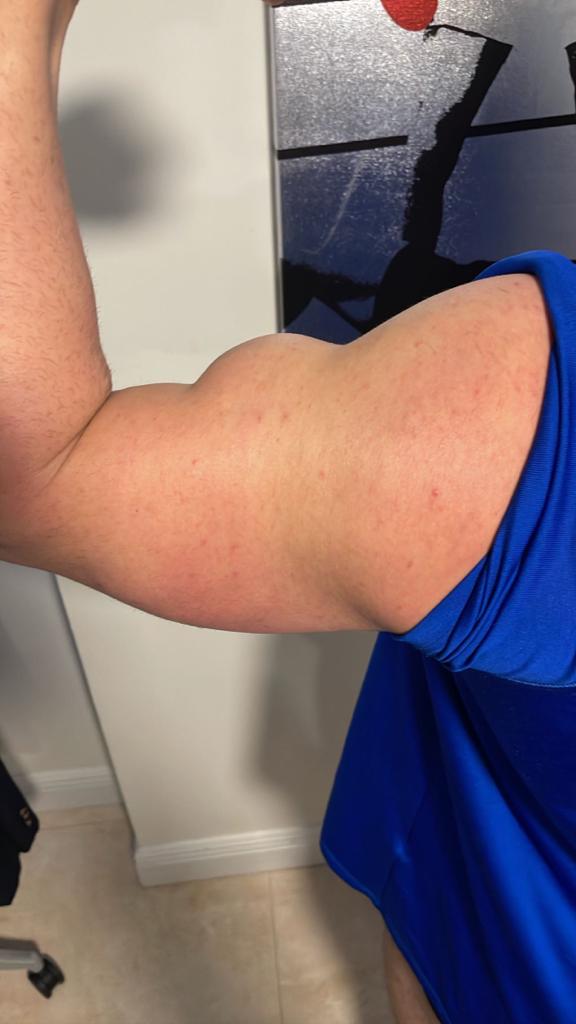
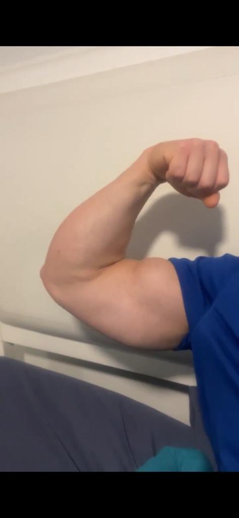
We present a clinical case of a bodybuilder patient with a delayed distal biceps tendon rupture with a severe frayed and retracted tissue, and one year follow-up clinical photos, showing a satisfied patient with complete range of motion.

Click here to learn about Pacira

Description of the issue

We describe the case of a 34-year-old male who presented to our practice with a right biceps tendon rupture. Thirty-five days prior to his visit, our patient was weightlifting, and while bearing a heavy load he heard a “pop” and felt an immediate inability to flex his elbow. He was first attended in a general emergency department, and after no solution was given to him, he sought help at an urgent care. No diagnosis nor surgical treatment was offered for the first month, and therefore nearly six weeks into the injury he came to our facilities.

The patient was a right-handed active male who had no relevant medical history. His BMI was 32 kg/m2, and his sports activity included professional heavy weightlifting. Socially, he was a non-smoker, occasional drinker, non-drug user and with no history of quinolone use nor corticosteroids in the last six months. He did have a positive history of testosterone cypionate injection, an anabolic and androgenic steroid which due to its long half-life and ability to produce large muscular growth improvements is one of the most popular injected steroids among athletes. No previous tendon injury was reported.

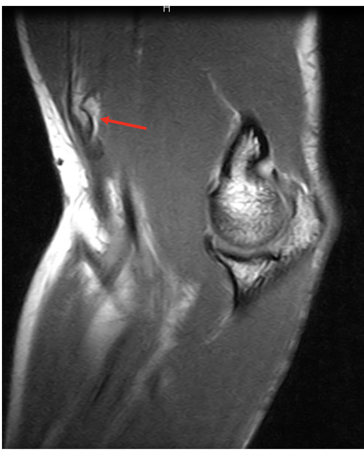
Concerning physical exam, he presented with weakness for elbow flexion and forearm supination. The stump was not palpated in the antecubital fossa but proximal to the elbow crease, and the “reverse Popeye’s sign” was present (Alentorn-Geli, Assenmacher, and Sánchez-Sotelo 2017). And MRI was ordered (Figure 1), and the patient was scheduled for surgical treatment.

Click here to learn more about Insignia Hip Stem
Figure 1
Figure 1.MRI showing tendon rupture and retraction of distal biceps tendon.

Results

The patient’s arm was sterilely prepped. Once anesthesia was obtained, and limb ischemia achieved, a longitudinal 3-cm incision just distal and radial to the antecubital fossa was performed. Before the incision, the stump could be palpated subcutaneously at 4-5 cm proximal to the elbow crease, which correlated with the appearance of the tendon’s MRI imaging. Therefore, it was felt that even though there was delayed presentation for this, we would be able to achieve primary repair.

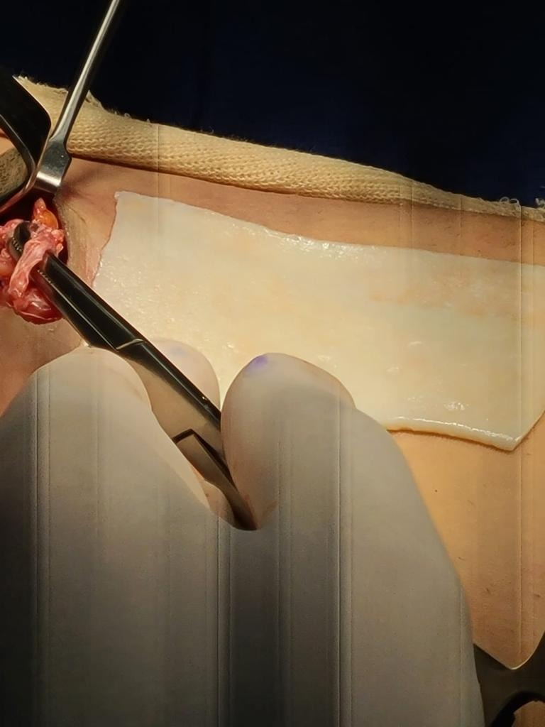
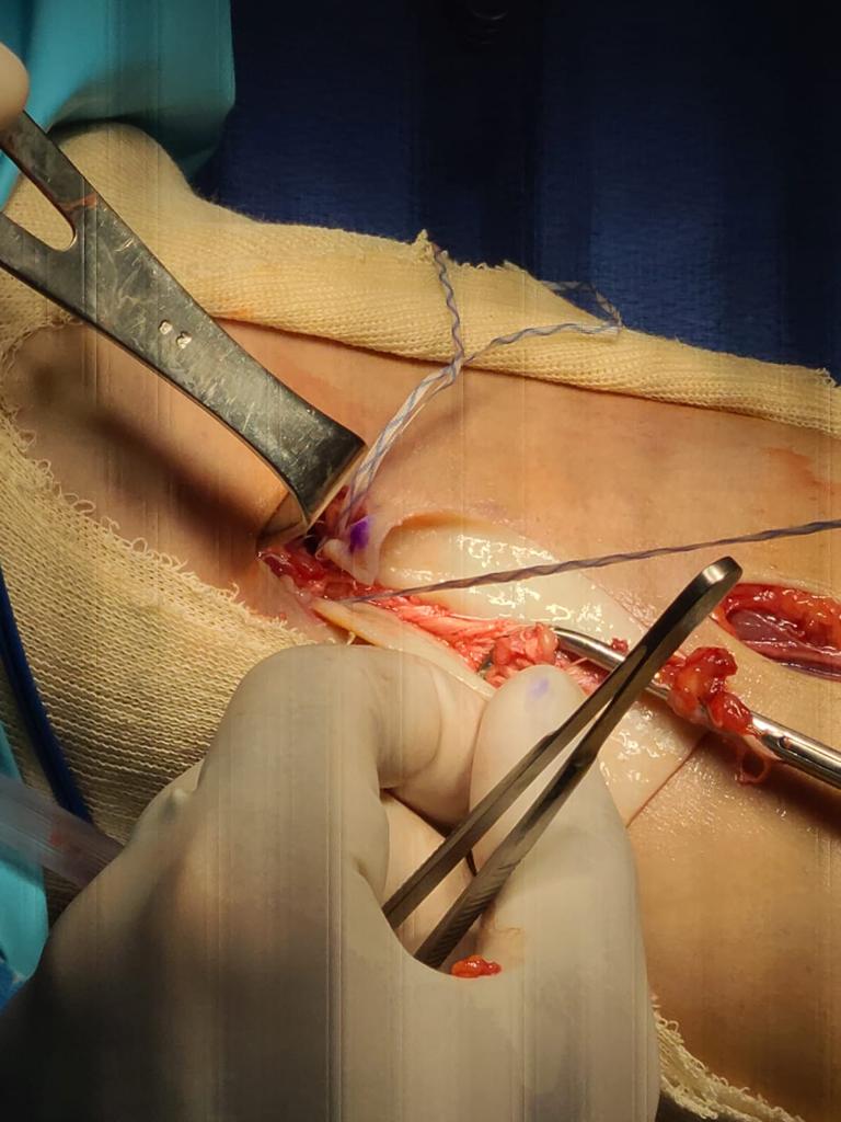
The brachialis and small vessels were identified and retracted. However, with digit exploration, biceps tendon could not be located. Incision was extended slightly and at this point, tendon was found, but unable to be pulled on with an Allis clamp, as it was noted to be quite frayed. Therefore, a second counter incision was made proximally at the level of the musculotendinous junction of the distal biceps, parallel to the antecubital crease for good cosmetic healing. The biceps tendon was noted to be markedly scarred, frayed, and rolled up in the coil (Figure 2). This is not the usual scenario where the tendon can be unwrapped and reattached to the bicep’s tuberosity. It was decided to proceed with tendon reconstruction in order to pull the biceps down distally and restore the contour of the upper arm. With this purpose, a 1.75mm thickness sterile acellular graft was used (Figure 3), entubulating the remaining frayed tendon and suturing it proximally at the musculotendinous junction with high resistance caliber-0 sutures (Figure 4). A Krackow suture was created distally in the acellular matrix (Figure 5), resulting in a final 8 millimeters width distal stump. This new tendon was then tunneled underneath the skin bridge between the proximal and distal incision in the same path of the anatomic normal biceps (Figure 6).

Figure 2
Figure 2.Frayed and scarred distal biceps tendon.
Figure 3
Figure 3.Preparation of dermal matrix allograft.
Figure 4
Figure 4.Entubulating the retracted frayed tendon and proximal suture.
Figure 5
Figure 5.Distal Krackow suture for reattachment.
Figure 6
Figure 6.Sterile acellular dermal matrix with the distal biceps tendon stump wrapped.

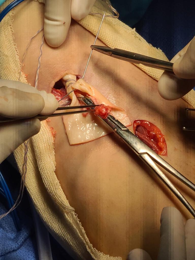
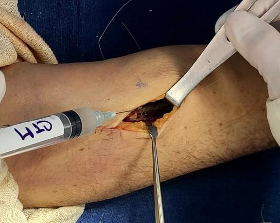
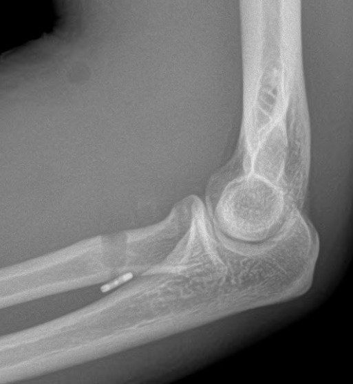
The scarred biceps tuberosity was identified while holding the forearm in supination. Debridement was carried out until bleeding was achieved. A central pin, followed with an 8-mm drill, was used unicortically, and a cortical button with a high resistance suture secured the reconstruction in its place. A 7-mm interference bioabsorbable screw was placed, with excellent purchase and grabbing the tendon strongly. A collagen tissue matrix was injected around the reconstruction for enhancing the healing environment (Figure 7). Hemostasis was achieved, and skin closed. The arm was held in 90 degrees of flexion with gentle supination. A postoperative control imaging was taken (Figure 8).

Figure 7
Figure 7.Collagen tissue matrix injection.
Figure 8
Figure 8.Postoperative x-ray control.

At this point, the elbow could completely be extended, however a thermoplastic cast was used to avoid extension further than 30º for four weeks, while early healing occurred. Wrist and hand-gripping exercises were encouraged from the beginning. In scheduled physical therapy, active passive flexion was authorized up to 90º and active extension as said, for the first six weeks. Later on, isometric exercises were introduced. Patient resumed his previous sports activity four months after surgery (Figure 9, 10).

Click here to learn more about The Orthopaedic Implant Company
Figure 9
Figure 9.Clinical image of one-year follow-up. Anterior view of elbow complete flexion.
Figure 10
Figure 10.Clinical image of one-year follow-up. Posterior view of elbow complete flexion.

Discussion

Early anatomical repair is the commonly accepted recommendation for distal biceps ruptures (Srinivasan, Pederson, and Morrey 2020). But, when the diagnosis is delayed, this anatomic repair becomes challenging, due to retraction, loosening of elasticity and atrophy of the stump (Darlis and Sotereanos 2006). At this point, use of allografts makes it possible. This augmentation has been published with a wide myriad of tendons, even with autogenous tendons (Hang, Bach, and Bojchuk 1996; Storti et al. 2021; Murthi et al. 2017). Cadaver allografts, mainly Achilles tendon, has shown good outcomes (Sanchez-Sotelo et al. 2002). Risk of infection has been described when using allografts in the knee (Bansal et al. 2018).

Recently, acellular dermal allografts have become a possible solution, diminishing risk infection and avoiding autograft morbidity, with a great biomechanical profile (Conroy et al. 2017; Barber, Herbert, and Coons 2006). These biological scaffolds allow vascular ingrowth, fibroblasts proliferation and host cells, while providing a good structural support (Barber et al. 2012). They have been used in ankle’s tendons ruptures (Rapley, Crates, and Barber 2010).

As has been indicated in our clinical case, this acellular dermal matrix achieves satisfactory suture strength, allowing complete range of motion and even performing heavy demanding sport practice.

Submitted: January 15, 2023 EDT

Accepted: February 27, 2023 EDT

References

Alentorn-Geli, Eduard, Andrew T. Assenmacher, and Joaquín Sánchez-Sotelo. 2017. “Distal Biceps Tendon Injuries: A Clinically Relevant Current Concepts Review.” EFORT Open Reviews 1 (9): 316–24. https:/​/​doi.org/​10.1302/​2058-5241.1.000053.
Google ScholarPubMed CentralPubMed
Bansal, Anchal, Joseph D. Lamplot, James VandenBerg, and Robert H. Brophy. 2018. “Meta-Analysis of the Risk of Infections After Anterior Cruciate Ligament Reconstruction by Graft Type.” The American Journal of Sports Medicine 46 (6): 1500–1508. https:/​/​doi.org/​10.1177/​0363546517714450.
Google Scholar
Barber, F. Alan, Joseph P. Burns, Allen Deutsch, Marc R. Labbé, and Robert B. Litchfield. 2012. “A Prospective, Randomized Evaluation of Acellular Human Dermal Matrix Augmentation for Arthroscopic Rotator Cuff Repair.” Arthroscopy 28 (1): 8–15. https:/​/​doi.org/​10.1016/​j.arthro.2011.06.038.
Google Scholar
Barber, F. Alan, Morley A. Herbert, and David A. Coons. 2006. “Tendon Augmentation Grafts: Biomechanical Failure Loads and Failure Patterns.” Arthroscopy 22 (5): 534–38. https:/​/​doi.org/​10.1016/​j.arthro.2005.12.021.
Google Scholar
Conroy, Christine, Paul Sethi, Craig Macken, David Wei, Marc Kowalsky, Raffy Mirzayan, Leo Pauzenberger, Felix Dyrna, Elifho Obopilwe, and Augustus D. Mazzocca. 2017. “Augmentation of Distal Biceps Repair With an Acellular Dermal Graft Restores Native Biomechanical Properties in a Tendon-Deficient Model.” The American Journal of Sports Medicine 45 (9): 2028–33. https:/​/​doi.org/​10.1177/​0363546517701426.
Google Scholar
Darlis, Nickolaos A., and Dean G. Sotereanos. 2006. “Distal Biceps Tendon Reconstruction in Chronic Ruptures.” Journal of Shoulder and Elbow Surgery 15 (5): 614–19. https:/​/​doi.org/​10.1016/​j.jse.2005.10.004.
Google Scholar
Hang, David W., Bernard R. Jr. Bach, and John Bojchuk. 1996. “Repair of Chronic Distal Biceps Brachii Tendon Rupture Using Free Autogenous Semitendinosus Tendon.” Clinical Orthopaedics and Related Research 323 (February):188–91. https:/​/​doi.org/​10.1097/​00003086-199602000-00025.
Google Scholar
Murthi, Anand M., Miguel A. Ramirez, Brent G. Parks, and Shannon R. Carpenter. 2017. “Lacertus Fibrosus Versus Achilles Allograft Reconstruction for Distal Biceps Tears: A Biomechanical Study.” The American Journal of Sports Medicine 45 (14): 3340–44. https:/​/​doi.org/​10.1177/​0363546517727511.
Google Scholar
O’Driscoll, Shawn W., Lucas B. J. Goncalves, and Patricio Dietz. 2007. “The Hook Test for Distal Biceps Tendon Avulsion.” The American Journal of Sports Medicine 35 (11): 1865–69. https:/​/​doi.org/​10.1177/​0363546507305016.
Google Scholar
Petrie, Kyla, Cameron T Cox, Benjamin C Becker, and Brendan J MacKay. 2022. “Clinical Applications of Acellular Dermal Matrices: A Review.” Scars, Burns & Healing 8 (January):205951312110383. https:/​/​doi.org/​10.1177/​20595131211038313.
Google ScholarPubMed CentralPubMed
Rapley, Jay H., John Crates, and F. Alan Barber. 2010. “Mid-Substance Peroneal Tendon Defects Augmented with an Acellular Dermal Matrix Allograft.” Foot & Ankle International 31 (2): 136–40. https:/​/​doi.org/​10.3113/​fai.2010.0136.
Google Scholar
Safran, Marc R., and Scott M. Graham. 2002. “Distal Biceps Tendon Ruptures: Incidence, Demographics, and the Effect of Smoking.” Clinical Orthopaedics and Related Research 404 (November):275–83. https:/​/​doi.org/​10.1097/​00003086-200211000-00042.
Google Scholar
Sanchez-Sotelo, J., B. F. Morrey, R. A. Adams, and S. W. OʼDRISCOLL. 2002. “Reconstruction of Chronic Ruptures of the Distal Biceps Tendon with Use of an Achilles Tendon Allograft.” The Journal of Bone and Joint Surgery-American Volume 84 (6): 999–1005. https:/​/​doi.org/​10.2106/​00004623-200206000-00015.
Google Scholar
Srinivasan, Ramesh C., William C. Pederson, and Bernard F. Morrey. 2020. “Distal Biceps Tendon Repair and Reconstruction.” The Journal of Hand Surgery 45 (1): 48–56. https:/​/​doi.org/​10.1016/​j.jhsa.2019.09.014.
Google Scholar
Storti, Thiago Medeiros, Rafael Gomes Dias, Gabriel Cesar Dias Dantas, Rafael Salomon Silva Faria, João Eduardo Simionatto, and Alexandre Firmino Paniago. 2021. “Clinical Evaluation of the Reconstruction of the Biceps Brachii using Triceps Graft.” Revista Brasileira de Ortopedia 56 (05): 656–63. https:/​/​doi.org/​10.1055/​s-0041-1729566.
Google ScholarPubMed CentralPubMed

This website uses cookies

We use cookies to enhance your experience and support COUNTER Metrics for transparent reporting of readership statistics. Cookie data is not sold to third parties or used for marketing purposes.

Powered by Scholastica, the modern academic journal management system