Loading [Contrib]/a11y/accessibility-menu.js
Skip to main content
null
J Orthopaedic Experience & Innovation
  • Menu
  • Articles
    • Brief Report
    • Case Report
    • Data Paper
    • Editorial
    • Hand
    • Meeting Reports/Abstracts
    • Methods Article
    • Product Review
    • Research Article
    • Review Article
    • Review Articles
    • Systematic Review
    • All
  • For Authors
  • Editorial Board
  • About
  • Issues
  • Blog
  • "Open Mic" Topic Sessions
  • Advertisers
  • Recorded Content
  • CME
  • JOEI KOL Connect
  • search

RSS Feed

Enter the URL below into your favorite RSS reader.

http://localhost:34284/feed
Case Report
Vol. 4, Issue 2, 2023July 08, 2023 EDT

Case Presentation for Arthroscopic Internal Fixation of Coracoid Fracture

Guy Ball, DO, Shariff K. Bishai, DO, Bodrie Jensen, DO, Michael Maceroni, DO, Samuel Howard, DO,
coracoid fractureshoulder arthroscopy
Copyright Logoccby-nc-nd-4.0 • https://doi.org/10.60118/001c.71397
J Orthopaedic Experience & Innovation
Ball, Guy, Shariff K. Bishai, Bodrie Jensen, Michael Maceroni, and Samuel Howard. 2023. “Case Presentation for Arthroscopic Internal Fixation of Coracoid Fracture.” Journal of Orthopaedic Experience & Innovation 4 (2). https:/​/​doi.org/​10.60118/​001c.71397.
Save article as...▾
Download all (13)
  • Click here to learn more about BioBrace
    Download
  • Figure 1. Grashey view of a right shoulder. A coracoid nonunion is clearly visible on this image and highlighted by the red arrow.
    Download
  • Figure 2. Axillary view of a right shoulder. A coracoid fracture nonunion is once again easily visualized and denoted by the red arrow. Of note is an asymptomatic Os Acromiale that is also easily visible on this x-ray film and highlighted by the yellow arrow.
    Download
  • Figure 3. Axial computed tomography scan of a right shoulder showing a coracoid nonunion. There is no evidence of any bony bridging at the fracture site, which is highlighted by the red arrow.
    Download
  • Figure 4. Axial T2 magnetic resonance imaging (MRI) scan of a right shoulder. This MRI scan demonstrates no enhancement at the fracture site as highlighted by the red arrow. This represents the chronic nature of this fracture. These types of MRI findings make it unlikely the fracture will eventually go on to union.
    Download
  • Click here to learn more about Iovera
    Download
  • Figure 5. An animation that depicts the arthroscopic Latarjet portals. Modified versions of the A, D, E, H, and I portals can be used for this procedure. Portals used for this procedure are highlighted by red circles.
    Download
  • Figure 6. A right shoulder in a patient in the beach chair position. The rotator interval is being viewed from a standard posterior portal (A portal). The rotator interval is being fully debrided to allow for maximal visualization of the coracoid process. Subscapularis tendon indicated by red arrow. Long head of biceps indicated by yellow arrow. Humeral head indicated by blue arrow. Glenoid indicated by green arrow.
    Download
  • Figure 7. A right shoulder in a patient in the beach chair position. The lateral edge of the coracoid process is being viewed from a standard posterior portal (A portal). A standard 30 scope is being used. The arthroscopic ablator is inserted through a standard anterior accessory portal (E portal). The coracoid is highlighted by the red arrow.
    Download
  • Figure 8. A right shoulder in a patient in the beach chair position. The coracoid nonunion is being visualized with the scope in an accessory anterolateral portal (D portal). The arthroscopic shaver is introduced through an accessory portal directly superior to the coracoid process (H portal). The arthroscopic shaver is being used to debride all the fibrous tissue away from the fracture site. The fibrous tissue is debrided back to fresh bleeding bone edges on the coracoid process.
    Download
  • Figure 9. A right shoulder in a patient in the beach chair position. The coracoid is being viewed through an accessory anterolateral portal (D portal). The drill guide is inserted through an accessory antero-inferolateral portal (I portal). The drill guide used in this case is a Mitek Latarjet coracoid guide (Mitek, Johnson and Johnson); however, any cannulated drill guide can be used. The drill is placed at the tip of the coracoid process to allow a k-wire to be placed down the center of the coracoid process.
    Download
  • Figure 10. A right shoulder in a patient in the beach chair position. The coracoid is being viewed through an accessory anterolateral portal (D portal). A 3.5 mm cannulated screw is inserted over a k-wire through an accessory anteroinferolateral portal (I portal). You can see a blunt trocar (red arrow) being used to hold the coracoid steady while the screw is fully tightened. The blunt trocar is inserted through an accessory portal directly superior to the coracoid process (H portal).
    Download
  • Figure 11. An anteroposterior (AP) and scapular Y radiograph of a right shoulder status post arthroscopy-aided coracoid fracture fixation. The x-ray film demonstrates good compression across the previous fracture site. Accurate screw length is assessed on the AP view. On the scapular-Y the screw is clearly visualized down the center of the coracoid process.
    Download

Sorry, something went wrong. Please try again.

If this problem reoccurs, please contact Scholastica Support

Error message:

undefined

View more stats

Abstract

This case presentation highlights the ability to fix coracoid fracture nonunion arthroscopically. A right hand dominant 27 year old male with 6 months of symptomatic nonunion of a coracoid fracture. After arthroscopic internal fixation the patient went on to have union at the fracture site and resolution of pain at the coracoid.

Case Presentation for Arthroscopic Internal Fixation of Coracoid Fractures

Case Description: Patient is a 27 year old right hand dominant male that presented initially with right shoulder pain. He denied any specific trauma or injury to the shoulder but has radiographic evidence of a Type II coracoid fracture that has progressed to symptomatic non-union. After 6 months of attempted non operative including physical therapy and activity restriction, treatment discussion of surgical options was initiated. Patients baseline activity includes frequent exercise activity but no formal sport or powerlifting. Historically coracoid fractures go on to uneventful union. Literature supports a very high union rate with a conservative estimate of 81% or more going on to union and returning to sport at an average of 2.8 months (6). Typically surgery is reserved for patients with greater than 1 cm of displacement, or less than 1 cm of displacement but concomitant shoulder instability/SSSC disruption. In discussing this patients atypical presentation of symptomatic non union, surgical intervention was deemed beneficial after failed conservative management. CT and MRI were both performed to fully evaluate whether healing was still actively taking place or if the fracture was truly a fibrous nonunion requiring intervention All surgical options were discussed with the patient and in the end the decision was made to perform the procedure arthroscopically. While this is a novel technique previously undescribed in the literature, it focused on arthroscopic principles utilized during the arthroscopic Latarjet, which made it a very comfortable surgical approach.

Click here to learn more about BioBrace

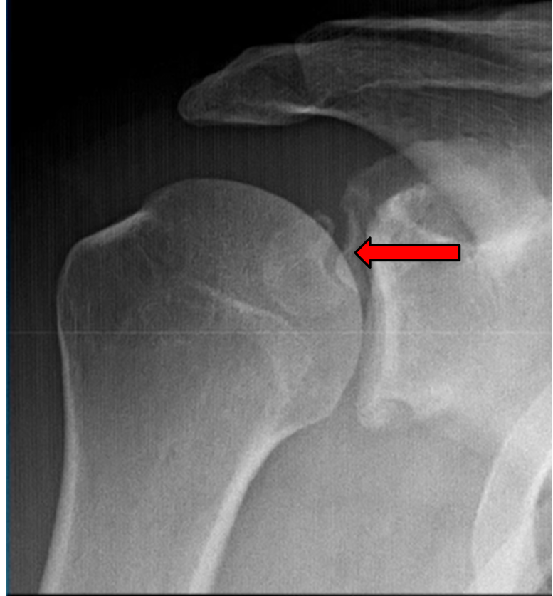
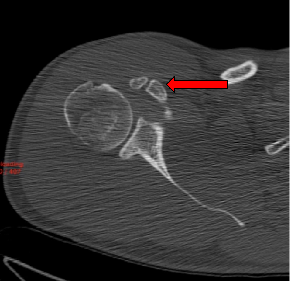
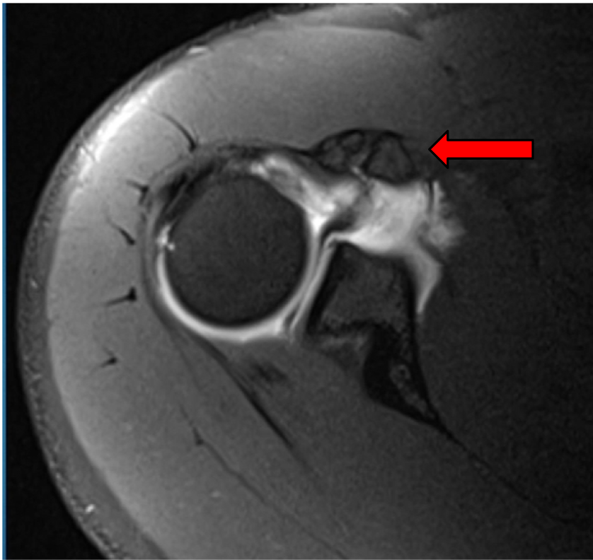
Imaging: Preoperative imaging included standard radiography using anteroposterior, Grashey, scapular Y and axillary views (Fig 1-2). After determining the presence of a coracoid fracture, advanced imaging was conducted via computed tomography with addition of 3-dimensional reconstruction. CT imaging was suggestive of a type 2 coracoid fracture nonunion (Fig 3) which was then further verified with no enhancement with T2 MRI sequences (Fig 4).

Figure 1
Figure 1.Grashey view of a right shoulder. A coracoid nonunion is clearly visible on this image and highlighted by the red arrow.
Figure 2
Figure 2.Axillary view of a right shoulder. A coracoid fracture nonunion is once again easily visualized and denoted by the red arrow. Of note is an asymptomatic Os Acromiale that is also easily visible on this x-ray film and highlighted by the yellow arrow.
Figure 3
Figure 3.Axial computed tomography scan of a right shoulder showing a coracoid nonunion. There is no evidence of any bony bridging at the fracture site, which is highlighted by the red arrow.
Figure 4
Figure 4.Axial T2 magnetic resonance imaging (MRI) scan of a right shoulder. This MRI scan demonstrates no enhancement at the fracture site as highlighted by the red arrow. This represents the chronic nature of this fracture. These types of MRI findings make it unlikely the fracture will eventually go on to union.

Preoperative plan: Initial preoperative plan included placing the patient in a standard beach chair configuration and completing a full diagnostic arthroscopy of the shoulder. The rotator interval would be taken down and utilization of arthroscopic Latarjet accessory portals would be used to fully identify the nonunion. If the fracture remained mobile, debridement of all fibrous tissue would be performed and a 4.5 mm cannulated screw would be used to compress across the fracture site.

Click here to learn more about Iovera

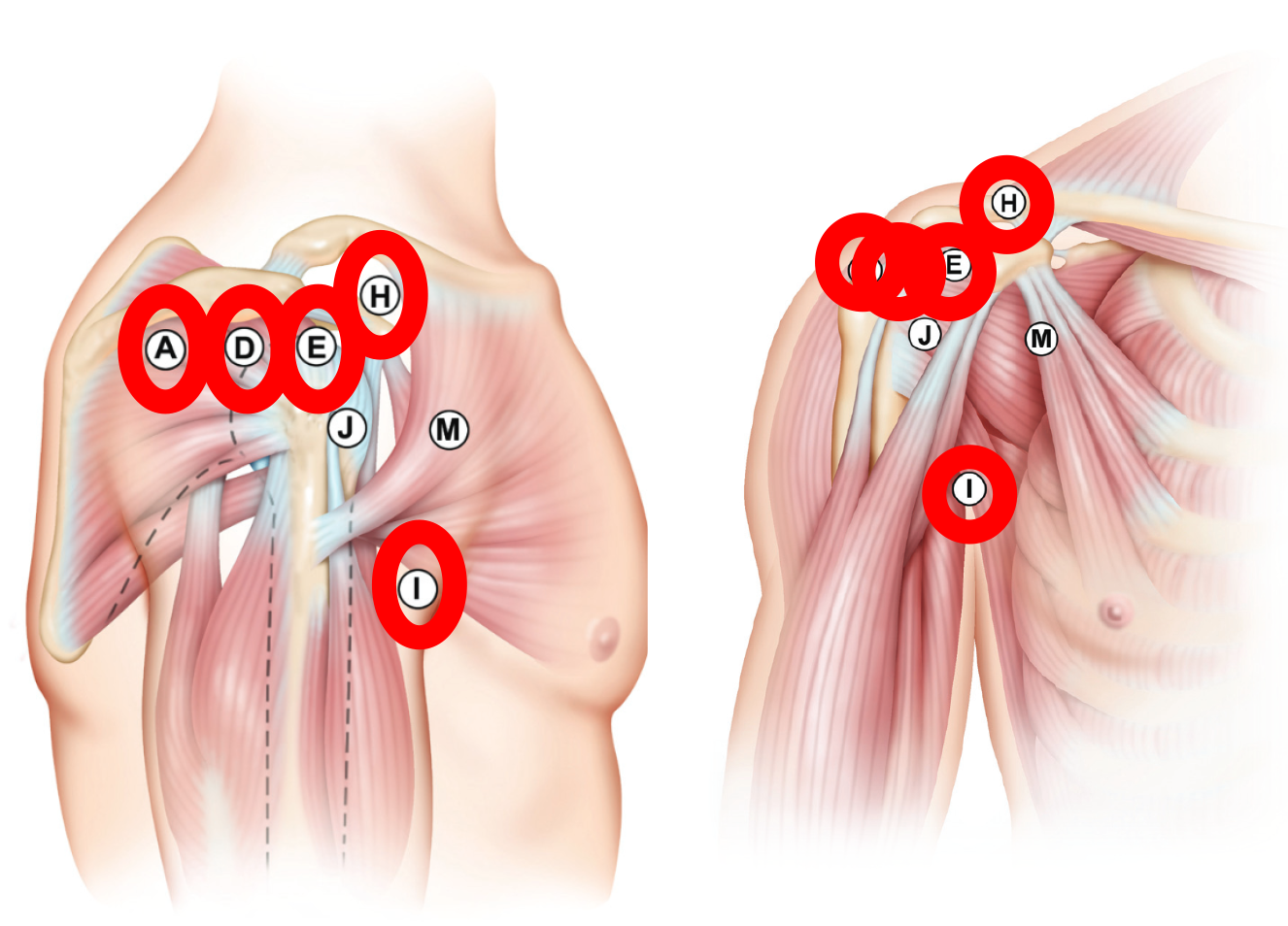
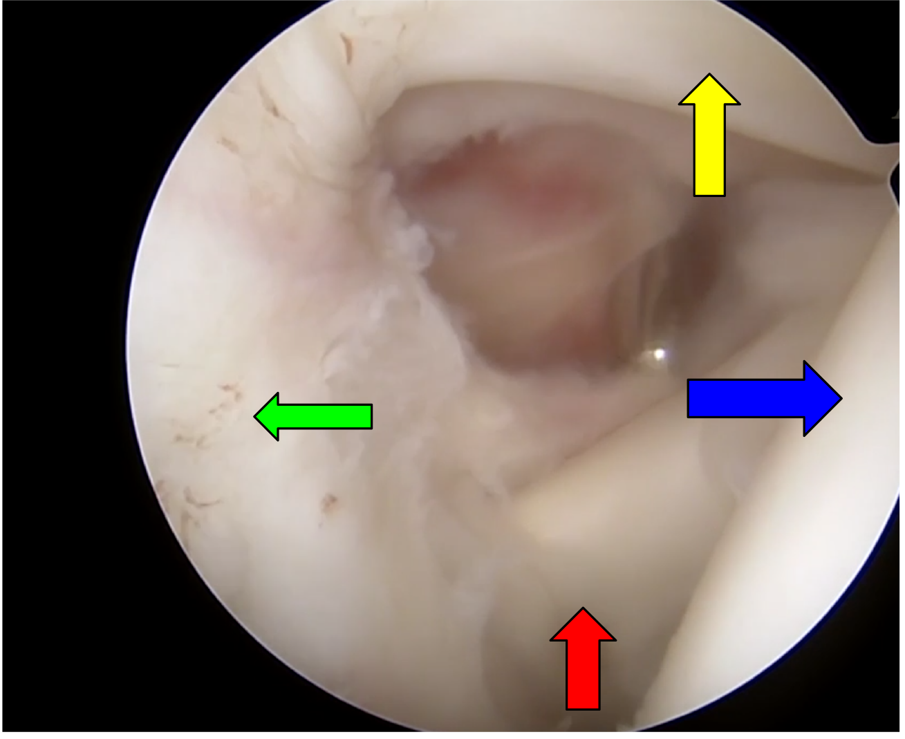
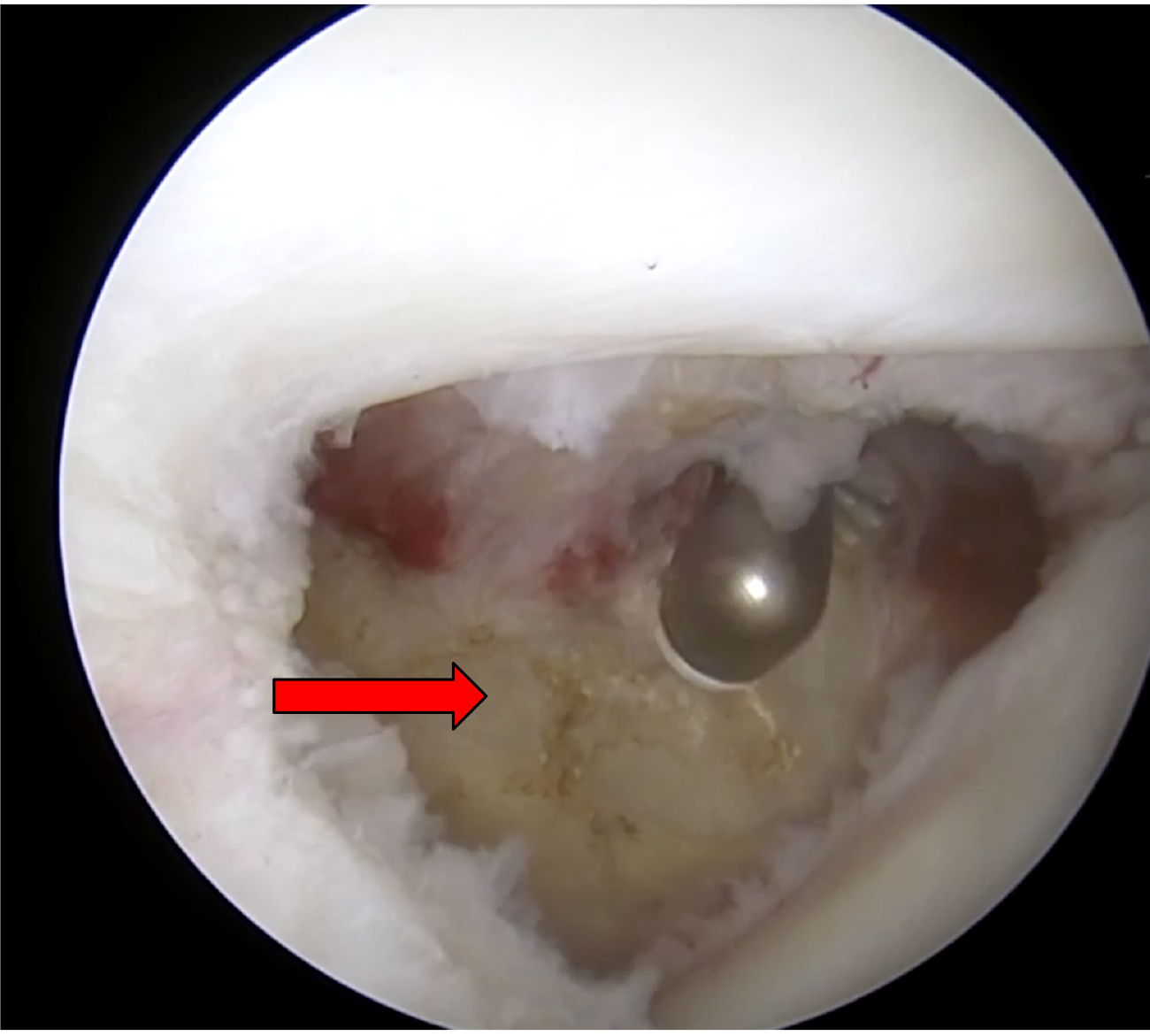
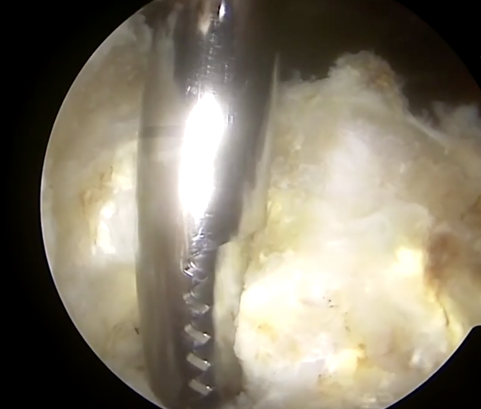
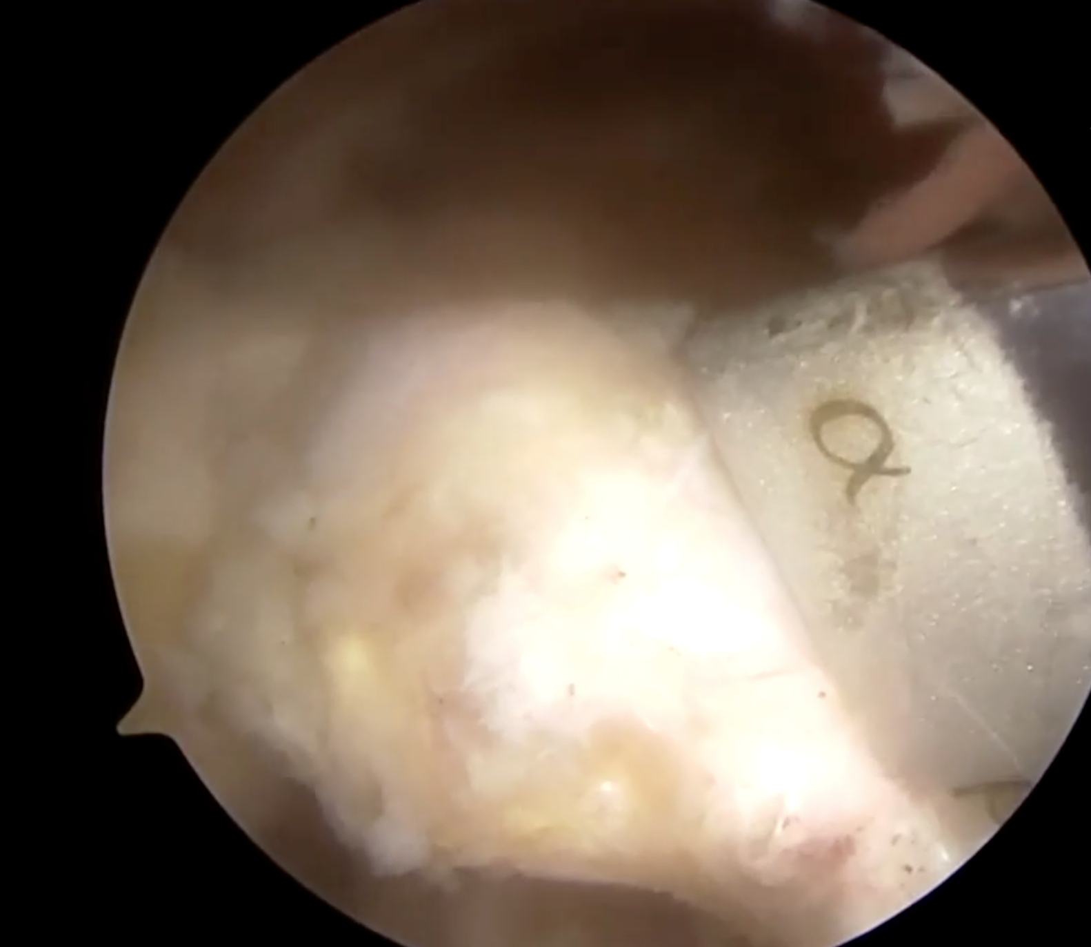
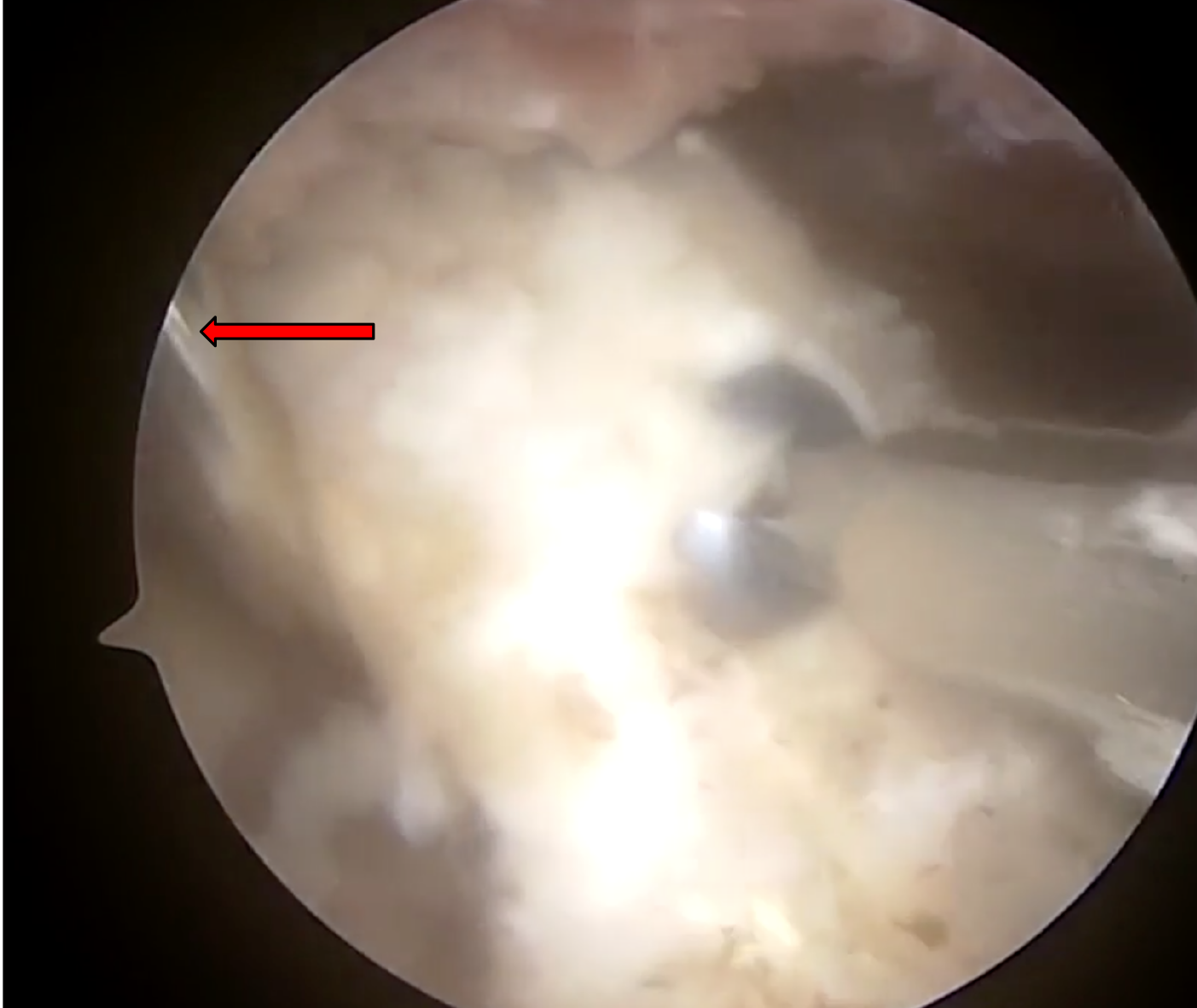
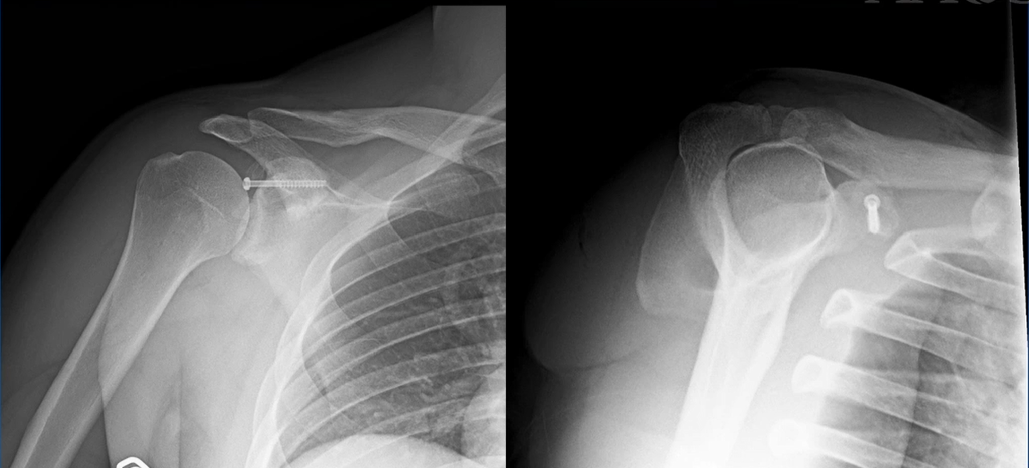
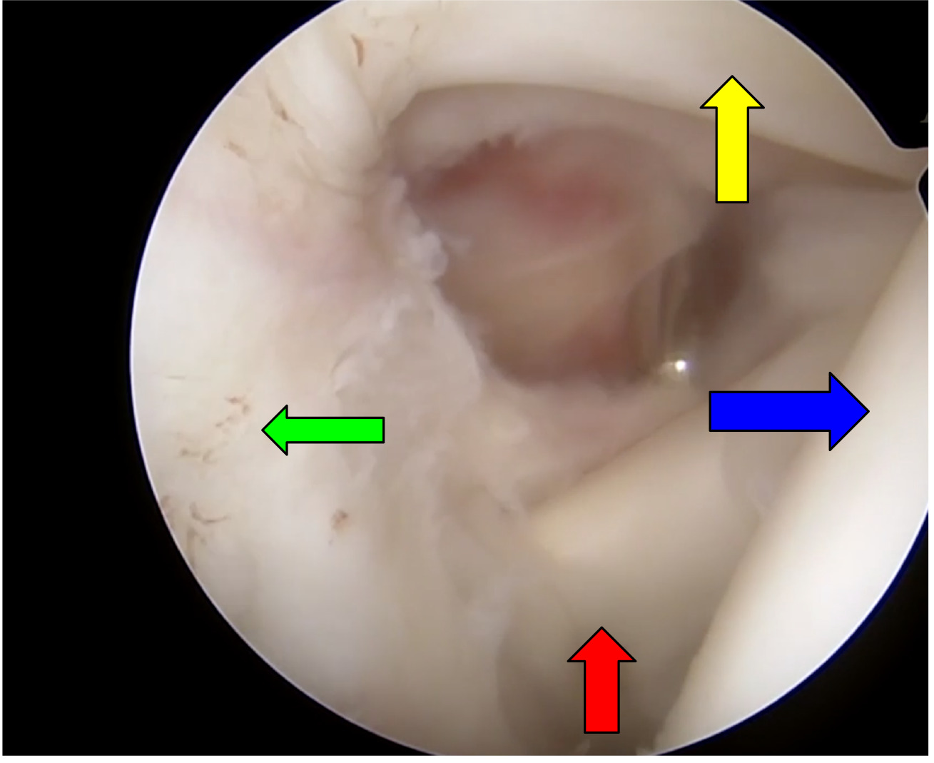
Operative Results: The patient was positioned supine in beach chair position with the assistance of a mechanical arm holder. A 30 degree scope was used during the entire procedure without need to switch to a 70 degree scope due to multiple portal use. The first portal was established as a standard posterior viewing portal (A) followed by a standard anterior portal (E) under direct visualization, Figure 5 demonstrates portal selection. From the anterior portal a thorough debridement via arthroscopic shaver and radiofrequency ablation was undergone of the rotator interval to the tip of the coracoid process (Figure 6). After visualization of the lateral coracoid was obtained, the radiofrequency ablator was used to clear the soft tissue until the coracoclavicular ligament is encountered at the anterolateral aspect of the coracoid (Figure 7). Unlike the arthroscopic Latarjet technique, the CC ligament does not need to be fully released but can be partially released for visualization. The D portal (anterolateral portal) is then made under visualization from the posterior portal and viewing is moved to the D portal. The ablator is then moved to the anterior portal to allow for clearing of the superior coracoid. The purpose of the switch of viewing to the D portal is increased visualization of the tip of the coracoid, conjoined tendon, and an en face view of the glenoid. Extra care should be undertaken when moving medially to the fracture site to avoid damage to the axillary nerve, blunt dissection can be done with the ablator to identify the axillary nerve. Next, through the D portal, the I portal (anterior inferolateral portal) can be established under direct visualization in line with the conjoined tendon for placement of the K-wire and cannulated screw. Through the I portal the ablator is inserted to remove a small portion of conjoined tendon for the K-wire insertion site and future cannulated screw. Placement of a blunt trocar is then inserted into portal E and fracture mobility is assessed. We then moved to creating portal H, insertion of a spinal needle under direct visualization in line and superior to the coracoid process. The ablator can then be used through portal H to debride the built up non union fibrous soft tissue (Figure 8). To get a full and adequate debridement of the fibrous tissue, a small amount of the pectoralis minor tendon may need to be released depending on the location of the fracture as it may act as a deforming force. After adequate debridement, a shaver or burr is used to debride bone edges to bleeding bone. A drill guide is then inserted through portal I at the tip of the coracoid process (Figure 9) while a probe is inserted through portal H to reduce and stabilize the fracture site for K wire insertion. The K-wire can be used to joystick the fracture as well while in the distal fragment. Once reduction is achieved the k wire is inserted into the proximal fragment, but not advancing too far to put the suprascapular nerve at the suprascapular notch. A cannulated drill is then passed over the K-wire in both proximal and distal fragments. A 4.5 cannulated screw is then inserted over the guidewire (Figure 10). A blunt trocar can be placed within the H portal to allow the operator to prevent/minimize rotation of the fragment during insertion of the cannulated screw. Post operative care included postoperative radiographs for reduction assessment and screw length (Figure 11). Patient placed within an abduction sling for 6 weeks, gentle range of motion was initiated at 2 weeks post op. After fracture union, the patient should return to normal activities at approximately 3 months post op.

Figure 5
Figure 5.An animation that depicts the arthroscopic Latarjet portals. Modified versions of the A, D, E, H, and I portals can be used for this procedure. Portals used for this procedure are highlighted by red circles.
Figure 6
Figure 6.A right shoulder in a patient in the beach chair position. The rotator interval is being viewed from a standard posterior portal (A portal). The rotator interval is being fully debrided to allow for maximal visualization of the coracoid process. Subscapularis tendon indicated by red arrow. Long head of biceps indicated by yellow arrow. Humeral head indicated by blue arrow. Glenoid indicated by green arrow.
Figure 7
Figure 7.A right shoulder in a patient in the beach chair position. The lateral edge of the coracoid process is being viewed from a standard posterior portal (A portal). A standard 30 scope is being used. The arthroscopic ablator is inserted through a standard anterior accessory portal (E portal). The coracoid is highlighted by the red arrow.
Figure 8
Figure 8.A right shoulder in a patient in the beach chair position. The coracoid nonunion is being visualized with the scope in an accessory anterolateral portal (D portal). The arthroscopic shaver is introduced through an accessory portal directly superior to the coracoid process (H portal). The arthroscopic shaver is being used to debride all the fibrous tissue away from the fracture site. The fibrous tissue is debrided back to fresh bleeding bone edges on the coracoid process.
Figure 9
Figure 9.A right shoulder in a patient in the beach chair position. The coracoid is being viewed through an accessory anterolateral portal (D portal). The drill guide is inserted through an accessory antero-inferolateral portal (I portal). The drill guide used in this case is a Mitek Latarjet coracoid guide (Mitek, Johnson and Johnson); however, any cannulated drill guide can be used. The drill is placed at the tip of the coracoid process to allow a k-wire to be placed down the center of the coracoid process.
Figure 10
Figure 10.A right shoulder in a patient in the beach chair position. The coracoid is being viewed through an accessory anterolateral portal (D portal). A 3.5 mm cannulated screw is inserted over a k-wire through an accessory anteroinferolateral portal (I portal). You can see a blunt trocar (red arrow) being used to hold the coracoid steady while the screw is fully tightened. The blunt trocar is inserted through an accessory portal directly superior to the coracoid process (H portal).
Figure 11
Figure 11.An anteroposterior (AP) and scapular Y radiograph of a right shoulder status post arthroscopy-aided coracoid fracture fixation. The x-ray film demonstrates good compression across the previous fracture site. Accurate screw length is assessed on the AP view. On the scapular-Y the screw is clearly visualized down the center of the coracoid process.

Discussion: This case highlights the exciting opportunity that exists for arthroscopic shoulder procedures. While most coracoid fractures will never require surgical stabilization it is also important to acknowledge that when they do, minimizing morbidity should be of the highest priority. We believe Arthroscopic shoulder surgery provides better visualization and detail of anatomy compared to open procedures. It also is important to note that literature supports decreased rates of infection with arthroscopic shoulder surgery versus open shoulder surgery (10). When you combine improved visualization with lower infection risk the argument against performing this procedure arthroscopically likely comes entirely down to comfort. Our comfort comes from performing many arthroscopic Latarjet procedures, but we believe that comfort is attainable. For this specific case not only were the same portals utilized but many of the proprietary tools specific to the Latarjet set were used to complete this surgery. While this case is not as complex as an arthroscopic Latarjet it exemplifies the possibilities that inevitably come to exist only when new surgical techniques are attempted and mastered.

Submitted: January 01, 2023 EDT

Accepted: February 19, 2023 EDT

References

Archik, S., S. N. Nanda, S. Tripathi, A. Choudhari, and H. Rajadhyaksha. 2016. “An Isolated Displaced Fracture of the Coracoid Process Treated with Open Reduction and Internal Fixation A Case Report and Review of Literature.” J Orthop Case Rep 6:37–39.
Google Scholar
Hill, Brian W., Aaron R. Jacobson, Jack Anavian, and Peter A. Cole. 2014. “Surgical Management of Coracoid Fractures: Technical Tricks and Clinical Experience [Published Correction Appears in J Orthop Trauma 2015;29(1):E23].” Journal of Orthopaedic Trauma 28 (5): e114–22. https:/​/​doi.org/​10.1097/​01.bot.0000435632.71393.bb.
Google Scholar
Imatani, Raymond J. 1975. “Fractures of the Scapula: A Review of 53 Fractures.” The Journal of Trauma 15 (6): 473–78. https:/​/​doi.org/​10.1097/​00005373-197506000-00002.
Google Scholar
Knapik, Derrick M., Sunny H. Patel, Robert J. Wetzel, and James E. Voos. 2017. “Prevalence and Management of Coracoid Fracture Sustained during Sporting Activities and Time to Return to Sport: A Systematic Review.” The American Journal of Sports Medicine 46 (3): 753–58. https:/​/​doi.org/​10.1177/​0363546517718513.
Google Scholar
Lafosse, Laurent, and Simon Boyle. 2010. “Arthroscopic Latarjet Procedure.” Journal of Shoulder and Elbow Surgery 19 (2): 2–12. https:/​/​doi.org/​10.1016/​j.jse.2009.12.010.
Google Scholar
Ogawa, K., A. Yoshida, M. Takahashi, and M. Ui. 1997. “Fractures of the Coracoid Process.” The Journal of Bone and Joint Surgery, British Volume 79-B (1): 17–19. https:/​/​doi.org/​10.1302/​0301-620x.79b1.0790017.
Google Scholar
Passaplan, Caroline, Silvan Beeler, Samy Bouaicha, and Karl Wieser. 2020. “Arthroscopic Management of a Coracoid Fracture Associated with Acromioclavicular Dislocation: Technical Note.” Arthroscopy Techniques 9 (11): e1767–71. https:/​/​doi.org/​10.1016/​j.eats.2020.07.019.
Google ScholarPubMed CentralPubMed
Schneider, Marco Michael, Maurice Balke, Paola Koenen, Bertil Bouillon, and Marc Banerjee. 2014. “Avulsion Fracture of the Coracoid Process in a Patient with Chronic Anterior Shoulder Instability Treated with the Latarjet Procedure: A Case Report.” Journal of Medical Case Reports 8 (1). https:/​/​doi.org/​10.1186/​1752-1947-8-394.
Google ScholarPubMed CentralPubMed
Wang, Kevin Y., Amil R. Agarwal, Amy L. Xu, Matthew J. Best, R. Timothy Kreulen, Meghana Jami, Edward G. McFarland, and Uma Srikumaran. 2022. “Increased Risk of Surgical-Site Infection and Need for Manipulation Under Anesthesia for Those Who Undergo Open Versus Arthroscopic Rotator Cuff Repair.” Arthroscopy, Sports Medicine, and Rehabilitation 4 (2): e527–33. https:/​/​doi.org/​10.1016/​j.asmr.2021.11.012.
Google ScholarPubMed CentralPubMed
Wilber, MC, and EB Evans. 1977. “Fractures of the Scapula. An Analysis of Forty Cases and a Review of the Literature.” The Journal of Bone & Joint Surgery 59 (3): 358–62. https:/​/​doi.org/​10.2106/​00004623-197759030-00012.
Google Scholar

This website uses cookies

We use cookies to enhance your experience and support COUNTER Metrics for transparent reporting of readership statistics. Cookie data is not sold to third parties or used for marketing purposes.

Powered by Scholastica, the modern academic journal management system