Loading [Contrib]/a11y/accessibility-menu.js
Skip to main content
null
J Orthopaedic Experience & Innovation
  • Menu
  • Articles
    • Brief Report
    • Case Report
    • Data Paper
    • Editorial
    • Hand
    • Meeting Reports/Abstracts
    • Methods Article
    • Product Review
    • Research Article
    • Review Article
    • Review Articles
    • Systematic Review
    • All
  • For Authors
  • Editorial Board
  • About
  • Issues
  • Blog
  • "Open Mic" Topic Sessions
  • Advertisers
  • Recorded Content
  • CME
  • JOEI KOL Connect
  • search

RSS Feed

Enter the URL below into your favorite RSS reader.

http://localhost:23905/feed
Case Report
Vol. 3, Issue 2, 2022December 27, 2022 EDT

Biologic Augmentation of the Subscapularis Tendon During Anatomic Shoulder Arthroplasty: A prospective case series utilizing dynamic ultrasound

Amit Nathani, M.D., MSc,
orthopedicsshoulder arthroplastyshoulder surgerybiologicsaugmentationsubscapularissports medicinecollagen
Copyright Logoccby-nc-nd-4.0 • https://doi.org/10.60118/001c.39782
J Orthopaedic Experience & Innovation
Nathani, Amit. 2022. “Biologic Augmentation of the Subscapularis Tendon During Anatomic Shoulder Arthroplasty: A Prospective Case Series Utilizing Dynamic Ultrasound.” Journal of Orthopaedic Experience & Innovation 3 (2). https:/​/​doi.org/​10.60118/​001c.39782.
Save article as...▾
Download all (12)
  • Click here to learn more about OIC
    Download
  • Click here to learn more about the Insignia Stem
    Download
  • Figure 1. TAPESTRY ® biointegrative implant with a shape specifically designed for subscapularis, 50x40x25mm
    Download
  • Figure 2. Subscapularis managed with a tenotomy technique.
    Download
  • Figure 3. The subscapularis tendon was repaired via stem-based transosseous drill tunnels and high-tensile strength sutures for a tendon-to-bone repair.
    Download
  • Figure 4. A collagen-based biointegrative implant was secured over top of the subscapularis repair.
    Download
  • Click here to learn more about Zilretta
    Download
  • Figure 5. Patient #1 representative 6-month post-operative ultrasound showing intact subscapularis tendon with normal fibrillar echogenic tendon architecture and no evidence of tendinosis or tear.
    Download
  • Figure 6. Patient #2 representative 6-month post-operative ultrasound demonstrating an intact subscapularis tendon with normal collagen fibrillar architecture with visibly aligned collagen fibers and anatomic size measurements.
    Download
  • Figure 7. Patient #3 representative 6-month post-operative ultrasound showing an intact subscapularis tendon with mild tendinosis.
    Download
  • Figure 8. Patient #4 representative 6-month post-operative ultrasound showing a mostly (> 75%) intact subscapularis tendon with normal fibrillar echogenic tendon architecture and thickness, with a low-grade partial-thickness tear (< 25%)
    Download
  • Figure 9. Patient #5 representative 6-month post-operative ultrasound showing intact subscapularis tendon with normal fibrillar echogenic tendon architecture and no evidence of tendinosis or tear.
    Download

Sorry, something went wrong. Please try again.

If this problem reoccurs, please contact Scholastica Support

Error message:

undefined

View more stats

Abstract

Five consecutively eligible patients with primary glenohumeral osteoarthritis underwent anatomic shoulder arthroplasty utilizing a collagen-based biointegrative implant (TAPESTRY®) to augment subscapularis tendon repairs. Evaluation of subscapularis healing was primarily assessed via dynamic ultrasound examinations at 6 months post-operatively. Subscapularis imaging was evaluated for tendon thickness, width, echotexture, and calcification. All five patients exhibited fully healed tendons at 6 months with no re-tears, representing a decrease in the 50% occurrence of subscapularis tears historically evidenced in ultrasound studies following shoulder arthroplasty. The subscapularis tendon in all patients was shown to be intact, four of which demonstrated macroscopically aligned collagen tendon architecture with anatomic size measurements, and one demonstrated mild tendinosis. Secondary clinical outcomes included validated shoulder outcome scores including the American Shoulder and Elbow Surgeons (ASES) assessment preoperatively and at 3 months and 6 months postoperatively. Improvements in ASES scores were greater than two-times (33 points at 3 months, and 35 points at 6 months) the published minimal clinically important difference (MCID) of 13.6 ± 2.3 for total shoulder arthroplasty (TSA). Additional data collected included graded belly press, lift-off, and range of motion assessments measured preoperatively and at 6 months postoperatively. Biologic augmentation of the subscapularis tendon with a collagen-based biointegrative implant in these 5 patients showed 100% healing rates assessed via dynamic ultrasound at 6 months post-operatively. All patients demonstrated tendon healing without postoperative complications or revisions. Further investigation is needed to validate the observations from this pilot study.

Click here to learn more about OIC

Introduction

Subscapularis management during shoulder arthroplasty continues to be an area of controversy (Collin et al. 2022). While catastrophic failure of the subscapularis tendon requiring revision surgery ranges between 5-6% of all cases (Boileau et al. 2006), loss of clinically detectable subscapularis function is present in 67.5% of patients following shoulder arthroplasty (Miller et al. 2003). Other complications include loss of motion, hematoma, weakness, anterosuperior escape, and instability. Revisions to reverse total shoulder arthroplasty are costly, estimated at $22,000 per revision procedure, and lead to worse outcomes compared to primary arthroplasty (Saltzman et al. 2014).

Many clinical studies have shown patients with subscapularis re-tears following primary arthroplasty surgery show lower satisfaction rates, lower shoulder function scores, and decreased internal rotation strength and range of motion. The healing of the subscapularis tendon is therefore critical to optimal shoulder arthroplasty results. Despite this, while shoulder arthroplasty has evolved to meet the early challenges of glenoid loosening and bony deformity, little progress has been made with understanding subscapularis healing and failure rates (Schrock et al. 2017). Ultrasound studies show that about 50% of patients undergoing shoulder arthroplasty experience a subscapularis re-tear that often is difficult to detect by physical exam or routine post-operative imaging (Miller et al. 2003). With the rapid increase in the number of annual shoulder arthroplasty procedures (100,000/year with 7% increase projected year-over-year) (Shoulder Replacement Market, n.d.), there remains a tremendous opportunity for surgeons to improve outcomes and decrease failure rates. The ability to improve subscapularis healing with biologic augmentation may represent a significant advancement in the treatment of the subscapularis tendon during shoulder arthroplasty.

As tendons are composed of collagen fibers that are uniaxially aligned and wavy, replicating the collagen composition and native structure (Lomas et al. 2015; O’Brien 2005) are critical in engineering a functional implant and healing of connective tissue. Organized surface structure, high porosity, and distribution of micro- and macropores are critical attributes in bioengineered implants to support neovascularization and tissue ingrowth, maturation, and integration into the surrounding native tissue (Loh and Choong 2013; Murphy, Haugh, and O’Brien 2010; O.’Brien, Harley, and Yannas, n.d.; Han et al. 2021; Mandal and Kundu 2009; Lien, Ko, and Huang 2009; Artel et al. 2011; Druecke et al. 2003; Cao et al. 2012; Baac et al. 2004; Gnavi et al. 2015; Simitzi, Ranella, and Stratakis 2017; Gigante et al. 2009; W. Wang et al. 2016; Yin et al. 2010; Salerno et al. 2011; Hollister 2005; Gomes et al. 2006; Wei et al. 2010; Oh et al. 2014; Boyan 1996; Z. Wang et al. 2018). To support biologic healing in the subscapularis, this study utilized a collagen-based biointegrative implant that was engineered to combine aligned surface topography and porosity to mimic native tendon and aid in tendon healing. Preclinical studies in a rabbit Achilles tendon defect model showed dense collagenous fibrous connective tissue ingrowth into and around the implant (NAMSA GLP Report, n.d.). This initial pilot study was intended to further demonstrate the safety and efficacy of a utilizing a novel collagen-based implant to augment subscapularis repairs in patients undergoing TSA.

Click here to learn more about the Insignia Stem
Figure 1
Figure 1.TAPESTRY ® biointegrative implant with a shape specifically designed for subscapularis, 50x40x25mm

Materials & Methods

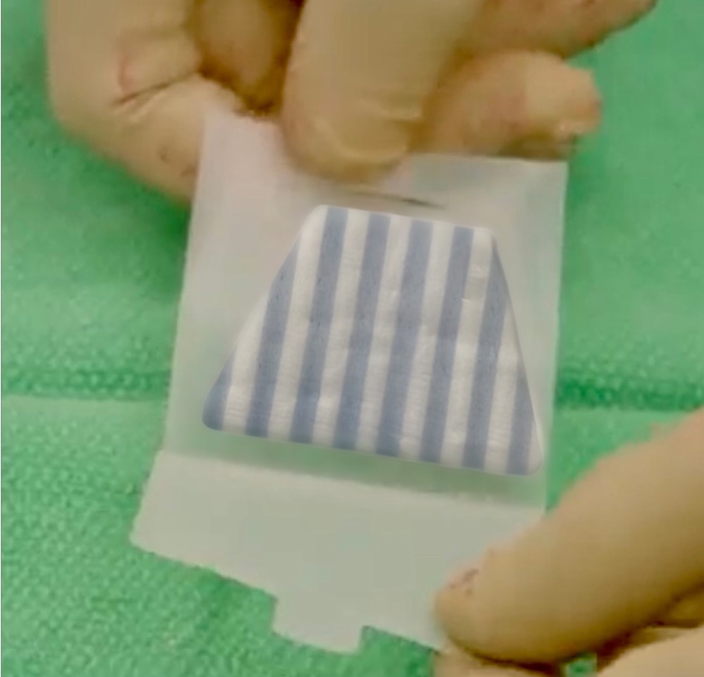
The implant selected was a collagen-based biointegrative implant, TAPESTRY® (Embody, Inc., Norfolk, VA) with a highly aligned and highly porous micro-architecture. It is composed of a co-polymer of type I collagen and poly(D,L-lactide) (PDLLA). It is manufactured using a novel fiber assembly process using electrospinning/pneumatospinning post-processing with low temperature annealing (Maghdouri-White et al. 2021).

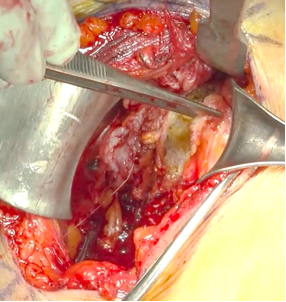
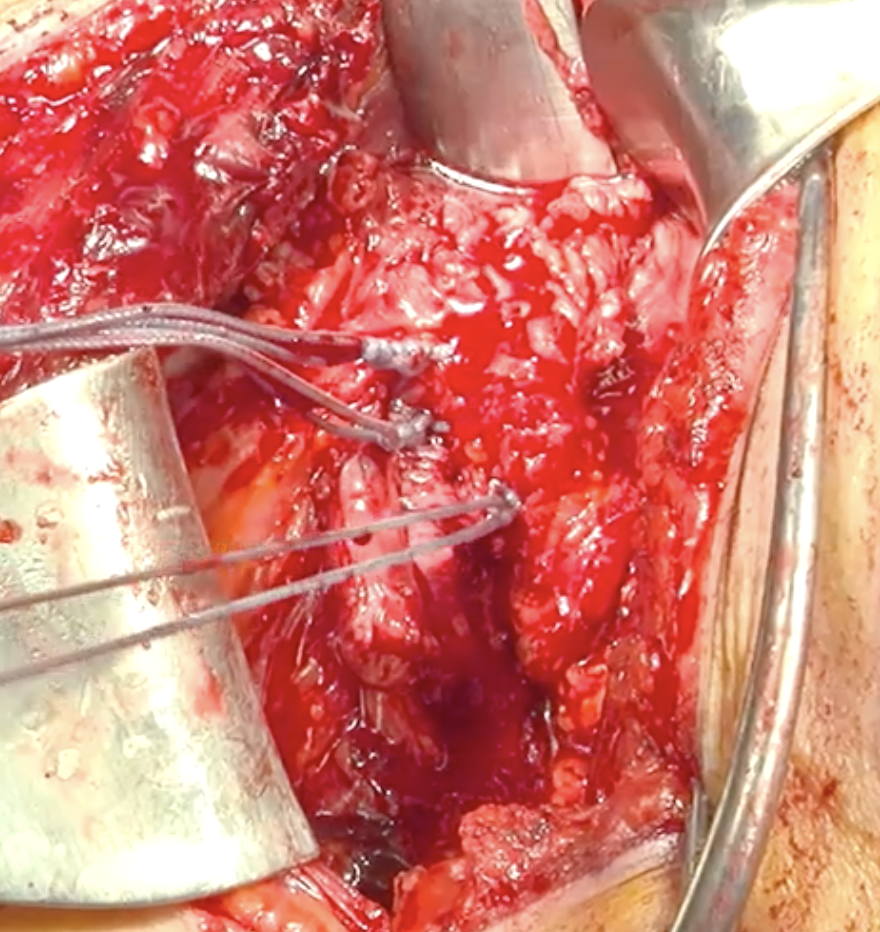
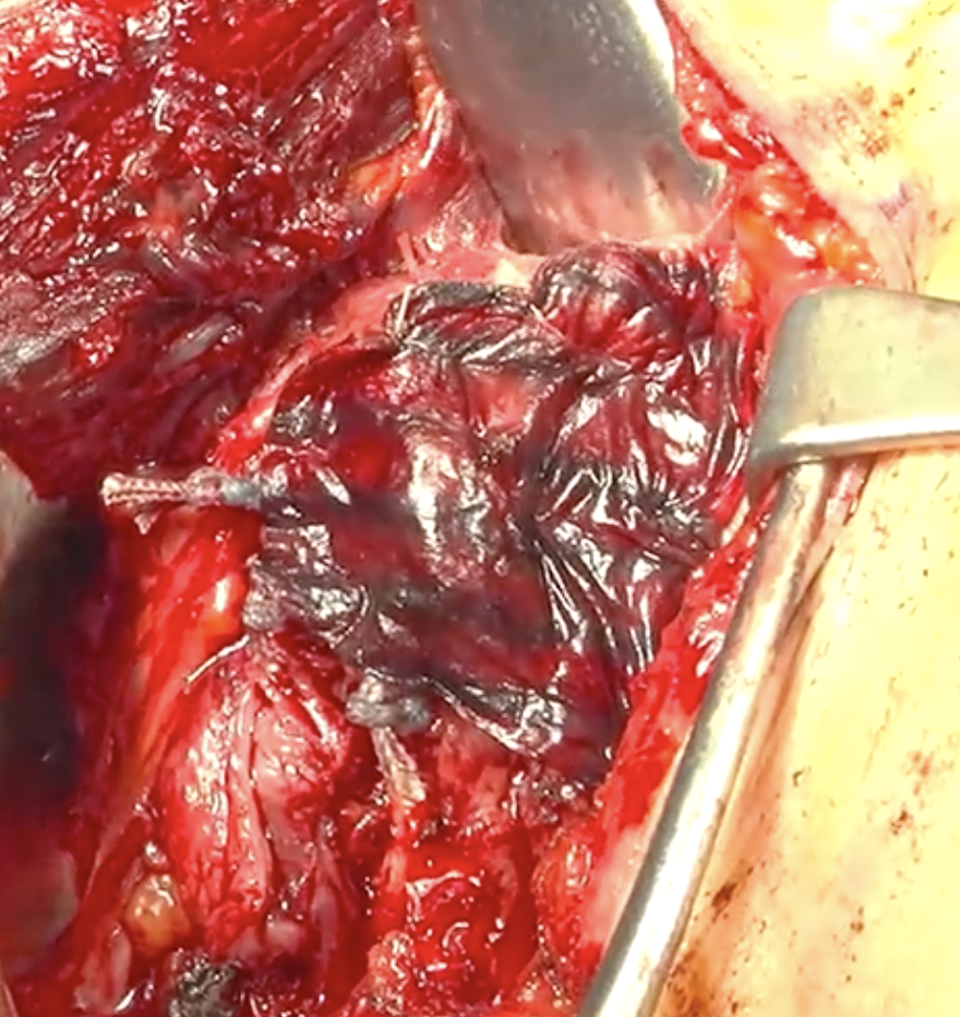
The subscapularis tendons in five consecutively eligible patients undergoing anatomic shoulder arthroplasty for primary glenohumeral arthritis were augmented with a 50x40x25mm collagen-based implant to promote subscapularis healing (Figure 1). The patient population had a mean age of 58 (range 40 to 76). Demographics are listed in Table 1. A deltopectoral approach was utilized and the subscapularis was managed with a tenotomy or a peel technique (Figure 2). The subscapularis tendon was repaired via stem-based transosseous drill tunnels and high-tensile strength sutures for a tendon-to-tendon repair (Figure 3). Afterwards, a collagen-based biointegrative implant was secured into place over top of the subscapularis repair with an absorbable monofilament suture, slightly overlapping the enthesis on the lesser tuberosity (Figure 4). Accelerated rehabilitation protocols were utilized including a sling and limiting external rotation to 0 degrees for 4 weeks.

Table 1.Baseline Patient Demographics
No. Shoulder Arthroplasty Type Comorbidities BMI Male/ Female Age (Years) Arm, Dominance (D/ND) Technique
1 Anatomic Smoker 22.38 Male 55 Left, ND Peel
2 Anatomic None 29.8 Male 60 Right, D Tenotomy
3 Anatomic Diabetic 27.3 Male 59 Left, ND Tenotomy
4 Anatomic None 29.8 Female 76 Right, D Tenotomy
5 Anatomic None 27.24 Male 40 Right, D Peel
Figure 2
Figure 2.Subscapularis managed with a tenotomy technique.
Figure 3
Figure 3.The subscapularis tendon was repaired via stem-based transosseous drill tunnels and high-tensile strength sutures for a tendon-to-bone repair.
Figure 4
Figure 4.A collagen-based biointegrative implant was secured over top of the subscapularis repair.

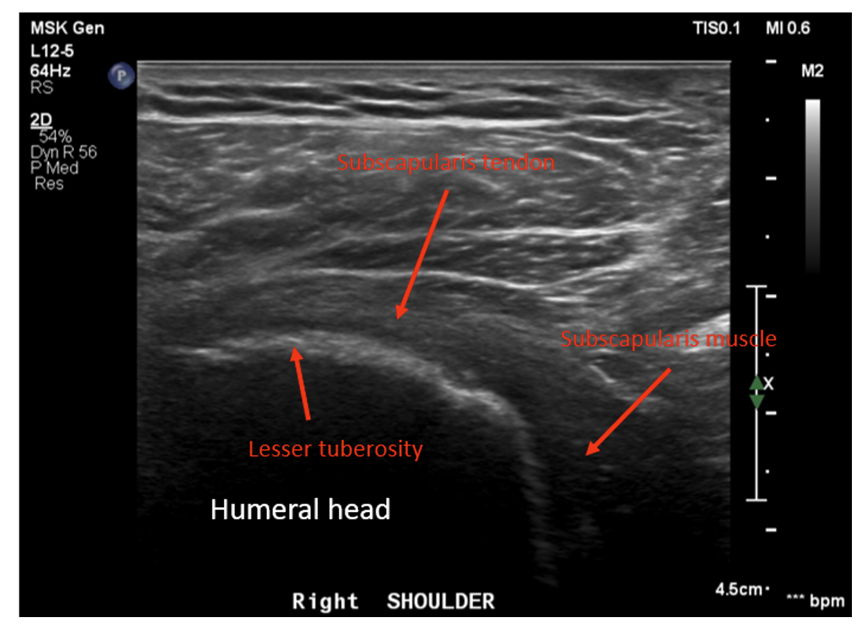
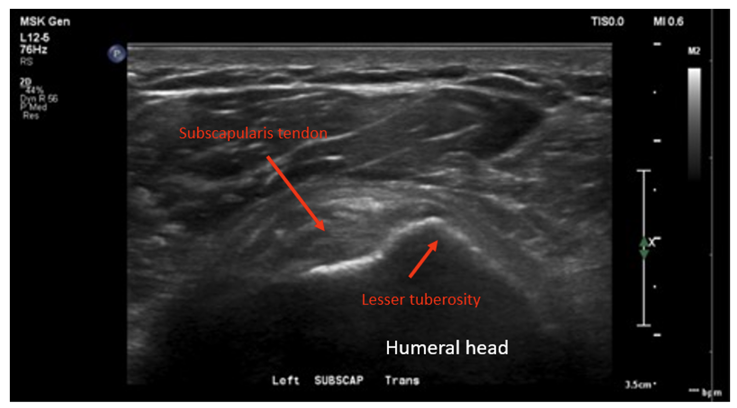
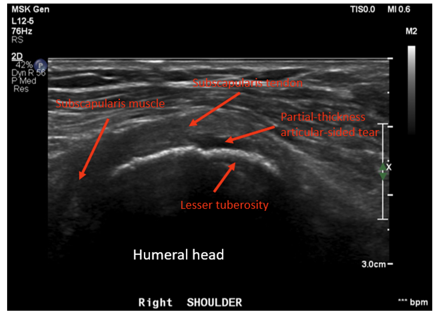
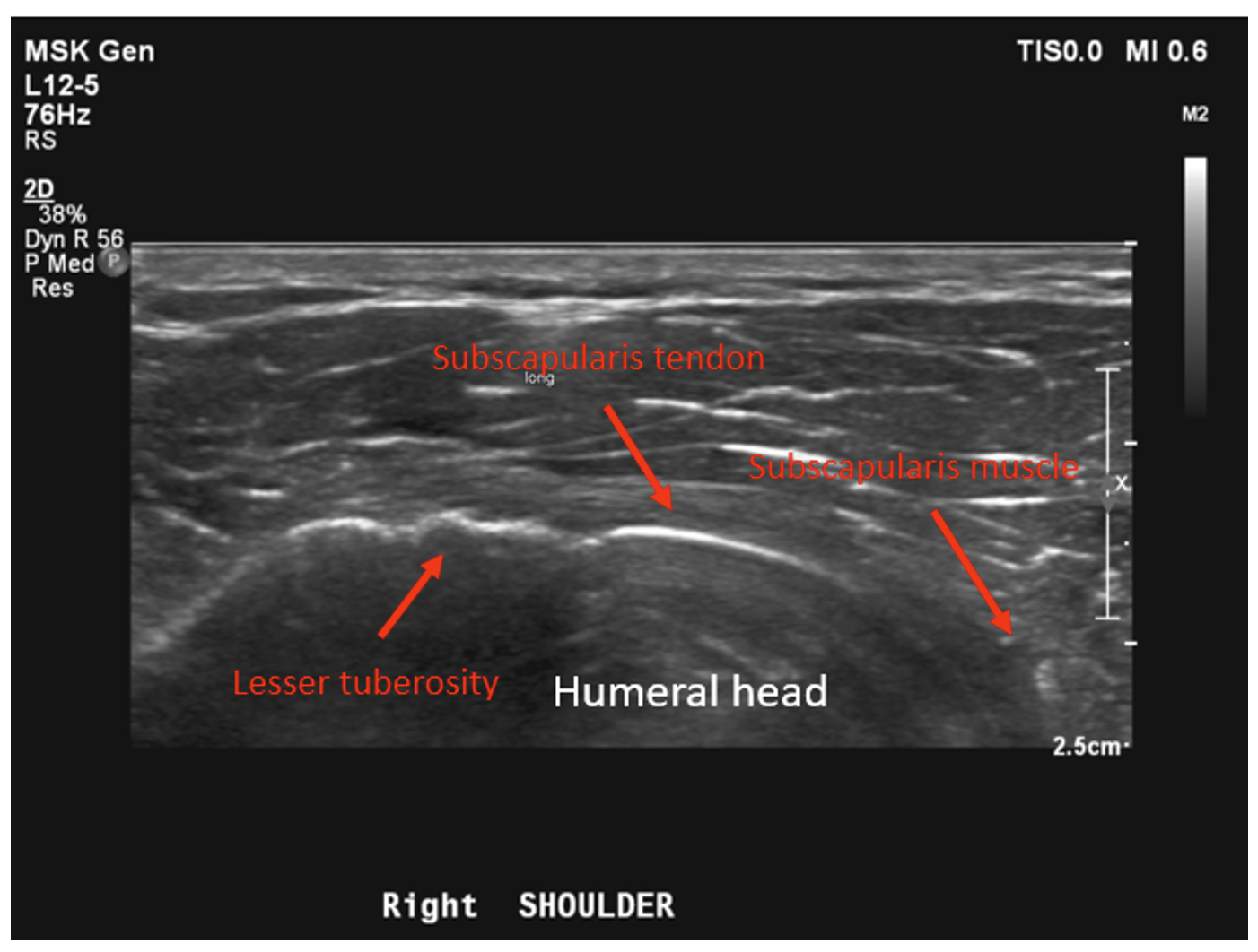
Dynamic ultrasounds performed by a fellowship trained musculoskeletal (MSK) radiologist were obtained 6 months post-surgery and evaluated for integrity of the subscapularis tendon as well as tendon thickness, width, echotexture, and calcification. Additional assessments included a standardized interview and physical examination of subscapularis function (graded belly press test and Gerber’s lift-off test), range of motion, and ASES scores were prospectively collected pre-operatively and post-operatively at 3 months and 6 months.

Click here to learn more about Zilretta

Results

The subscapularis tendon was intact (healed) in all shoulders as determined by the musculoskeletal radiologist. No tendon tears were observed. Four of 5 patients showed no evidence of tendinosis, while 1 patient showed mild tendinosis. Overall, the echogenic fibrillar architecture of the healed subscapularis tendon appeared normal and aligned. Six-month ultrasounds images for all patients are presented in Figures 5-9. The width of the subscapularis tendon in all patients showed a mean of 0.48 cm maximal tendon thickness and 2.72 cm mean tendon width (Table 2), similar to native tendon (Saltzman et al. 2014). The patients’ ASES shoulder scores showed an average improvement of 33 total points at 3 months and 35 points at 6 months post-operatively (MCID 13.6 +/- 2.3) (O.’Brien, Harley, and Yannas, n.d.). Results of secondary outcome measurements, including subscapularis function and range of motion, are presented in Table 3. All patients exhibited tendon healing without complications or revisions. At the 6-month ultrasound, there was no evidence of the collagen-based implant.

Figure 5
Figure 5.Patient #1 representative 6-month post-operative ultrasound showing intact subscapularis tendon with normal fibrillar echogenic tendon architecture and no evidence of tendinosis or tear.
Figure 6
Figure 6.Patient #2 representative 6-month post-operative ultrasound demonstrating an intact subscapularis tendon with normal collagen fibrillar architecture with visibly aligned collagen fibers and anatomic size measurements.
Figure 7
Figure 7.Patient #3 representative 6-month post-operative ultrasound showing an intact subscapularis tendon with mild tendinosis.
Figure 8
Figure 8.Patient #4 representative 6-month post-operative ultrasound showing a mostly (> 75%) intact subscapularis tendon with normal fibrillar echogenic tendon architecture and thickness, with a low-grade partial-thickness tear (< 25%)
Figure 9
Figure 9.Patient #5 representative 6-month post-operative ultrasound showing intact subscapularis tendon with normal fibrillar echogenic tendon architecture and no evidence of tendinosis or tear.
Table 2.Subscapularis Tendon Measurements
No. Maximal Tendon Thickness (anterior-posterior) Tendon Width (cranial-caudal) superior-inferior
1 0.5 cm 3.1 cm
2 0.5 cm 3.0 cm
3 0.7 cm 2.8 cm
4 0.4 cm 2.3 cm
5 0.3 cm 2.4 cm
Table 3.Preoperative Assessment and Postoperative Secondary Clinical Outcomes
No. Subscapularis Function
(graded belly press, lift-off, internal rotation)
Range of Motion
(forward flexion/abduction/ external rotation)
ASES Score
Pre-Op 6-Month Post-Op Pre-Op 6-Month Post-Op Pre-Op 3-Month Post-Op 6-Month Post-Op
1 0, 4+, back pocket 0/5-/L4 140/60/5 160/110/40 5.01 59.9 60
2 0, 5-, L5/S1 0/5/L1 150/75/35 160/100/60 49.9 56.66 56.6
3 0, 4+, L4/5 0/5/L2 150/55/35 155/100/55 38.3 71.6 63.3
4 0, 5-, L5/S1 1/5/L1 140/45/20 165/110/60 34.9 96.6 96.6
5 0, 5-, L4/5 0/5/L2 150/45/30 150/100/60 36.6 45 66.6

Conclusions

In this five patient case series, the subscapularis tendon in all patients was shown to be intact, with four showing normal fibrillar tendon architecture; one patient showed mild tendinosis and < 25% partial tear. All repairs demonstrate aligned new tissue formation rather than disorganized scar tissue. As evident in Figures 5-9, augmentation of the subscapularis tendon with the collagen-based implant was shown to be safe and effective with excellent healing outcomes at 6 months. Improvements in ASES scores, internal rotation strength and range of motion were also seen with all patients, suggesting the importance of subscapularis tendon healing after anatomic shoulder arthroplasty for optimal outcomes. This pilot study suggests subscapularis tendon augmentation has the potential to improve subscapularis healing rates and function following shoulder arthroplasty. This study is limited by a small sample size, non-randomization, and some heterogeneity in subscapularis management technique. Expansion to a multi-center, multi-surgeon clinical trial would further validate findings.


Disclosure

Amit Nathani, MD, MSc is a consultant and shareholder of Embody, Inc.’

Submitted: September 30, 2022 EDT

Accepted: October 30, 2022 EDT

References

Artel, Arsun, Hamidreza Mehdizadeh, Yu-Chieh Chiu, Eric M. Brey, and Ali Cinar. 2011. “An Agent-Based Model for the Investigation of Neovascularization within Porous Scaffolds.” Tissue Engineering Part A 17 (17–18): 2133–41. https:/​/​doi.org/​10.1089/​ten.tea.2010.0571.
Google Scholar
Baac, Hyoungwon, Jeong-Hyun Lee, Jong-Mo Seo, Tai Hyun Park, Hum Chung, Sin-Doo Lee, and Sung June Kim. 2004. “Submicron-Scale Topographical Control of Cell Growth Using Holographic Surface Relief Grating.” Materials Science and Engineering: C 24 (1–2): 209–12. https:/​/​doi.org/​10.1016/​j.msec.2003.09.009.
Google Scholar
Boileau, P., R. J. Sinnerton, C. Chuinard, and G. Walch. 2006. “Arthroplasty of the Shoulder.” The Journal of Bone and Joint Surgery. British Volume 88-B (5): 562–75. https:/​/​doi.org/​10.1302/​0301-620x.88b5.16466.
Google Scholar
Boyan, B. 1996. “Role of Material Surfaces in Regulating Bone and Cartilage Cell Response.” Biomaterials 17 (2): 137–46. https:/​/​doi.org/​10.1016/​0142-9612(96)85758-9.
Google Scholar
Cao, Haoqing, Guillaume Marcy, Eyleen Lay Keow Goh, Feng Wang, Jun Wang, and Sing Yian Chew. 2012. “The Effects of Nanofiber Topography on Astrocyte Behavior and Gene Silencing Efficiency.” Macromolecular Bioscience 12 (5): 666–74. https:/​/​doi.org/​10.1002/​mabi.201100436.
Google Scholar
Collin, Philippe, Morgane Rol, Moganadass Muniandy, Solenn Gain, Alexandre Lädermann, and Gabriella Ode. 2022. “Relationship between Postoperative Integrity of Subscapularis Tendon and Functional Outcome in Reverse Shoulder Arthroplasty.” Journal of Shoulder and Elbow Surgery 31 (1): 63–71. https:/​/​doi.org/​10.1016/​j.jse.2021.05.024.
Google Scholar
Druecke, Daniel, Stefan Langer, Evert Lamme, Jeroen Pieper, Marija Ugarkovic, Hans Ulrich Steinau, and Heinz Herbert Homann. 2003. “Neovascularization of Poly(Ether Ester) Block-Copolymer Scaffoldsin Vivo: Long-Term Investigations Using Intravital Fluorescent Microscopy.” Journal of Biomedical Materials Research 68A (1): 10–18. https:/​/​doi.org/​10.1002/​jbm.a.20016.
Google Scholar
Gigante, Antonio, Eugenio Cesari, Alberto Busilacchi, Sandra Manzotti, Kyriaki Kyriakidou, Francesco Greco, Roberto Di Primio, and Monica Mattioli-Belmonte. 2009. “Collagen I Membranes for Tendon Repair: Effect of Collagen Fiber Orientation on Cell Behavior.” Journal of Orthopaedic Research 27 (6): 826–32. https:/​/​doi.org/​10.1002/​jor.20812.
Google Scholar
Gnavi, Sara, Benedetta Fornasari, Chiara Tonda-Turo, Rossella Laurano, Marco Zanetti, Gianluca Ciardelli, and Stefano Geuna. 2015. “The Effect of Electrospun Gelatin Fibers Alignment on Schwann Cell and Axon Behavior and Organization in the Perspective of Artificial Nerve Design.” International Journal of Molecular Sciences 16 (12): 12925–42. https:/​/​doi.org/​10.3390/​ijms160612925.
Google ScholarPubMed CentralPubMed
Gomes, Manuela E., Heidi L. Holtorf, Rui L. Reis, and Antonios G. Mikos. 2006. “Influence of the Porosity of Starch-Based Fiber Mesh Scaffolds on the Proliferation and Osteogenic Differentiation of Bone Marrow Stromal Cells Cultured in a Flow Perfusion Bioreactor.” Tissue Engineering 12 (4): 801–9. https:/​/​doi.org/​10.1089/​ten.2006.12.801.
Google Scholar
Han, Yu, Meifei Lian, Qiang Wu, Zhiguang Qiao, Binbin Sun, and Kerong Dai. 2021. “Effect of Pore Size on Cell Behavior Using Melt Electrowritten Scaffolds.” Frontiers in Bioengineering and Biotechnology 9 (July): 1–13. https:/​/​doi.org/​10.3389/​fbioe.2021.629270.
Google ScholarPubMed CentralPubMed
Hollister, Scott J. 2005. “Porous Scaffold Design for Tissue Engineering.” Nature Materials 4 (7): 518–24. https:/​/​doi.org/​10.1038/​nmat1421.
Google Scholar
Lien, Sio-Mei, Liang-Yu Ko, and Ta-Jen Huang. 2009. “Effect of Pore Size on ECM Secretion and Cell Growth in Gelatin Scaffold for Articular Cartilage Tissue Engineering.” Acta Biomaterialia 5 (2): 670–79. https:/​/​doi.org/​10.1016/​j.actbio.2008.09.020.
Google Scholar
Loh, Qiu Li, and Cleo Choong. 2013. “Three-Dimensional Scaffolds for Tissue Engineering Applications: Role of Porosity and Pore Size.” Tissue Engineering Part B: Reviews 19 (6): 485–502. https:/​/​doi.org/​10.1089/​ten.teb.2012.0437.
Google ScholarPubMed CentralPubMed
Lomas, A.J., C.N.M. Ryan, A. Sorushanova, N. Shologu, A.I. Sideri, V. Tsioli, G.C. Fthenakis, et al. 2015. “The Past, Present and Future in Scaffold-Based Tendon Treatments.” Advanced Drug Delivery Reviews 84 (April):257–77. https:/​/​doi.org/​10.1016/​j.addr.2014.11.022.
Google Scholar
Maghdouri-White, Yas, Nardos Sori, Stella Petrova, Hilary Wriggers, Nathan Kemper, Amrita Dasgupta, Kelly Coughenour, et al. 2021. “Biomanufacturing Organized Collagen-Based Microfibers as a Tissue ENgineered Device (TEND) for Tendon Regeneration.” Biomedical Materials 16 (2): 025025. https:/​/​doi.org/​10.1088/​1748-605x/​abb875.
Google Scholar
Mandal, Biman B., and Subhas C. Kundu. 2009. “Cell Proliferation and Migration in Silk Fibroin 3D Scaffolds.” Biomaterials 30 (15): 2956–65. https:/​/​doi.org/​10.1016/​j.biomaterials.2009.02.006.
Google Scholar
Miller, Suzanne L., Yassamin Hazrati, Steven Klepps, Alexis Chiang, and Evan L. Flatow. 2003. “Loss of Subscapularis Function after Total Shoulder Replacement: A Seldom Recognized Problem.” Journal of Shoulder and Elbow Surgery 12 (1): 29–34. https:/​/​doi.org/​10.1067/​mse.2003.128195.
Google Scholar
Murphy, Ciara M., Matthew G. Haugh, and Fergal J. O’Brien. 2010. “The Effect of Mean Pore Size on Cell Attachment, Proliferation and Migration in Collagen–Glycosaminoglycan Scaffolds for Bone Tissue Engineering.” Biomaterials 31 (3): 461–66. https:/​/​doi.org/​10.1016/​j.biomaterials.2009.09.063.
Google Scholar
NAMSA GLP Report. n.d. “ISO Evaluation of Systemic Toxicity, Local Tissue Response and Article Degradation in Calcaneal Tendon Implantation in Rabbits—2, 8, 16, 32, 52, and 72 Weeks. Data on File at Embody, Inc.”
O.’Brien, F., B.A. Harley, and I.V. Yannas. n.d. “The Effect of Pore Size on Cell Adhesion in Collagen-GAG Scaffolds,” no. 627.
Google Scholar
O’Brien, Moira. 2005. “The Anatomy of the Achilles Tendon.” Foot and Ankle Clinics 10 (2): 225–38. https:/​/​doi.org/​10.1016/​j.fcl.2005.01.011.
Google Scholar
Oh, Daniel S., Young Joon Kim, Min-Ho Hong, Myung-Ho Han, and Kyungsoo Kim. 2014. “Effect of Capillary Action on Bone Regeneration in Micro-Channeled Ceramic Scaffolds.” Ceramics International 40 (7): 9583–89. https:/​/​doi.org/​10.1016/​j.ceramint.2014.02.033.
Google Scholar
Salerno, A., E. Di Maio, S. Iannace, and P. A. Netti. 2011. “Tailoring the Pore Structure of PCL Scaffolds for Tissue Engineering Prepared via Gas Foaming of Multi-Phase Blends.” Journal of Porous Materials 19 (2): 181–88. https:/​/​doi.org/​10.1007/​s10934-011-9458-9.
Google Scholar
Saltzman, Bryan M., Peter N. Chalmers, Anil K. Gupta, Anthony A. Romeo, and Gregory P. Nicholson. 2014. “Complication Rates Comparing Primary with Revision Reverse Total Shoulder Arthroplasty.” Journal of Shoulder and Elbow Surgery 23 (11): 1647–54. https:/​/​doi.org/​10.1016/​j.jse.2014.04.015.
Google Scholar
Schrock, John B., Matthew J. Kraeutler, Charles T. Crellin, Eric C. McCarty, and Jonathan T. Bravman. 2017. “How Should I Fixate the Subscapularis in Total Shoulder Arthroplasty? A Systematic Review of Pertinent Subscapularis Repair Biomechanics.” Shoulder & Elbow 9 (3): 153–59. https:/​/​doi.org/​10.1177/​1758573217700833.
Google ScholarPubMed CentralPubMed
Shoulder Replacement Market. n.d. Procedure: Total Shoulder Replacement, Reverse Shoulder Replacement, Partial Shoulder Replacement, Shoulder Resurfacing, and Revision Shoulder Arthroplasty; End User: Hospitals, Ambulatory Surgical Centers, and Orthopedic Clinics - Global Industry Analysis, Size, Share, Growth, Trends, and Forecast, 2019 – 2027.
Google Scholar
Simitzi, C., A. Ranella, and E. Stratakis. 2017. “Controlling the Morphology and Outgrowth of Nerve and Neuroglial Cells: The Effect of Surface Topography.” Acta Biomaterialia 51 (March):21–52. https:/​/​doi.org/​10.1016/​j.actbio.2017.01.023.
Google Scholar
Wang, Wenbo, Jing He, Bei Feng, Zhiyong Zhang, Wenjie Zhang, Guangdong Zhou, Yilin Cao, Wei Fu, and Wei Liu. 2016. “Aligned Nanofibers Direct Human Dermal Fibroblasts to Tenogenic Phenotype in Vitro and Enhance Tendon Regeneration in Vivo.” Nanomedicine 11 (9): 1055–72. https:/​/​doi.org/​10.2217/​nnm.16.24.
Google Scholar
Wang, Z., W. J. Lee, B. T. H. Koh, M. Hong, W. Wang, P. N. Lim, J. Feng, L. S. Park, M. Kim, and E. S. Thian. 2018. “Functional Regeneration of Tendons Using Scaffolds with Physical Anisotropy Engineered via Microarchitectural Manipulation.” Science Advances 4 (10): 1–13. https:/​/​doi.org/​10.1126/​sciadv.aat4537.
Google ScholarPubMed CentralPubMed
Wei, Jie, Junfeng Jia, Fan Wu, Shicheng Wei, Huanjun Zhou, Hongbo Zhang, Jung-Woog Shin, and Changsheng Liu. 2010. “Hierarchically Microporous/Macroporous Scaffold of Magnesium–Calcium Phosphate for Bone Tissue Regeneration.” Biomaterials 31 (6): 1260–69. https:/​/​doi.org/​10.1016/​j.biomaterials.2009.11.005.
Google Scholar
Yin, Zi, Xiao Chen, Jia Lin Chen, Wei Liang Shen, Thi Minh Hieu Nguyen, Ling Gao, and Hong Wei Ouyang. 2010. “The Regulation of Tendon Stem Cell Differentiation by the Alignment of Nanofibers.” Biomaterials 31 (8): 2163–75. https:/​/​doi.org/​10.1016/​j.biomaterials.2009.11.083.
Google Scholar

This website uses cookies

We use cookies to enhance your experience and support COUNTER Metrics for transparent reporting of readership statistics. Cookie data is not sold to third parties or used for marketing purposes.

Powered by Scholastica, the modern academic journal management system