Loading [Contrib]/a11y/accessibility-menu.js
Skip to main content
null
J Orthopaedic Experience & Innovation
  • Menu
  • Articles
    • Brief Report
    • Case Report
    • Data Paper
    • Editorial
    • Hand
    • Meeting Reports/Abstracts
    • Methods Article
    • Product Review
    • Research Article
    • Review Article
    • Review Articles
    • Systematic Review
    • All
  • For Authors
  • Editorial Board
  • About
  • Issues
  • Blog
  • "Open Mic" Topic Sessions
  • Advertisers
  • Recorded Content
  • CME
  • JOEI KOL Connect
  • search

RSS Feed

Enter the URL below into your favorite RSS reader.

http://localhost:54963/feed
Research Article
Vol. 3, Issue 1, 2022February 14, 2022 EDT

Mini-Open Carpal Tunnel Release with a Pediatric Nasal Speculum: A 10-Year Retrospective Case Series

Brandon Lung, MD, Lauren Grossman, MD, Matthew Kim, BS, David Komatsu, PhD, Jie Yang, PhD, Edward Wang, MD,
Carpal Tunnel ReleasePediatric Nasal Speculumretrospective case seriesCarpal tunnelFunctional statusSymptom severityNasal SpeculumMini Open
Copyright Logoccby-nc-nd-4.0 • https://doi.org/10.60118/001c.32361
J Orthopaedic Experience & Innovation
Lung, Brandon, Lauren Grossman, Matthew Kim, David Komatsu, Jie Yang, and Edward Wang. 2022. “Mini-Open Carpal Tunnel Release with a Pediatric Nasal Speculum: A 10-Year Retrospective Case Series.” Journal of Orthopaedic Experience & Innovation 3 (1). https:/​/​doi.org/​10.60118/​001c.32361.
Save article as...▾
Download all (4)
  • Figure 1. Surgical Procedure
    Download
  • Figure 2. Surgical Instruments
    Download
  • Figure 3. Patient Accrual Flow Chart
    Download
  • Figure 4. Symptom Severity and Functional Status Scores
    Download

Sorry, something went wrong. Please try again.

If this problem reoccurs, please contact Scholastica Support

Error message:

undefined

View more stats

Abstract

Background

Nasal speculum assisted carpal tunnel release (CTR) is performed using smaller incisions than are normally achieved with open procedures. We performed a retrospective case series analysis to evaluate short and long-term symptom and functional outcomes of a single surgeon’s results with this technique.

Methods

During the period of 2005-2011, 678 cases met the inclusion criteria. Of these, 198 cases representing 152 unique individuals were contacted and consented to participate in the study. A chart review was performed to identify demographics, pre-operative questionnaire results, and post-operative characteristics. Post-operative Symptom Severity Scale and Functional Status Scale results from the Carpal Tunnel Questionnaire were then obtained via telephone interviews.

Results

The mean (StDev) time to follow up was 4.08 (2.1) years with the mean age at follow up 61.94 (14) years. More females (56%) were included than males (44%). Using linear mixed models, patients with nasal speculum assisted CTR had significant improvements in both Symptom Severity and Functional Status Scales following surgery. In addition, 97.7% of patients reported their incisions were aesthetically pleasing and only 78.7% reported their incisions were still visible. Several pre-operative factors had a significant association with good surgical outcomes, including: Positive Compression Test/Durkin’s; Severe/very severe difficulties on the Functional Status Scale; High scores on the Symptom Severity and Functional Status Scales.

Conclusions

This surgeon’s experience with nasal speculum assisted CTR shows that this technique results in not only aesthetically pleasing results, but also excellent symptomatic and functional outcomes.

Level of Evidence

Level IV Case Series

Background

Carpal tunnel syndrome (CTS) is one of the most common neuropathies with an estimated US prevalence of 3-5% (Atroshi et al. 1999). CTS is caused by compression of the median nerve as it travels through the carpal tunnel bounded superiorly by the transverse carpal ligament (TCL) and inferiorly by the carpal bones. The exact mechanism of injury is unclear. The prevailing explanation is that increased intra-carpal pressure from inflammation and scarring of the flexor tendons leads to compression of the microcirculation in the perineurium which then causes nerve ischemia (Amirfeyz, Gozzard, and Leslie 2005; Gelberman et al. 1981). There are several risk factors for CTS, including; increased BMI (Hlebs, Majhenic, and Vidmar 2014; de Krom et al. 1990; Werner et al. 1994), female gender (Atroshi et al. 1999; Stevens et al. 2001, 1988), genetic predisposition (Alford, Weiss, and Akelman 2004; Hakim et al. 2002), pregnancy (Meems et al. 2015), diabetes mellitus (Oktayoglu 2015), and hypothyroidism (Oktayoglu 2015). In addition, occupational exposure requiring highly repetitive manual tasks and hand/arm vibrational work have been identified as independent risk factors (Kozak et al. 2015). CTS is diagnosed through the characteristic symptoms of dull aching pain, paresthesia, and numbness on the thumb, index, and middle fingers. Symptoms are typically worse at night or after use of the hand. Usually, patients will have partial resolution of symptoms if they change hand positions (Dawson 1993). This may be accompanied by a decreased functional status and thenar atrophy; thus the diagnosis can be confirmed with nerve conduction abnormalities.

Conservative management of CTS involves splinting, exercises, oral medications, and corticosteroid injections. While some short-term benefits have been shown for conservative management of mild-moderate CTS (Meems et al. 2014), only surgery has been shown to result in long-term positive effects (Jarvik et al. 2009). Surgical management of CTS succeeds by relieving intra-carpal pressure by transecting the transverse carpal ligament (TCL) (Aroori and Spence 2008). Historically, open carpal tunnel release (OCTR) was considered the gold standard and most reliable procedure for relieving symptoms (Badger et al. 2008). OCTR is typically performed via a longitudinal incision starting from Kaplan’s cardinal line and extended in a curvilinear manner just ulnar to the thenar crease. It is thought that the increased visualization and access to the carpal canal can decrease both the risk of neurovascular injury and the rate of incomplete transection of the carpal tunnel ligament (Steinberg 2002). In recent years, modifications to OCTR and endoscopic techniques have been developed in order to minimize post-operative pain, scarring, and improve recovery times (Sabesan et al. 2012; Nazerani et al. 2014; Atroshi et al. 2006; Cellocco et al. 2005; Tessitore, Schonauer, and Moraci 2004; Elsharif, Papanna, and Helm 2014). The proponents of open procedures often cite higher complication rates and incomplete transection of the TCL as to the reason why they perform the procedure in an open manner (Atroshi et al. 2006). However, when endoscopic releases began to be described, their safety and efficacy was clearly demonstrated (Okutsu et al. 1987; Tsai et al. 1995). Endoscopic techniques have continued to improve, and a recent analysis comparing endoscopic, open, and mini-open procedures showed open and mini-open techniques have near equal efficacy for long-term outcomes compared to traditional OCTR (Sabesan et al. 2012; Nazerani et al. 2014; Elsharif, Papanna, and Helm 2014; Gerritsen et al. 2001; Holmgren and Rabow 1987; Huisstede et al. 2010).

We have been performing a CTS using a mini-open technique that utilizes a pediatric nasal speculum for visualization of the TCL. This study is a 7-year retrospective analysis of our experience with this procedure. The primary goal is to evaluate the short- and long- term safety, efficacy, and aesthetic results of this technique. The secondary goal is to determine if there are any patient specific factors that are associated with positive surgical outcomes. Our hypothesis is that our mini-open technique utilizing a pediatric nasal speculum is safe and provides good outcomes to patients particularly with high scores on the Symptom Severity and Functional Status Scales.

Materials and Methods

Subjects

A retrospective review and telephone follow-up of patients having undergone this procedure was approved by our University’s Institutional Review Board prior to study initiation and all procedures conformed to the ethical guidelines of the 1975 Declaration of Helsinki. Participating patients’ consent was obtained verbally and recoded by members of the study team. Consent and HIPAA notification forms are on file.

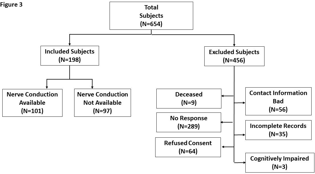
During a six year time-period, a single orthopedic hand surgeon performed mini-open carpal tunnel release surgeries on 654 subjects. Three attempts were made to contact each of the subjects by telephone in order to consent them into the study and interview them. A total of 198 unique subjects were successfully consented to the study (Fig. 3).

Each case had a chart review performed to identify demographics (gender, age, height, weight, race, and dominant hand), comorbidities (hypothyroidism, obesity, pregnancy, diabetes, osteoarthritis, rheumatoid arthritis, and neck, wrist or UE injury), patient history (wake up at night from pain/numbness, pain, numbness, paresthesias, weakness, difficulty with fine motor skills), documented pre-operative Boston Carpal Tunnel Questionnaire, physical exam (light touch, Tinel’s, Compression Test/Durkin’s, thenar atrophy, thenar strength; Atroshi et al. 1999; Amirfeyz, Gozzard, and Leslie 2005; Gelberman et al. 1981; Hlebs, Majhenic, and Vidmar 2014; de Krom et al. 1990), post-operative information (surgery completed, surgical date, surgical complications, release successful), and follow-up findings including pain, numbness, and paresthesia.

During the phone interviews, the patients answered exclusion criteria questions (i.e., trauma to the hand since surgery, stroke affecting limb operated on since surgery, neuropathy other than CTS, cognitive impairment, and nerve injury to the affected hand). They were then asked yes or no questions on whether they still experience pain, numbness/paresthesias, whether the incision was still visible, if the incision was aesthetically pleasing, and if their hand was better or worse since the surgery. Finally, participants were given the Boston Carpal Tunnel Questionnaire to assess their current symptoms compared to the documented preoperative questionnaire in the chart (Levine et al. 1993).

Surgical Procedure

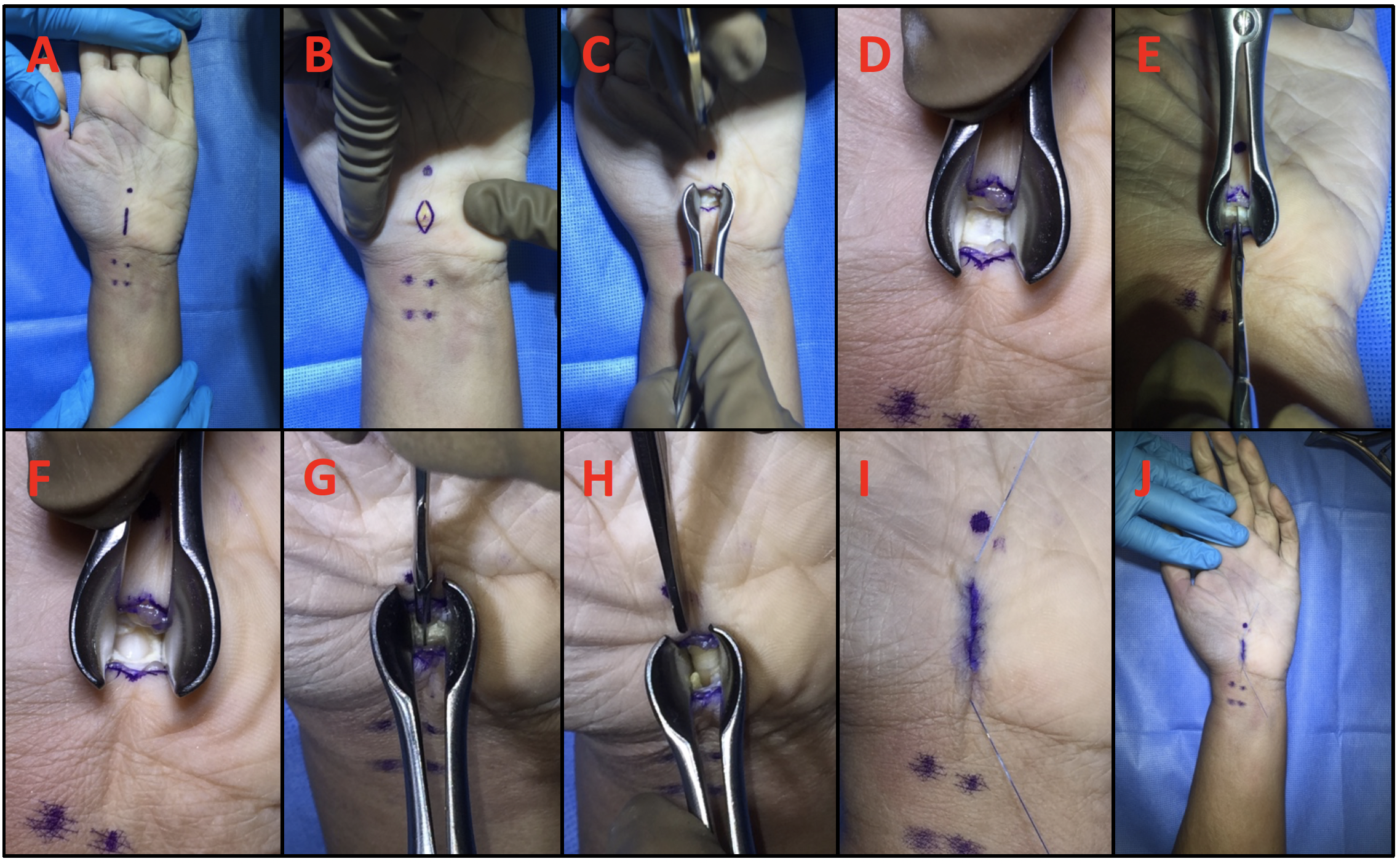
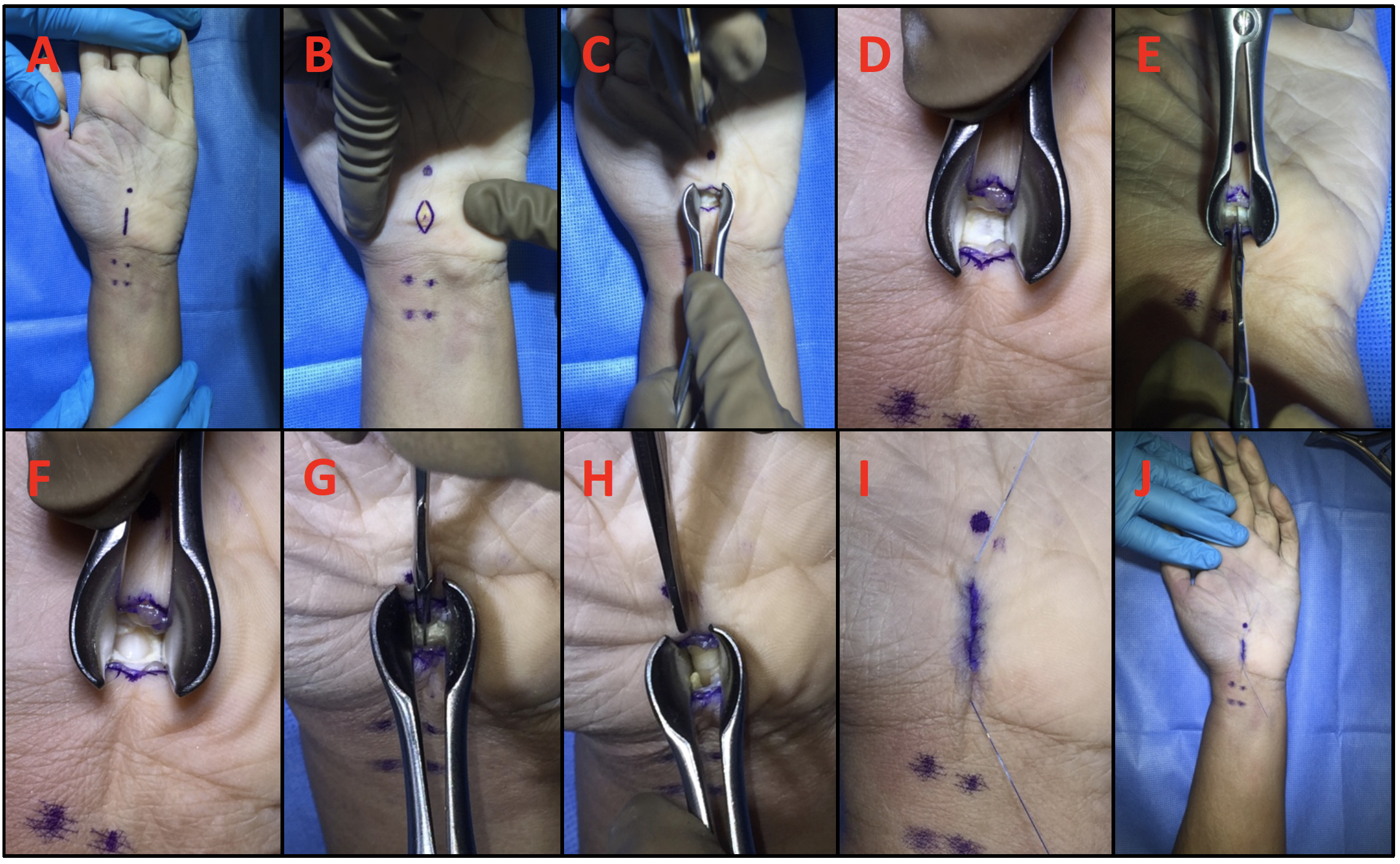
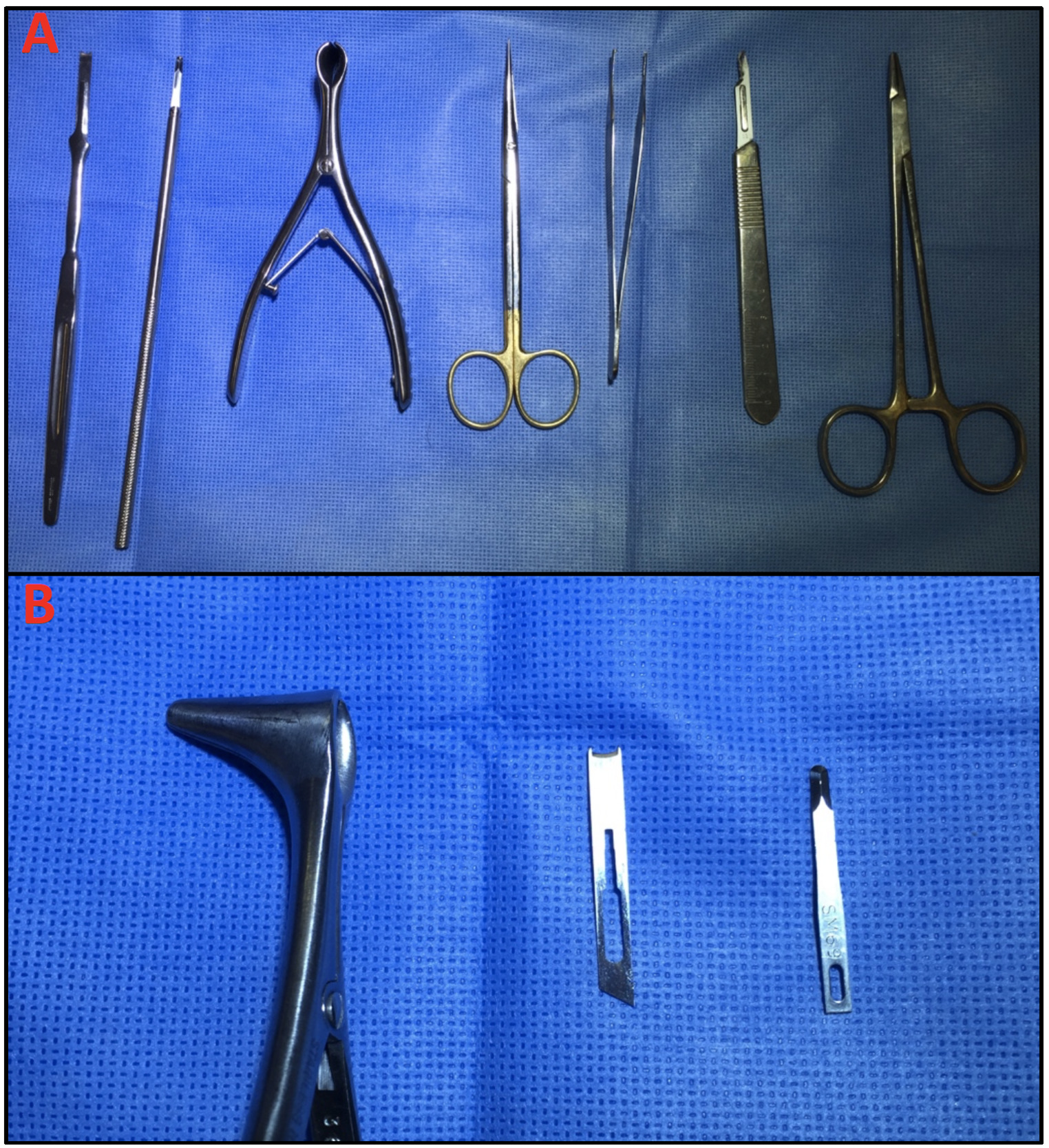
The palmaris longus (PL) and the flexor carpi radialis (FCR) are identified and their locations indicated with a surgical marker (Fig 1A). The superficial palmar arch is identified and marked distally. A ~10mm incision line over the transverse carpal ligament (TCL) is then drawn. The skin is next incised using a #15 blade and then blunt dissected to expose the palmar fascia (Fig1B). A pediatric nasal speculum is then introduced to the incision (Fig 1CD) and a mini-blade (69, Beaver) is used to open the palmar fascia and visualize the TCL (Fig 1EF). The TCL is then released using the Hans Lee CT blade (Fig 1GH) and care is made to release the volar antebrachial fascia. After ensuring complete release, the incision is then closed with poliglecaprone sutures (MONOCRYL, Johnson and Johnson) (Fig 1IJ). The complete set of surgical instruments used in this procedure is shown in Figure 2.

Figure 1
Figure 1.Surgical Procedure

This presents a series of intraoperative photographs from a cadaveric demonstration of the surgical procedure. A) Volar surface with landmarks indicated (as described in methods). B) Skin incision completed. C) Speculum introduced. D) Close up of speculum in position. E) Cutting the palmar fascia. F) Close up showing the TCL. G) Cutting the TCL. H) Close up of released TCL. I) Suturing completed. J) View of completed repair.

Figure 2
Figure 2.Surgical Instruments

Photograph showing the instruments used in this procedure. A) From left to right: Hans Lee blade with handle; Beaver blade with handle; pediatric nasal speculum; iris scissors; Adson forceps; #15 blade; needle driver. B) Zoomed in photograph showing, from left to right: pediatric nasal speculum; Hans Lee blade; mini-blade.

Figure 3
Figure 3.Patient Accrual Flow Chart

This figure breaks down the number of patients included and excluded from the study. Of the 654 subjects that underwent carpal tunnel surgery during the study period, 198 were included in the analysis (with 101 having nerve conduction studies available for review) and 456 were excluded for the stated reasons.

Statistical Analyses

In the multivariable regression analysis, answers to all of the questions from the two questionnaires were re-organized, i.e., the first two of 5 ordinal categories were grouped together as one level, and the rest were re-grouped together as another level. For example, for answers to question 1 of the Symptom Severity Questionnaire, no pain or mild pain were grouped together as not severe pain, and answers for moderate pain to very severe pain were grouped together as severe pain.

An excellent surgical outcome was defined as having at least a 60% decrease in either the Symptom Severity Scale sum score (SSS) or the Functional Status Scale sum score (FSS) after surgery. All other results were defined as good outcomes. Welch’s T-tests were utilized to compare the continuous variables in the excellent surgical outcome group to the rest. Fisher’s exact tests and Chi-square tests with exact p-values based on the Monte Carlo simulation were utilized to test the association between the excellent surgical outcomes and categorical variables with binary outcomes or multi-level outcomes, respectively. Generalized Estimating Equation (GEE) models were employed to investigate variables associated with excellent surgical outcomes, considering the correlations of observations from the same patients. Exchangeable working correlation matrices proved to be better than the independent or autoregressive working correlation matrices, and were used in all of the GEE models. The associations between excellent surgical outcome and patients’ demographic information, pre-operative measures, SSS, and FSS before surgery were examined one by one in univariate analysis. Then any variables that had a p-value <0.05 in univariate GEE models were further put into a multiple GEE models to study each factor’s association with excellent surgical outcomes after controlling for the rest factors using stepwise selection due to the limited event size (Atroshi et al. 1999). Odds ratios and their corresponding 95% confidence intervals were reported. The c-statistic was calculated to demonstrate the performance of the final predictive model. Statistical significance was set at 0.05 and analysis was done using SAS 9.3 (SAS Institute, Inc., Cary, NC) (Peduzzi et al. 1996).

Results

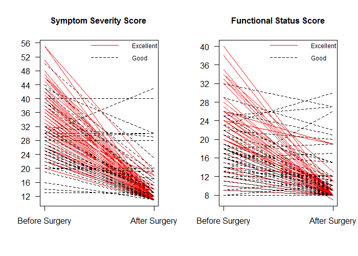
One hundred ninety-eight cases met study criteria and were included in the analysis. Chart review of demographics and clinic visits found no significant difference in age, pre-operative severity of symptoms as measured in the Boston Carpal Tunnel Questionnaire, and incidences of immediate 3-month postoperative complications, wound breakdown, or pillar pain between the study patients and those who could not be reached by telephone. The mean surgical to interview follow-up time was 4.01 years, mean age at time of surgery was 58 years, 55.7% were female, and 44.3% were male. There were no cases that required revision CTS and all cases had full transection of the transverse carpal ligament without complications. The pre and post-operative SSS scores were 32.17 and 13.08, respectively. The pre and post-operative FSS scores were 20.17 and 9.71, respectively (Table 1). In total, 113 (57%) of the cases met criteria for excellent outcome (Fig. 4).

Table 1.Pre- and Post-Operative Symptom Severity and Functional Status Scale
Scale Pre-Operative
Mean ± StDev
Post-Operative
Mean ± StDev
p-value
Symptom Severity 2.92 ± 0.78 1.19 ± 0.45 < 0.01
Functional Status 2.52 ± 0.85 1.21 ± 0.52 < 0.01

This table presents the pre- and post-operative results ± standard deviations for the Boston Carpal Tunnel Questionnaire Symptom Severity and Functional Status Scales. Pre- and post-operative values were compared using student’s t-test and the arising p-values (bolded if significant) are listed in the last column.

Figure 4
Figure 4.Symptom Severity and Functional Status Scores

These graphs show the changes in Symptom Severity and Functional Status Scores from before to after carpal tunnel surgery. Individual lines are plotted for each patient with those having ‘Excellent’ outcomes presented in solid red lines and those having ‘Good’ outcomes presented in dashed black lines.

Using univariate analysis with patients’ demographics and clinical variables separated based upon surgical outcomes, we found that patients’ age at surgery, rheumatoid arthritis, latency at the wrist, latency at the elbow, amplitude at the wrist, amplitude at the elbow, conduction velocity at the elbow, and thenar atrophy were significantly associated with surgical outcomes (all p-values <0.04). In addition, patients with excellent outcomes had longer follow up time (4.24 vs 3.59 years) and younger age (55.29 vs 64.77 years old). Complete patients’ demographics and clinical variables are shown in Table 2.

Table 2.Subject Characteristics and Pre-Operative Clinical Information
Variable Value Good
n (%) or
Mean ± StDev
Excellent
n (%) or
Mean ± StDev
p-value
Subject Characteristics
Time to follow up as of December 2013 - 3.59±2.06 4.24±2.04 0.08
Age at surgery - 64.77±15.31 55.29±12.12 <0.01
Weight (kg) - 85.20±21.18 86.95±20.86 0.64
Height (cm) - 167.90±12.18 167.27±9.60 0.75
BMI - 30.02±6.39 31.04±6.97 0.42
Sex F 32 (52.46) 65 (57.52) 0.56
M 29 (47.54) 48 (42.48)
Race Caucasian 58 (95.08) 105 (93.75) 0.72
Other 3 (4.92) 7 (6.25)
Dominant Hand Right 46 (85.19) 91 (84.26) 0.89
Left 8 (14.81) 17 (15.74)
Affected Hand Right 41 (68.33) 67 (59.29) 0.18
Left 19 (31.67) 46 (40.71)
Hypothyroidism (on Treatment) has condition 7 (11.86) 8 (7.14) 0.36
no condition 52 (88.14) 104 (92.86)
Obesity (BMI>30) has condition 22 (37.29) 52 (46.43) 0.31
no condition 37 (62.71) 60 (53.57)
Pregnancy (Self Report) has condition 2 (3.39) 0 (0.00) 0.12
no condition 57 (96.61) 112 (100.00)
Diabetes has condition 6 (10.17) 13 (11.71) 0.79
no condition 53 (89.83) 98 (88.29)
Osteoarthritis no condition 59 (100.00) 112 (100.00) .
Rhematoid Arthritis has condition 8 (13.56) 4 (3.57) 0.03
no condition 51 (86.44) 108 (96.43)
Wrist has condition 0 (0.00) 1 (0.89) 1.00
no condition 59 (100.00) 111 (99.11)
Neck no condition 59 (100.00) 112 (100.00) .
Other Upper extremity no condition 59 (100.00) 112 (100.00) .
Medical History
Wake up at night from pain/numbness Yes 46 (92.00) 90 (87.38) 0.41
No 4 (8.00) 13 (12.62)
Pain Yes 47 (85.45) 94 (87.85) 0.69
No 8 (14.55) 13 (12.15)
Reports Numbness Yes 57 (100.00) 108 (98.18) 0.54
No 0 (0.00) 2 (1.82)
Report Parasthesia Yes 30 (88.24) 77 (95.06) 0.21
No 4 (11.76) 4 (4.94)
Reports Weakness Yes 7 (77.78) 46 (93.88) 0.14
No 2 (22.22) 3 (6.12)
Reports Difficulty with fine motor skills Yes 8 (80.00) 49 (96.08) 0.09
No 2 (20.00) 2 (3.92)
Physical Exam
Light Touch Normal 24 (41.38) 53 (47.32) 0.29
Decreased 34 (58.62) 56 (50.00)
Absent 0 (0.00) 3 (2.68)
Tinel's Yes 51 (87.93) 88 (78.57) 0.15
No 7 (12.07) 24 (21.43)
Compression Test/Durkin's Yes 47 (81.03) 95 (84.82) 0.56
No 11 (18.97) 17 (15.18)
Thenar Atrophy Yes 18 (31.03) 17 (15.18) 0.03
No 40 (68.97) 95 (84.82)
Nerve Conduction
Thenar Strength (0; 1; 2; 3; 4; 5) 0-3 6 (10.34) 4 (3.57) 0.09
4-5 52 (89.66) 108 (96.43)
Lat (ms) wrist - 7.23±2.83 5.96±1.81 <0.01
Lat (ms) elbow - 11.97±3.31 10.67±2.35 0.03
Amp (mv) wrist - 4.74±3.28 7.20±3.39 <0.01
Amp (mv) elbow - 4.25±2.95 6.88±3.30 <0.01
C.V. (m/s) elbow - 48.84±7.87 52.91±9.24 0.02

This table presents the patient characteristics and clinical information (categorized by medical history, physical exam, and nerve conduction studies) obtained from a review of their medical records. Variable are listed in the first column followed by the possible values. Mean values ± standard deviations for patients deemed to have ‘Good’ or ‘Excellent’ surgical results are then presented along with p-values (bolded if significant) calculated from Wald tests on the univariate GEE models.

The surgical outcomes from the Boston Carpal Tunnel Questionnaire are presented in Tables 3 and 4. Pre-operative SSS and FSS scores by surgical outcome initially had 5 answer options and were categorized as either severe or not (Responses to the patient’s answers were categorized as either severe or not severe). Univariate analysis showed that all pre-operative SSS questions and most of pre-operative FSS questions were significantly associated with surgical outcomes.

Table 3.Pre-Operative Symptom Severity Score
Variable Value Good
n (%)
Excellent
n (%)
p-value
Hand or wrist pain at night Not Severe 45 (73.77) 59 (52.21) 0.01
Severe 16 (26.23) 54 (47.79)
How often did hand or wrist pain wake you up Less than Once 43 (70.49) 66 (58.41) 0.12
More than Once 18 (29.51) 47 (41.59)
Hand or wrist pain during daytime Not Severe 50 (81.97) 59 (52.21) <0.01
Severe 11 (18.03) 54 (47.79)
How often do you have hand or wrist pain during daytime Less than twice 53 (86.89) 72 (63.72) <0.01
More than Twice 8 (13.11) 41 (36.28)
How long an episode of pain last during daytime Less than 10 min 46 (75.41) 51 (45.13) <0.01
More than 10 min 15 (24.59) 62 (54.87)
Numbness in hand Not Severe 48 (78.69) 63 (55.75) 0.01
Severe 13 (21.31) 50 (44.25)
Weakness in hand or wrist Not Severe 49 (80.33) 71 (62.83) 0.04
Severe 12 (19.67) 42 (37.17)
Tingling sensations in hand Not Severe 52 (85.25) 72 (63.72) 0.02
Severe 9 (14.75) 41 (36.28)
How severe is numbness or tingling at night Not Severe 20 (32.79) 17 (15.04) 0.02
Severe 41 (67.21) 96 (84.96)
How often hand numbness or tingling wake you up Less than once 36 (59.02) 47 (41.59) 0.05
More than once 25 (40.98) 66 (58.41)
Difficulty with the grasping and use of small objects No Severe Difficulty 39 (63.93) 37 (32.74) <0.01
Severe Difficulty 22 (36.07) 76 (67.26)

This table presents the results for each item in the pre-operative Symptom Severity Scale. Questions are listed in the first column followed by the possible values. Mean values ± standard deviations for patients deemed to have ‘Good’ or ‘Excellent’ surgical results are then presented along with p-values (bolded if significant) calculated from Wald tests on the univariate GEE models.

Table 4.Pre-operative Functional Status Score
Variable Value Good
n (%)
Excellent
n (%)
p-value3
Writing No Severe Difficulty 25 (41.67) 39 (34.51) 0.35
Severe Difficulty 35 (58.33) 74 (65.49)
Buttoning of clothes No Severe Difficulty 19 (31.15) 12 (10.62) 0.01
Severe Difficulty 42 (68.85) 101 (89.38)
Holding a book while reading No Severe Difficulty 28 (45.90) 13 (11.50) <0.01
Severe Difficulty 33 (54.10) 100 (88.50)
Gripping a telephone handle No Severe Difficulty 38 (62.30) 19 (16.81) <0.01
Severe Difficulty 23 (37.70) 94 (83.19)
Opening of jars No Severe Difficulty 18 (29.51) 6 (5.31) <0.01
Severe Difficulty 43 (70.49) 107 (94.69)
Household chores No Severe Difficulty 27 (44.26) 20 (17.86) <0.01
Severe Difficulty 34 (55.74) 92 (82.14)
Carrying of grocery bags No Severe Difficulty 33 (54.10) 23 (20.35) <0.01
Severe Difficulty 28 (45.90) 90 (79.65)
Bathing and dressing No Severe Difficulty 44 (72.13) 38 (33.63) <0.01
Severe Difficulty 17 (27.87) 75 (66.37)

This table presents the results for each item in the pre-operative Functional Status Scale. Questions are listed in the first column followed by the possible values. Mean values ± standard deviations for patients deemed to have ‘Good’ or ‘Excellent’ surgical results are then presented along with p-values (bolded if significant) calculated from Wald tests on the univariate GEE models.

The odds ratio estimates and the corresponding 95% CI from the fitted multiple GEE model (C-statistic=0.83) are shown in Table 5. The final multiple GEE model included these four variables: gripping a telephone handle before surgery, how long an episode of pain last during daytime before surgery, how severe is numbness or tingling at night before surgery, and difficulty with bathing and dressing before surgery (all p-values <0.03), indicating that these four variables had the strongest association with surgical outcomes. For instance, patients with no severe difficulty in gripping a telephone handle before hand operations had 83.56% lower odds of having excellent surgical outcomes compared to patients with severe difficulty (95% CI: 61.34%-93.01%, p-value <0.01), i.e., patients with severe difficulty in gripping a telephone handle before surgery tended to have a higher chance of achieving excellent improvement.

Table 5.Estimated Odds Ratio and the Corresponding 95% CIs from the Fitted Multiple GEE Model
Variable Value Odds Ratio Estimate 95% CI p-value
Gripping a telephone handle No Severe Difficulty vs Severe Difficulty 0.1644 0.0699 0.3866 <0.01
How long an episode of pain last during daytime Less than 10 min vs More than 10 min 0.3618 0.1428 0.9167 0.03
How severe is numbness or tingling at night Not Severe vs Severe 0.3299 0.1231 0.8844 0.03
Bathing and dressing No Severe Difficulty vs Severe Difficulty 0.2948 0.1202 0.7231 0.01

This table presents the results of the final multiple GEE model. With the four variables that had the strongest association with surgical outcomes listed in the first column, followed by the possible values, estimated odds ratios, 95% confidence intervals, and p-values (bolded if significant) calculated from Wald tests on the univariate GEE models.

Discussion

CTS is the most common neuropathy and many surgical techniques have been developed for its treatment (Atroshi et al. 1999). Historically, OCTR was the most reliable procedure for relieving the symptoms of CTS (Badger et al. 2008). However, mini-open and endoscopic procedures have been developed to minimize post-operative pain, scarring and shorter recovery times (Sabesan et al. 2012; Nazerani et al. 2014; Atroshi et al. 2006; Cellocco et al. 2005; Tessitore, Schonauer, and Moraci 2004; Elsharif, Papanna, and Helm 2014). Endoscopic procedures have been criticized for their increased cost due to the need for additional instrumentation along with high rates of vessel injury and incomplete release (Lorgelly et al. 2005). Proponents of the endoscopic approach claim that the shorter recovery time allows for earlier return to work, which cancels out these higher costs (Trumble et al. 2002; Vasiliadis et al. 2014). The nasal speculum assisted approach has the benefit of a single small incision, which gives the same benefits of faster recovery time without the need for expensive instrumentation and training. However, the mini-open approach has been criticized because this procedure does not allow full visualization of the carpal tunnel ligament (CTL) leading to incomplete transections of the CTL and increased complications (Wong et al. 2003). We therefore performed a retrospective analysis to see if this criticism was valid.

In our nasal speculum assisted carpal tunnel release (CTR) study, we had 100% success in transecting the CTL and no operative complications. A similar nasal speculum approach of 101 patients confirms the safety and efficacy of this approach (Muppavarapu et al. 2015). While the major disadvantage of the mini-open technique includes limited visualization, no cases of blood vessel and nerve damage or incomplete transection occurred in order to preserve a small incision site. Mini-open release has been shown to increase the flattening ratio of the median nerve while decreasing pathologic swelling, allowing for fewer wound complications and shorter postoperative pain. In addition, 97.7% of patients reported their mini incisions were aesthetically pleasing and only 78.7% reported their incisions were still visible. However, it is important for surgeons to understand the difficulty of inserting the nasal speculum into a small incision site while avoiding damage to the median nerve.

Prior studies have found poor surgical outcomes with high Boston pre-operative scores, delay of surgery, and poor EMG results (Rege and Sher 2001; Aksekili, Bicici, Isik, et al. 2015). However, no study has ever analyzed all the pre-operative risk factors and symptoms associated with good surgical outcomes. Through this study, we found that only four questions are needed to predict whether the patient would have a good surgical outcome or not. While previous studies have reported improved objective measurements of sensation, pinch, and grip strength using nasal instrumentation, our study focuses on subjective evaluation, which may more accurately reflect functional improvements as judged by the patient (Sever et al. 2010; Karnezis and Fragkiadakis 2002). As seen in our results, patients with more severe preoperative symptom severity had a higher likelihood of excellent surgical outcome compared with patients with less severe symptoms. These results may reflect the idea that patients with more severe preoperative symptoms may experience greater improved physical and emotional changes postoperatively not always correlated with motor tests.

The reliability of clinical signs, motor tests, outcome questionnaires, and electrophysiological studies for outcome measures after CTS surgery remains unknown. Past studies have shown severity scoring using the Boston Questionnaire to be predictive of outcome measures and in identifying patients likely to respond to surgery (Yucel and Seyithanoglu 2015). Improvements in Boston Questionnaire scores correlate with a decrease in the carpal tunnel inlet cross sectional area suggesting reduced pathologic swelling and perceived symptom severity are associated (Oh et al. 2017). While most aspects of symptom severity and functional status were associated with excellent outcomes on univariate analysis, our fitted multiple GEE model predicted duration of pain episode and severity of numbness or tingling at night to be symptoms most predictive of surgical outcome. Routine daily activities, such as gripping a telephone handle, bathing, and dressing were functional measures that if severely impaired would be predictive of favorable postoperative results. Compared to other components of the functional status scale, telephone gripping and bathing are daily routine tasks where improved limitation and discomfort is most noticeable on follow up. It is important for surgeons to be aware of demographic, history, and physical exam components associated with excellent surgical outcomes in order to communicate appropriate expectations and return to functioning with patients. Clinicians should consider mini-open incision as a safe, satisfactory procedure in patients with CTS coming to clinic who present with severe pain, prolonged daytime numbness, and inability to bathe and use the telephone.

Because this study is a retrospective case series, limitations include prolonged mean follow-up time and recall bias. Patient reported outcomes measurements are subject to recall bias as prior studies have found retrospective subjective outcomes produce more favorable results than prospectively monitored health status data (Gotlin et al. 2019). Review of literature suggests many subjective outcomes after CTS are similar three months postoperatively (Wong et al. 2003). However, the excellent surgical outcomes recorded from our mean time to follow up of 4.08 years may be explained by expected symptom relief from extended recovery from natural healing processes. Many subjects were excluded due to no responses on telephone interview attempts, thus limiting our study to selection bias. Finally, from a surgical technique perspective, the insertion of the nasal speculum into the carpal tunnel is challenging and makes for further tunnel exploration and synovectomy difficult.

Conclusion

Overall, this study provides evidence that surgeons should consider nasal-speculum assisted mini-open carpal tunnel release as a safe, satisfactory procedure in patients presenting with CTS. Patients with more severe preoperative symptom severity had a higher likelihood of excellent surgical outcome compared with patients with less severe symptoms. Duration and severity of pain episodes along with difficulties in bathing and gripping a telephone were found to be most predictive of improved subjective evaluation. As healthcare economics prioritizes patient satisfaction outcomes, it is important for surgeons to consider this mini-open technique as a way for patients with difficulties in routine daily activities to experience improved self-reported assessments, which may more closely reflect functional improvements as judged by the patient.


List of Abbreviations

CTR: Carpal Tunnel Release

CTS: Carpal Tunnel Syndrome

CTL: Carpal Tunnel Ligament

GEE: Generalized Estimating Equation

EMG: Electromyography

OCTR: Open Carpal Tunnel Release

SSS: Symptom Severity Scale

FSS: Functional Status Scale

TCL: Transverse Carpal Ligament

Ethics approval and consent to participate

A retrospective review and telephone follow-up of patients having undergone this procedure was approved by our University’s Institutional Review Board prior to study initiation and all procedures conformed to the ethical guidelines of the 1975 Declaration of Helsinki.

Consent for publication

Participating patients’ consents were obtained verbally and recoded by members of the study team. Consent and HIPAA notification forms are on file.

Availability of data and materials

The datasets used and analyzed during the current study are available from the corresponding author on reasonable request.

Competing Interests

The authors declare that they have no competing interests.

Funding

The authors report no external funding for this study.

Submitted: December 27, 2021 EDT

Accepted: January 27, 2022 EDT

References

Aksekili, M.A., V. Bicici, C. Isik, et al. 2015. “Comparison of Early Postoperative Period Electrophysiological and Clinical Findings Following Carpal Tunnel Syndrome: Is EMG Necessary?” Int J Clin Exp Med 8 (6): 10011–15.
Google Scholar
Alford, J. W., A. P. Weiss, and E. Akelman. 2004. “The Familial Incidence of Carpal Tunnel Syndrome in Patients with Unilateral and Bilateral Disease.” Am J Orthop 33 (8): 397–400.
Google Scholar
Amirfeyz, R., C. Gozzard, and I. J. Leslie. 2005. “Hand Elevation Test for Assessment of Carpal Tunnel Syndrome.” Journal of Hand Surgery 30 (4): 361–64. https:/​/​doi.org/​10.1016/​j.jhsb.2005.04.007.
Google Scholar
Aroori, S., and R. A. Spence. 2008. “Carpal Tunnel Syndrome.” Ulster Med J 77 (1): 6–17.
Google Scholar
Atroshi, Isam, C. Gummesson, R. Johnsson, E. Ornstein, J. Ranstam, and I. Rosen. 1999. “Prevalence of Carpal Tunnel Syndrome in a General Population.” JAMA 282 (2): 153–58. https:/​/​doi.org/​10.1001/​jama.282.2.153.
Google Scholar
Atroshi, Isam, Gert-Uno Larsson, Ewald Ornstein, Manfred Hofer, Ragnar Johnsson, and Jonas Ranstam. 2006. “Outcomes of Endoscopic Surgery Compared with Open Surgery for Carpal Tunnel Syndrome among Employed Patients: Randomised Controlled Trial.” BMJ 332 (7556): 1473. https:/​/​doi.org/​10.1136/​bmj.38863.632789.1f.
Google ScholarPubMed CentralPubMed
Badger, S. A., M. E. O’Donnell, J. M. Sherigar, P. Connolly, and R. A. Spence. 2008. “Open Carpal Tunnel Release--Still a Safe and Effective Operation.” Ulster Med J 77 (1): 22–24.
Google Scholar
Cellocco, Paolo, Constantino Rossi, Francesco Bizzarri, Luigi Patrizio, and Giuseppe Costanzo. 2005. “Mini-Open Blind Procedure versus Limited Open Technique for Carpal Tunnel Release: A 30-Month Follow-up Study.” The Journal of Hand Surgery 30 (3): 493–99. https:/​/​doi.org/​10.1016/​j.jhsa.2005.02.007.
Google Scholar
Dawson, David M. 1993. “Entrapment Neuropathies of the Upper Extremities.” New England Journal of Medicine 329 (27): 2013–18. https:/​/​doi.org/​10.1056/​nejm199312303292707.
Google Scholar
Elsharif, M., M. Papanna, and R. Helm. 2014. “Long-Term Follow up Outcome Results of Knifelight Carpal Tunnel Release and Conventional Open Release Following a Departmental Randomized Controlled Trial. A Prospective Study.” Pol Orthop Traumatol 79:67–70.
Google Scholar
Gelberman, R. H., P. T. Hergenroeder, A. R. Hargens, G. N. Lundborg, and W. H. Akeson. 1981. “The Carpal Tunnel Syndrome. A Study of Carpal Canal Pressures.” Journal of Bone & Joint Surgery 63 (3): 380–83. https:/​/​doi.org/​10.2106/​00004623-198163030-00009.
Google Scholar
Gerritsen, A. A. M., B. M. J. Uitdehaag, D. van Geldere, R. J. P. M. Scholten, H. C. W. de Vet, and L. M. Bouter. 2001. “Systematic Review of Randomized Clinical Trials of Surgical Treatment for Carpal Tunnel Syndrome.” British Journal of Surgery 88 (10): 1285–95. https:/​/​doi.org/​10.1046/​j.0007-1323.2001.01858.x.
Google Scholar
Gotlin, Matthew J., Samuel Baron, Matthew T. Kingery, Joseph McCafferty, Laith M. Jazrawi, and Robert J. Meislin. 2019. “Recall Bias in Retrospective Assessment of Preoperative Patient Reported Outcomes.” Presented at the AOSSM Annual Meeting. https:/​/​doi.org/​10.1177/​2325967119s00339.
Hakim, Alan J., Lynn Cherkas, Sameh El Zayat, Alexander J. MacGregor, and Tim D. Spector. 2002. “The Genetic Contribution to Carpal Tunnel Syndrome in Women: A Twin Study.” Arthritis & Rheumatism 47 (3): 275–79. https:/​/​doi.org/​10.1002/​art.10395.
Google Scholar
Hlebs, S., K. Majhenic, and G. Vidmar. 2014. “Body Mass Index and Anthropometric Characteristics of the Hand as Risk Factors for Carpal Tunnel Syndrome.” Coll Antropol 38 (1): 219–26.
Google Scholar
Holmgren, Helen, and L. Rabow. 1987. “Internal Neurolysis or Ligament Division Only in Carpal Tunnel Syndrome. II. A 3 Year Follow-up with an Evaluation of Various Neurophysiological Parameters for Diagnosis.” Acta Neurochirurgica 87 (1–2): 44–47. https:/​/​doi.org/​10.1007/​bf02076014.
Google Scholar
Huisstede, Bionka M., Manon S. Randsdorp, J. Henk Coert, Suzanne Glerum, Marienke van Middelkoop, and Bart W. Koes. 2010. “Carpal Tunnel Syndrome. Part II: Effectiveness of Surgical Treatments—A Systematic Review.” Archives of Physical Medicine and Rehabilitation 91 (7): 1005–24. https:/​/​doi.org/​10.1016/​j.apmr.2010.03.023.
Google Scholar
Jarvik, Jeffrey G., Bryan A. Comstock, Michel Kliot, Judith A. Turner, Leighton Chan, Patrick J. Heagerty, William Hollingworth, Carolyn L. Kerrigan, and Richard A. Deyo. 2009. “Surgery versus Non-Surgical Therapy for Carpal Tunnel Syndrome: A Randomised Parallel-Group Trial.” The Lancet 374 (9695): 1074–81. https:/​/​doi.org/​10.1016/​s0140-6736(09)61517-8.
Google Scholar
Karnezis, I. A., and E. G. Fragkiadakis. 2002. “Association between Objective Clinical Variables and Patient-Rated Disability of the Wrist.” The Journal of Bone and Joint Surgery. British Volume 84 (7): 967–70. https:/​/​doi.org/​10.1302/​0301-620x.84b7.0840967.
Google Scholar
Kozak, Agnessa, Grita Schedlbauer, Tanja Wirth, Ulrike Euler, Claudia Westermann, and Albert Nienhaus. 2015. “Association between Work-Related Biomechanical Risk Factors and the Occurrence of Carpal Tunnel Syndrome: An Overview of Systematic Reviews and a Meta-Analysis of Current Research.” BMC Musculoskeletal Disorders 16 (1): 231. https:/​/​doi.org/​10.1186/​s12891-015-0685-0.
Google ScholarPubMed CentralPubMed
Krom, M. C. de, A. D. Kester, P. G. Knipschild, and F. Spaans. 1990. “Risk Factors for Carpal Tunnel Syndrome.” American Journal of Epidemiology 132 (6): 1102–10. https:/​/​doi.org/​10.1093/​oxfordjournals.aje.a115753.
Google Scholar
Levine, D. W., B. P. Simmons, M. J. Koris, L. H. Daltroy, G. G. Hohl, A. H. Fossel, and J. N. Katz. 1993. “A Self-Administered Questionnaire for the Assessment of Severity of Symptoms and Functional Status in Carpal Tunnel Syndrome.” The Journal of Bone & Joint Surgery 75 (11): 1585–92. https:/​/​doi.org/​10.2106/​00004623-199311000-00002.
Google Scholar
Lorgelly, Paula K., Joseph J. Dias, Mary J. Bradley, and Frank D. Burke. 2005. “Carpal Tunnel Syndrome, the Search for a Cost-Effective Surgical Intervention: A Randomised Controlled Trial.” Annals of The Royal College of Surgeons of England 87 (1): 36–40. https:/​/​doi.org/​10.1308/​1478708051469.
Google ScholarPubMed CentralPubMed
Meems, Margreet, Brenda Den Oudsten, Berend-Jan Meems, and Victor Pop. 2014. “Effectiveness of Mechanical Traction as a Non-Surgical Treatment for Carpal Tunnel Syndrome Compared to Care as Usual: Study Protocol for a Randomized Controlled Trial.” Trials 15 (1): 180. https:/​/​doi.org/​10.1186/​1745-6215-15-180.
Google ScholarPubMed CentralPubMed
Meems, Margreet, S. E. M. Truijens, V. Spek, L. H. Visser, and V. J. M. Pop. 2015. “Prevalence, Course and Determinants of Carpal Tunnel Syndrome Symptoms during Pregnancy: A Prospective Study.” BJOG 122 (8): 1112–18. https:/​/​doi.org/​10.1111/​1471-0528.13360.
Google Scholar
Muppavarapu, R. C., S. S. Rajaee, D. E. Ruchelsman, and M. R. Belsky. 2015. “Open Carpal Tunnel Release With Use of a Nasal Turbinate Speculum.” Am J Orthop 44 (11): 495–98.
Google Scholar
Nazerani, Shahram, Mohamad Hossein Kalantar Motamedi, Tina Nazerani, Amir Saraii, and M. R. Keramati. 2014. “Endoscopic Carpal Tunnel Release: A 5-Year Experience.” Trauma Monthly 19 (4): e18058. https:/​/​doi.org/​10.5812/​traumamon.18058.
Google ScholarPubMed CentralPubMed
Oh, Won-Taek, Ho-Jung Kang, Il-Hyun Koh, Jin-Young Jang, and Yun-Rak Choi. 2017. “Morphologic Change of Nerve and Symptom Relief Are Similar after Mini-Incision and Endoscopic Carpal Tunnel Release: A Randomized Trial.” BMC Musculoskeletal Disorders 18 (1): 65. https:/​/​doi.org/​10.1186/​s12891-017-1438-z.
Google ScholarPubMed CentralPubMed
Oktayoglu, Pelin. 2015. “Assessment of the Presence of Carpal Tunnel Syndrome in Patients with Diabetes Mellitus, Hypothyroidism and Acromegaly.” Journal of Clinical and Diagnostic Research 9 (6): OC14-18. https:/​/​doi.org/​10.7860/​jcdr/​2015/​13149.6101.
Google ScholarPubMed CentralPubMed
Okutsu, I., S. Ninomiya, Y. Ugawa, N. Kuroshima, H. Inanami, and S. Hiraki. 1987. “New Operative Procedure for Caparl Tunnel Syndrome: Endoscopic Operation and Clinical Results.” Society Surgery Hand 4:117–20.
Google Scholar
Peduzzi, Peter, John Concato, Elizabeth Kemper, Theodore R. Holford, and Alvan R. Feinstein. 1996. “A Simulation Study of the Number of Events per Variable in Logistic Regression Analysis.” Journal of Clinical Epidemiology 49 (12): 1373–79. https:/​/​doi.org/​10.1016/​s0895-4356(96)00236-3.
Google Scholar
Rege, A. J., and J. L. Sher. 2001. “Can the Outcome of Carpal Tunnel Release Be Predicted?” Journal of Hand Surgery 26 (2): 148–50. https:/​/​doi.org/​10.1054/​jhsb.2000.0544.
Google Scholar
Sabesan, Vani J., Dawn Pedrotty, James R. Urbaniak, and J. Mack Aldridge. 2012. “An Evidence-Based Review of a Single Surgeon’s Experience with Endoscopic Carpal Tunnel Release.” Journal of Surgical Orthopaedic Advances 21 (3): 117–21. https:/​/​doi.org/​10.3113/​jsoa.2012.0117.
Google Scholar
Sever, Celalettin, Yalcin Kulahci, Sinan Oksuz, and Cihan Sahin. 2010. “The Mini Incision Technique for Carpal Tunnel Decompression Using Nasal Instruments.” Turkish Neurosurgery 20 (3): 353–57. https:/​/​doi.org/​10.5137/​1019-5149.jtn.2526-09.0.
Google Scholar
Steinberg, David R. 2002. “Surgical Release of the Carpal Tunnel.” Hand Clinics 18 (2): 291–98. https:/​/​doi.org/​10.1016/​s0749-0712(01)00011-7.
Google Scholar
Stevens, J. Clarke, S. Sun, C. M. Beard, W. M. O’Fallon, and L. T. Kurland. 1988. “Carpal Tunnel Syndrome in Rochester, Minnesota, 1961 to 1980.” Neurology 38 (1): 134–134. https:/​/​doi.org/​10.1212/​wnl.38.1.134.
Google Scholar
Stevens, J. Clarke, J. C. Witt, B. E. Smith, and A. L. Weaver. 2001. “The Frequency of Carpal Tunnel Syndrome in Computer Users at a Medical Facility.” Neurology 56 (11): 1568–70. https:/​/​doi.org/​10.1212/​wnl.56.11.1568.
Google Scholar
Tessitore, Enrico, Claudio Schonauer, and Aldo Moraci. 2004. “Mini-Open Carpal Tunnel Decompression.” Neurosurgery 55 (4): 1010. https:/​/​doi.org/​10.1227/​01.neu.0000139577.13399.d9.
Google Scholar
Trumble, Thomas E., Edward Diao, Reid A. Abrams, and Mary M. Gilbert-Anderson. 2002. “Single-Portal Endoscopic Carpal Tunnel Release Compared with Open Release: A Prospective, Randomized Trial.” The Journal of Bone & Joint Surgery 84 (7): 1107–15. https:/​/​doi.org/​10.2106/​00004623-200207000-00003.
Google Scholar
Tsai, T. M., T. Tsuruta, S. A. Syed, and H. Kimura. 1995. “A New Technique for Endoscopic Carpal Tunnel Decompression.” British Journal of Hand Surgery 17 (August):987–95. https:/​/​doi.org/​10.1016/​s0266-7681(05)80154-8.
Google Scholar
Vasiliadis, Haris S., Petros Georgoulas, Ian Shrier, Georgia Salanti, and Rob J. P. M. Scholten. 2014. “Endoscopic Release for Carpal Tunnel Syndrome.” Cochrane Database of Systematic Reviews 1 (January):CD008265. https:/​/​doi.org/​10.1002/​14651858.cd008265.pub2.
Google Scholar
Werner, Robert A., James W. Albers, Alfred Franzblau, and Thomas J. Armstrong. 1994. “The Relationship between Body Mass Index and the Diagnosis of Carpal Tunnel Syndrome.” Muscle & Nerve 17 (6): 632–36. https:/​/​doi.org/​10.1002/​mus.880170610.
Google Scholar
Wong, K. C., L. K. Hung, P. C. Ho, and J. M. W. Wong. 2003. “Carpal Tunnel Release. A Prospective, Randomised Study of Endoscopic versus Limited-Open Methods.” The Journal of Bone and Joint Surgery. British Volume 85 (6): 863–68. https:/​/​doi.org/​10.1302/​0301-620x.85b6.13759.
Google Scholar
Yucel, Hulya, and H. Seyithanoglu. 2015. “Choosing the Most Efficacious Scoring Method for Carpal Tunnel Syndrome.” Acta Orthop Traumatol Turc 49 (1): 23–29. https:/​/​doi.org/​10.3944/​aott.2015.13.0162.
Google Scholar

This website uses cookies

We use cookies to enhance your experience and support COUNTER Metrics for transparent reporting of readership statistics. Cookie data is not sold to third parties or used for marketing purposes.

Powered by Scholastica, the modern academic journal management system