Loading [Contrib]/a11y/accessibility-menu.js
Skip to main content
null
J Orthopaedic Experience & Innovation
  • Menu
  • Articles
    • Brief Report
    • Case Report
    • Data Paper
    • Editorial
    • Hand
    • Meeting Reports/Abstracts
    • Methods Article
    • Product Review
    • Research Article
    • Review Article
    • Review Articles
    • Systematic Review
    • All
  • For Authors
  • Editorial Board
  • About
  • Issues
  • Blog
  • "Open Mic" Topic Sessions
  • Advertisers
  • Recorded Content
  • CME
  • JOEI KOL Connect
  • search

RSS Feed

Enter the URL below into your favorite RSS reader.

https://journaloei.scholasticahq.com/feed
Methods Article
Vol. 2, Issue 2, 2021October 17, 2021 EDT

The Next Frontier for Rotator Cuff Augmentation? Strength + Bio-Induction

Sean McMillan, DO, Robert Arciero, MD, Elizabeth Ford, DO,
rotator cuff augmentationbio-inductivebio-compositerotator cuff strengthshoulder
Copyright Logoccby-nc-nd-4.0 • https://doi.org/10.60118/001c.28081
J Orthopaedic Experience & Innovation
McMillan, Sean, Robert Arciero, and Elizabeth Ford. 2021. “The Next Frontier for Rotator Cuff Augmentation?  Strength + Bio-Induction.” Journal of Orthopaedic Experience & Innovation 2 (2). https:/​/​doi.org/​10.60118/​001c.28081.
Save article as...▾
Download all (6)
  • Figure 1. The BioBrace™ implant for rotator cuff augmentation is pictured with dimensions of 23mm wide, 30mm in length, and 3mm in thickness. The implant can be trimmed to the physicians desired specifications without weakening the implant.
    Download
  • Figure 2. The BioBrace™ is 80% porous with PLLA microfilament reinforcement to allow for induction of host tissue, maturation, and strength.
    Download
  • Figure 3. (Courtesy of Michael Russonella, DO) The BioBrace™ has the ability to act as a wick during hydration. Picture above is a BioBrace™ (arrow) that has been pre-soaked in platelet rich plasma (PRP) for augmentation of an anterior cruciate ligament reconstruction (ACLR).
    Download
  • Figure 4. After passage of the RCR sutures through the native tendon arthroscopically, the repair sutures are brought through the lateral cannula where there were passed through the BioBrace™ implant using a commercial passing gun.
    Download
  • Figure 5. The BioBrace™ was marked on the superior surface for orientation purposes and delivered into the shoulder using a “back-grasper”. Once delivered, a knot pusher was placed on sutures to bring the implant flush to the tissue.
    Download
  • Figure 6. The posterior and central-posterior limbs were tied together in the above construct, followed by the anterior and central-anterior limbs. After the medial row compression was complete, the suture limbs that passed through the native tendon and the BioBrace™ were brought lateral in a compression bridge configuration with two self-punching PEEK anchors. Note the blood from the surrounding tissues and bone is adhering to the implant even in an aqueous environment.
    Download

Sorry, something went wrong. Please try again.

If this problem reoccurs, please contact Scholastica Support

Error message:

undefined

View more stats

Abstract

Rotator cuff repair (RCR) is one of the most common arthroscopic orthopedic procedures performed worldwide. A high number of clinical and subclinical re-tears exist post procedure, particularly in the subset group of large (>3cm) to massive tears and revision tears (Agrawal 2012; Galatz et al. 2004; Djurasovic et al. 2001; Burkhart et al. 2007). Advances in RCR over the past two decades have nearly eliminated failures related to implant failure. Currently the most common mechanism of failure lies in suture “pull through” at the suture-tendon interface (Mirzayan et al. 2019; Ponce et al. 2013; Ma et al. 2006). The addition of rotator cuff augmentation has seen significant growth, with improved outcomes rates compared to non-augmented repairs. However, each of the most commonly used synthetic and biologic augmentation options have drawbacks. We describe the current overview of RCR augmentation and introduce a novel bio-composite scaffold featuring a highly porous type I collagen scaffold reinforced with bio-resorbable poly(L-Lactide) (PLLA) microfilaments. This scaffold is intended for the reinforcement of soft-tissue repair and uniquely combines bio-inductive properties with load-sharing strength.

INTRODUCTION

Rotator cuff repair (RCR) is one of the most common arthroscopic orthopedic procedures performed worldwide. On average, over 400,000 RCR’s are performed annually in the United States (“PearlDiver,” n.d.). However, a high number of clinical and subclinical re-tears exist post procedure. These reported rates vary tremendously within the literature, however the highest rates have been seen in large (>3cm) to massive tears and revision tears (Agrawal 2012; Galatz et al. 2004; Burkhart et al. 2007). A multitude of variables have been attributed to the risk of failure. These include: size of the tear, time to surgery, patient comorbidities, poor surgical technique, and fatty atrophy of the rotator cuff tissue (Gladstone et al. 2007).

Miller et al. looked at 22 consecutive large (>3cm) double row RCR’s and reported a 41% failure rate based upon ultrasound evaluation. 78% of these (9/22) occurred within the first 3 months (Miller et al. 2011). Chona et al. reviewed 13 articles examining re-tear rates in medium, large, and massive rotator cuff tears. Their findings demonstrated an approximate 20% re-tear rate for medium tears over the course of 15 months. Large tears had an approximate 40% re-tear rate and massive tears ranged from 20% to 60% (Chona et al. 2017).

Advances in the RCR over the past 2 decades have nearly eliminated failures related to implant (Mirzayan et al. 2019). Currently the most common mechanism of RCR failure appears to be suture “pull through” at the suture-tendon interface (Ponce et al. 2013; Ma et al. 2006). As such, the use of biologics and rotator cuff augmentation have seen significant growth in interest, both in the literature and amongst industry. As a method of decreasing RCR failure rates, the use of bio-inductive implants and dermal grafting have presented attractive surgical options for surgeons to re-inforce repairs and decrease the risk of “failure”.

The use of a type I bovine, bio-inductive implant (REGENETEN™) has shown promising results in both partial and full thickness rotator cuff repairs through the induction new tendon-like tissue (Bishai et al. 2021a; Bushnell et al. 2021). While biologically successful at the induction of new tissue, the bio-inductive implant lacks structural strength. As such the benefit of this type of implant is not necessarily realized at time zero of implantation for additional strength of the repair, but rather in the induction of new tissue to prevent future re-tear.

On the contrary, dermal allografts, (acellular dermal matrices (ADM)), when used to reinforce rotator cuff repairs, increase the load to failure across the construct at time zero of implantation (Mirzayan et al. 2019; Barber, Herbert, and Boothby 2008; Barber et al. 2012). In patients with large tears (>3cm), an 85% successful healing rate via gadolinium enhanced MRI was noted with human ADM augmentation versus a 40% healing rate in the non-augmented group (Barber et al. 2012). However, drawbacks exist with human ADM, including increased time to tissue incorporation, cost, technical difficulty with implantation, and the inherent risks of allograft tissue. The purpose of this review is to examine the potential fit for a bio-composite scaffold in rotator cuff repair augmentation in order to bridge the gaps between the currently available options.

BIO-BRACE™

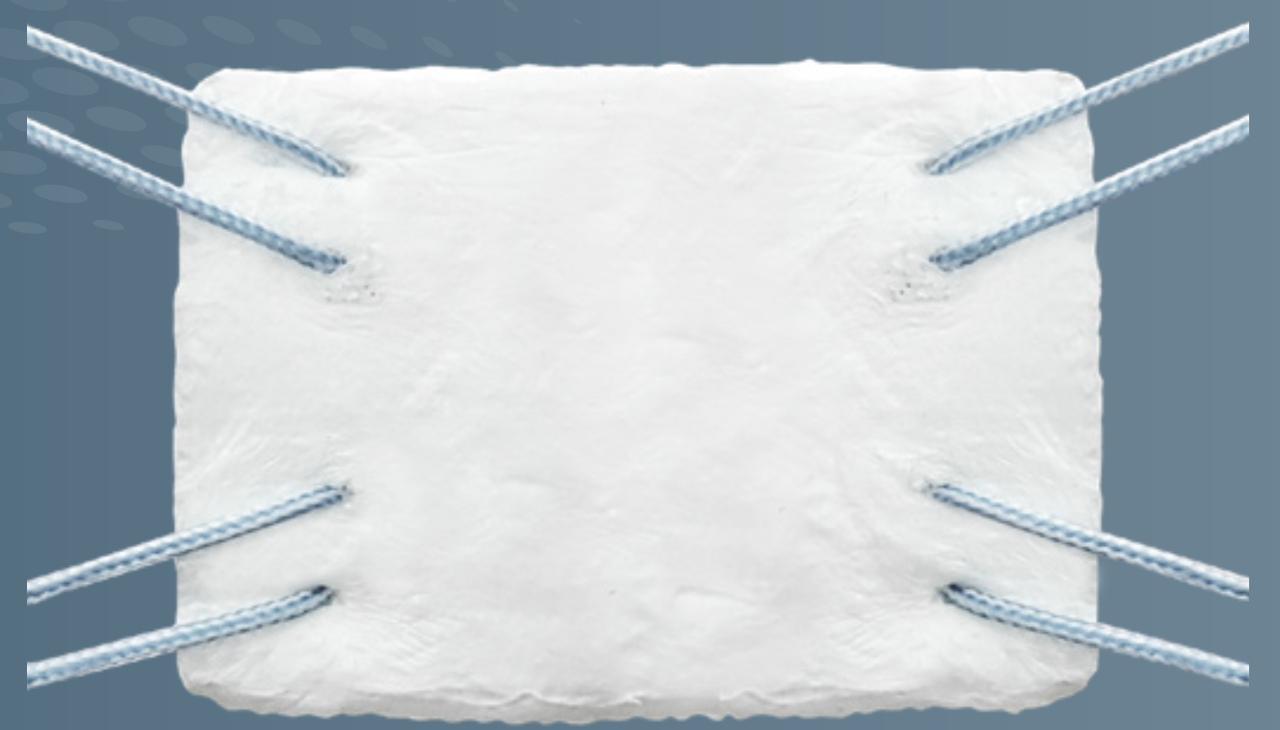
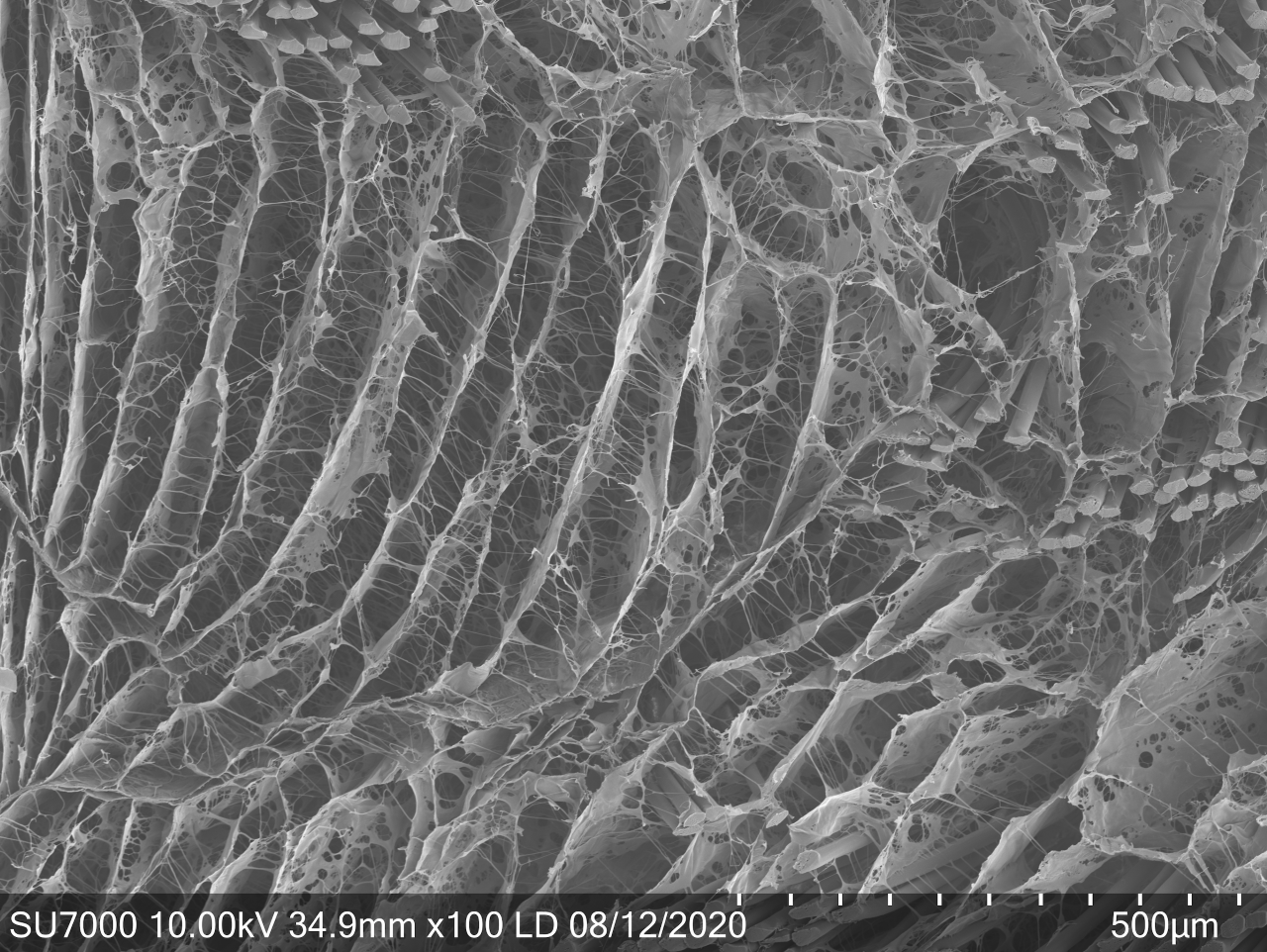
The BioBrace™ (Biorez Inc, New Haven, CT, USA) is a novel bio-inductive scaffold composed of highly porous type I collagen and bio-resorbable poly(L-Lactide) (PLLA) microfilaments intended for the augmentation of tendon and ligament repair (Figure 1). It’s open 3-D scaffold allows for induction, maturation, and remodeling of new host tissue while providing load sharing strength (141N) at the time of implantation (Figure 2) (Carter et al. 2021). In turn, gapping and re-tears prevention may occur by increasing the thickness of the tendon at time zero and beyond (Bokor et al. 2019a, 2019b; Thon et al. 2019).

Figure 1
Figure 1.The BioBrace™ implant for rotator cuff augmentation is pictured with dimensions of 23mm wide, 30mm in length, and 3mm in thickness. The implant can be trimmed to the physicians desired specifications without weakening the implant.
Figure 2
Figure 2.The BioBrace™ is 80% porous with PLLA microfilament reinforcement to allow for induction of host tissue, maturation, and strength.

The large open pores (80% porosity) of the implant allow for native soft tissue ingrowth, while maintaining structural strength provided by the bio-resorbable PLLA micro-filaments (15 micron) reinforcement.(Carter et al. 2021) The degradation curve of the PLLA micro-filaments permits the functional maturation and remodeling of host-generated repair tissue while maintaining strength for up to 24 months (Walsh et al. 2021). This provides more than sufficient time as a structural scaffold, without the concern of being a permanent implant.

The BioBrace™ for rotator cuff augmentation uniquely meets the need for the next generation of biologic rotator cuff solutions by combining the benefits from bio-inductive xenograft implants and ADM. The highly porous bio-inductive/bio-composite implant allows for a resorbable rotator cuff augment that can provide strength at time zero, rapid incorporation, and ultimate resorption of the implant while new native tissue remains. The BioBrace™ has been shown to incite a robust native healing response and the formation of regularly oriented connective tissue fibers in large animal models.(Walsh et al. 2021; Carter et al. 2021) These characteristics may lend themselves to enhanced graft/repair healing incorporation. In turn this can potentially lead to enhanced rehabilitation protocols and ultimately decreased failures.

CASE APPLICATION ALGORITHM

The lead authors’ current indication for the use of the BioBrace™ in RCR are: revision full thickness RCR, large (>3cm) and massive tears, chronic full thickness tears with Goutier classification grade 3 or 4 fatty infiltration, and full thickness tears in patients with multiple medical co-morbidities (smoking, diabetes, etc.). This is not used as a replacement for an ADM sheet in superior capsular reconstructions (SCR).

Xenograft bio-inductive implants (REGENETEN™) are reserved for high grade articular sided partial thickness RCR’s and patients with recalcitrant rotator cuff tendinopathies. ADM strips (DERMIS ON DEMAND™) have become a very efficient RCR reinforcement in medium to large RCR’s and are utilized as “rebar” and reinforcement when appropriate.

TECHNICAL PEARLS

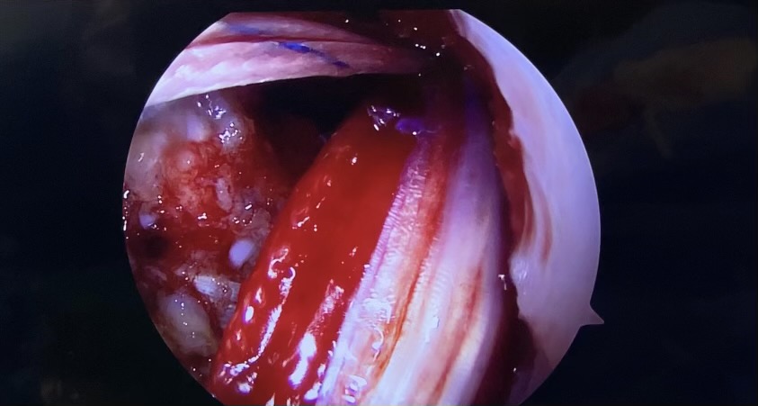
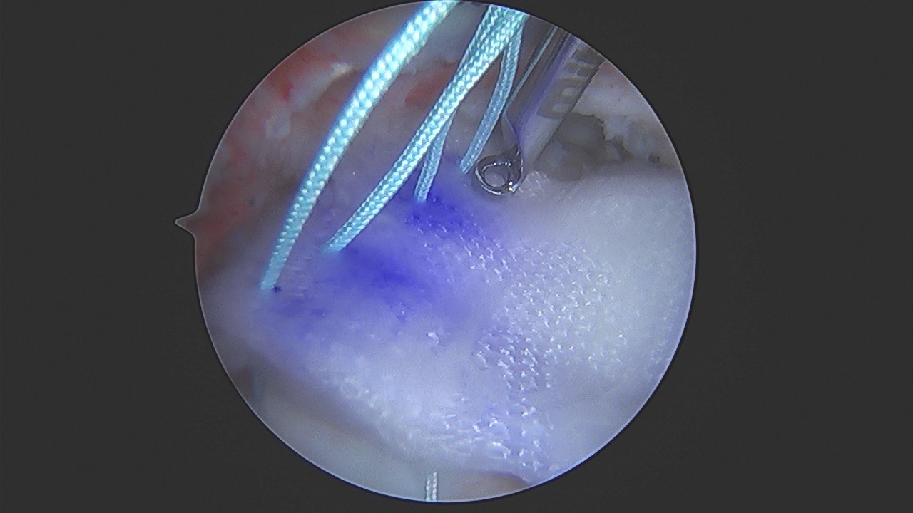
The BioBrace™ implant for RCR comes available as a 23mm wide by 30mm length implant, with a 3 mm thickness. The implant can be trimmed if needed to allow for appropriate rotator cuff coverage. The implant is easy to work with, however the lead author recommends wetting the implant prior to passing suture through it, as this appears to decrease the suture friction. Interestingly, the BioBrace™ has the ability to “wick” when reconstituted (Figure 3). As such the individual surgeon maybe choose to soak the implant in either saline or a biologic of their choosing such as platelet rich plasma (PRP) or bone marrow aspirate concentrate (BMAC).

Figure 3
Figure 3.(Courtesy of Michael Russonella, DO) The BioBrace™ has the ability to act as a wick during hydration. Picture above is a BioBrace™ (arrow) that has been pre-soaked in platelet rich plasma (PRP) for augmentation of an anterior cruciate ligament reconstruction (ACLR).

Suture Passing and Delivery

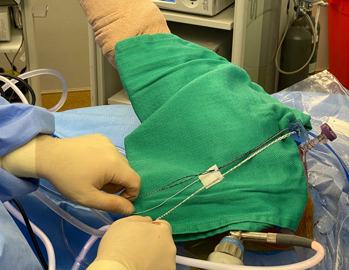
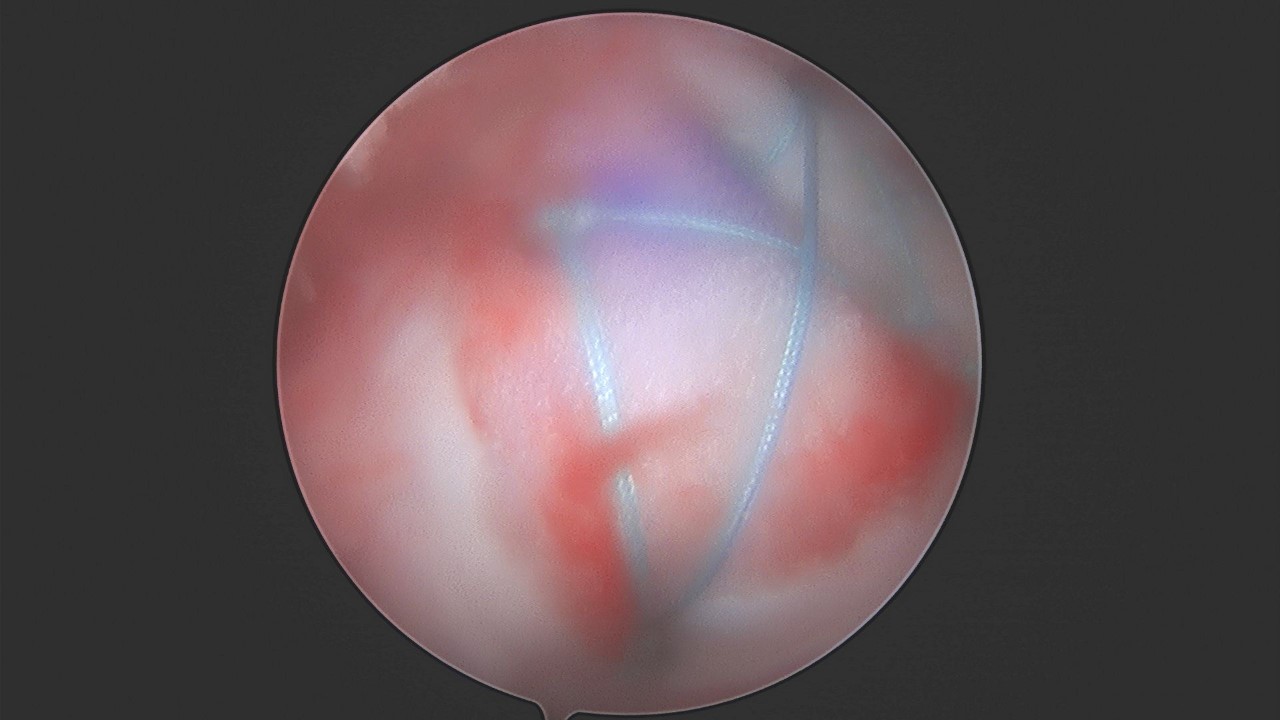
The BioBrace™ can be incorporated into either a single row, double row, or trans-osseous equivalent RCR repair. Once the RCR sutures are passed through the native tissue, the lead authors’ preference is to pass these sutures through the BioBrace™ outside the body through the lateral portal (Figure 4). Passing guns (ESPRESEW®, SCORPION™ etc.) or a simple 18G spinal needle and passing wire (CHIA®) may be used to facilitate suture passage through the scaffold. Given the strength of the implant, the sutures can be placed along the edges as desired. Once suture passing is completed along the medial margin of the implant, it can be delivered into the body by either using a “parachute technique”, a “back grasper” or any standard non-traumatic rotator cuff grasper. A 10mm or 12mm diameter cannula is recommended for passage. Marking the top-side of the implant can prove beneficial for orientation purposes (Figure 5). Once delivered the lateral aspect of the implant can be secured to the greater tuberosity through the lateral anchors Figure 6). In instances of massive cuff tears that require medialization of the footprint to the medial aspect of the tuberosity, the BioBrace™ can be used to both reinforce the repair as well as cover the bare greater tuberosity. In these cases, bone marrow venting of the greater tuberosity will allow for biologic seepage into the implant to potentially allow for enhancement of a “neo-tendon”, as has been shown by Savoie et al. in their work with xenografts (Thon et al. 2019).

Figure 4
Figure 4.After passage of the RCR sutures through the native tendon arthroscopically, the repair sutures are brought through the lateral cannula where there were passed through the BioBrace™ implant using a commercial passing gun.
Figure 5
Figure 5.The BioBrace™ was marked on the superior surface for orientation purposes and delivered into the shoulder using a “back-grasper”. Once delivered, a knot pusher was placed on sutures to bring the implant flush to the tissue.
Figure 6
Figure 6.The posterior and central-posterior limbs were tied together in the above construct, followed by the anterior and central-anterior limbs. After the medial row compression was complete, the suture limbs that passed through the native tendon and the BioBrace™ were brought lateral in a compression bridge configuration with two self-punching PEEK anchors. Note the blood from the surrounding tissues and bone is adhering to the implant even in an aqueous environment.

DISCUSSION

Rotator cuff repairs are one of the most challenging, yet rewarding, procedures for an orthopedic surgeon. Despite good clinical successes, repeat imaging of these repairs often show sub-optimal healing and or failures (Agrawal 2012; Galatz et al. 2004; Djurasovic et al. 2001; Burkhart et al. 2007). In particular, patients undergoing revision RCR or large to massive repairs, are highly susceptible to recurrent tearing (Agrawal 2012; Galatz et al. 2004; Djurasovic et al. 2001; Burkhart et al. 2007; Gladstone et al. 2007; Miller et al. 2011; Chona et al. 2017).

The addition of RCR’s with graft augmentation has been shown to be beneficial in the literature for these challenging patients (Mirzayan et al. 2019; Bishai et al. 2021a; Bushnell et al. 2021; Barber, Herbert, and Boothby 2008; Barber et al. 2012; Bishai et al. 2021b). Nevertheless, each of the currently available augmentation options has pros and cons. Xenograft bio-inductive implants have demonstrated significant success in the partial thickness RCR cohort (Bishai et al. 2021b). However, the lack of structural strength of the implant is a drawback in the high risk full-thickness tear subset due to its inability to share tensile load at the time of implantation. ADM’s have been shown to provide load sharing ability and decreased failure rates in the literature for high risk patients (Barber, Herbert, and Boothby 2008; Barber et al. 2012). Pre-packaged ADM strips have helped reduce the challenge of delivery of the graft into the shoulder. Despite this, drawbacks exist with ADM’s, including delayed incorporation time to host, inability to differentiate into native host tissue, and the inherent risk of utilizing allograft dermis.

The BioBrace™ is a bio-composite open porous 3D scaffold that allows for augmenting RCR’s through the induction, maturation, and remodeling of new host tissue. This can prevent tissue gapping or re-tears by increasing the thickness of the tendon. Given the strength of the implant, it is able to act as rebar for the suture and help mitigate the risk of suture-tendon pull through. This has long been a known advantage of ADM’s. The ability to combine the benefits of biology and strength into a single implant, while still having the implant resorb overtime, is very appealing. Concerns over the risk of inflammatory response due to the PLLA microfilaments should be tempered at this time due to the relatively low total mass and size within the scaffold. The resorb-able nature of the PLLA microfilaments does induce a mild, localized, foreign body response. However it is minimal and typical of the normal biological response to the degradation of biodegradable material.(Hollinger and Battistone 1986) Walsh et al. similarly found this minimal inflammatory response in their animal model testing without negative outcomes or concern.(Walsh et al. 2021)

The ability to improve RCR outcomes with a bio-composite reinforced implant which allows for increased healing potential and reinforcement of surgically treated tendon holds appeal for many surgeons. The BioBrace™ construct provides a unique environment for soft tissue regeneration and mechanical support which was previously not available for surgeons. Further studies and outcome collections are of paramount importance as this technology moves forward.


Conflict of Interest

The author Ford E declares they have no conflicts of interest. Arciero RA receives educational and research funding from Arthrex and Don-Joy; is a consultant with Biorez and Smith & Nephew. McMillan S is on the scientific advisory board with Biorez, and is a consultant with Depuy Mitek, Arthrex, Smith & Nephew and Trice Medical.

Funding

None

Submitted: August 16, 2021 EDT

Accepted: September 04, 2021 EDT

References

Agrawal, Vivek. 2012. “Healing Rates for Challenging Rotator Cuff Tears Utilizing an Acellular Human Dermal Reinforcement Graft.” International Journal of Shoulder Surgery 6 (2): 36–44. https:/​/​doi.org/​10.4103/​0973-6042.96992.
Google ScholarPubMed CentralPubMed
Barber, F. Alan, Joseph P. Burns, Allen Deutsch, Marc R. Labbé, and Robert B. Litchfield. 2012. “A Prospective, Randomized Evaluation of Acellular Human Dermal Matrix Augmentation for Arthroscopic Rotator Cuff Repair.” Arthroscopy 28 (1): 8–15. https:/​/​doi.org/​10.1016/​j.arthro.2011.06.038.
Google Scholar
Barber, F. Alan, Morley A. Herbert, and Michael H. Boothby. 2008. “Ultimate Tensile Failure Loads of a Human Dermal Allograft Rotator Cuff Augmentation.” Arthroscopy: The Journal of Arthroscopic & Related Surgery 24 (1): 20–24. https:/​/​doi.org/​10.1016/​j.arthro.2007.07.013.
Google Scholar
Bishai, Shariff, Ryan Krupp, Sean McMillan, Brian Schofield, Scott Trenhaile, Louis McIntyre, and Brandon Bushnell. 2021a. “Treatment of Partial-Thickness Rotator Cuff Repairs With A Resorbable Bioinductive Bovine Collagen Implant: 1-Year Results From A Prospective Multi-Center Registry.” Orthopaedic Journal of Sports Medicine 9 (7_suppl4): 2325967121S0020. https:/​/​doi.org/​10.1177/​2325967121s00209.
Google ScholarPubMed Central
———. 2021b. “Treatment of Partial-Thickness Rotator Cuff Repairs With A Resorbable Bioinductive Bovine Collagen Implant: 1-Year Results From A Prospective Multi-Center Registry.” Orthopaedic Journal of Sports Medicine 9 (7_suppl4): 2325967121S0020. https:/​/​doi.org/​10.1177/​2325967121s00209.
Google ScholarPubMed Central
Bokor, D.J., D. Sonnabend, L. Deady, B. Cass, A. Young, C. Van Kampen, and S. Arnoczky. 2019a. “Preliminary Investigation of a Biological Augmentation of Rotator Cuff Repairs Using a Collagen Implant: A 2-Year MRI Follow-Up.” Muscle Ligaments and Tendons Journal 05 (03): 144–50. https:/​/​doi.org/​10.32098/​mltj.03.2015.01.
Google ScholarPubMed CentralPubMed
Bokor, D.J., D.H. Sonnabend, L. Deady, B. Cass, A.A. Young, C.L. Van Kampen, and S.P. Arnoczky. 2019b. “Healing of Partial-Thickness Rotator Cuff Tears Following Arthroscopic Augmentation with a Highly-Porous Collagen Implant: A 5-Year Clinical and MRI Follow-Up.” Muscle Ligaments and Tendons Journal 9 (3): 338. https:/​/​doi.org/​10.32098/​mltj.03.2019.07.
Google Scholar
Burkhart, Stephen S., Johannes R.H. Barth, David P. Richards, Michael B. Zlatkin, and Mitchell Larsen. 2007. “Arthroscopic Repair of Massive Rotator Cuff Tears With Stage 3 and 4 Fatty Degeneration.” Arthroscopy 23 (4): 347–54. https:/​/​doi.org/​10.1016/​j.arthro.2006.12.012.
Google Scholar
Bushnell, Brandon D., Patrick M. Connor, Howard W. Harris, Charles P. Ho, Scott W. Trenhaile, and Jeffrey S. Abrams. 2021. “Retear Rates and Clinical Outcomes at 1 Year after Repair of Full-Thickness Rotator Cuff Tears Augmented with a Bioinductive Collagen Implant: A Prospective Multicenter Study.” JSES International 5 (2): 228–37. https:/​/​doi.org/​10.1016/​j.jseint.2020.10.020.
Google ScholarPubMed CentralPubMed
Carter, AJ, V Lovric, P Morberg, J Ott, J Bendigo, J Komenda, M Aronson, K Rocco, S Arnoczky, and WR Walsh. 2021. “Characterization of a Novel Bio-Inductive Biocomposite Scaffold for Tendon and Ligament Healing.” Presented at the Orthopaedic Research Society (ORS) 2021 Annual Meeting; February 12-16, 2021, Virtual.
Chona, Deepak V., Nikita Lakomkin, Ariana Lott, Alan D. Workman, Aneel C. Henry, Andrew F. Kuntz, G. Russell Huffman, and David L. Glaser. 2017. “The Timing of Retears after Arthroscopic Rotator Cuff Repair.” Journal of Shoulder and Elbow Surgery 26 (11): 2054–59. https:/​/​doi.org/​10.1016/​j.jse.2017.07.015.
Google Scholar
Djurasovic, Mladen, Guido Marra, Julian S. Arroyo, Roger G. Pollock, Evan L. Flatow, and Louis U. Bigliani. 2001. “Revision Rotator Cuff Repair: Factors Influencing Results.” The Journal of Bone and Joint Surgery-American Volume 83 (12): 1849–55. https:/​/​doi.org/​10.2106/​00004623-200112000-00013.
Google Scholar
Galatz, Leesa M., Craig M. Ball, Sharlene A. Teefey, William D. Middleton, and Ken Yamaguchi. 2004. “The Outcome and Repair Integrity of Completely Arthroscopically Repaired Large and Massive Rotator Cuff Tears.” The Journal of Bone & Joint Surgery 86 (2): 219–24. https:/​/​doi.org/​10.2106/​00004623-200402000-00002.
Google Scholar
Gladstone, James N., Julie Y. Bishop, Ian K.Y. Lo, and Evan L. Flatow. 2007. “Fatty Infiltration and Atrophy of the Rotator Cuff Do Not Improve after Rotator Cuff Repair and Correlate with Poor Functional Outcome.” The American Journal of Sports Medicine 35 (5): 719–28. https:/​/​doi.org/​10.1177/​0363546506297539.
Google Scholar
Hollinger, Jeffrey O., and Gino C. Battistone. 1986. “Biodegradable Bone Repair Materials. Synthetic Polymers and Ceramics.” Clinical Orthopaedics and Related Research, no. 207 (June), 290–306. https:/​/​doi.org/​10.1097/​00003086-198606000-00046.
Google Scholar
Ma, C. Benjamin, Lyn Comerford, Joseph Wilson, and Christian M. Puttlitz. 2006. “Biomechanical Evaluation of Arthroscopic Rotator Cuff Repairs: Double-Row Compared with Single-Row Fixation.” The Journal of Bone & Joint Surgery 88 (2): 403–10. https:/​/​doi.org/​10.2106/​jbjs.d.02887.
Google Scholar
Miller, Bruce S., Brian K. Downie, Robert B. Kohen, Theresa Kijek, Bryson Lesniak, Jon A. Jacobson, Richard E. Hughes, and James E. Carpenter. 2011. “When Do Rotator Cuff Repairs Fail? Serial Ultrasound Examination after Arthroscopic Repair of Large and Massive Rotator Cuff Tears.” The American Journal of Sports Medicine 39 (10): 2064–70. https:/​/​doi.org/​10.1177/​0363546511413372.
Google Scholar
Mirzayan, Raffy, Alexander E. Weber, Frank A. Petrigliano, and Jorge Chahla. 2019. “Rationale for Biologic Augmentation of Rotator Cuff Repairs.” Journal of the American Academy of Orthopaedic Surgeons 27 (13): 468–78. https:/​/​doi.org/​10.5435/​jaaos-d-18-00281.
Google Scholar
“PearlDiver.” n.d. http:/​/​www.pearldiverinc.com.
Ponce, Brent A., Chad D. Hosemann, Parthasarathy Raghava, Janet P. Tate, Evan D. Sheppard, and Alan W. Eberhardt. 2013. “A Biomechanical Analysis of Controllable Intraoperative Variables Affecting the Strength of Rotator Cuff Repairs at the Suture-Tendon Interface.” The American Journal of Sports Medicine 41 (10): 2256–61. https:/​/​doi.org/​10.1177/​0363546513499228.
Google Scholar
Thon, Stephen G., Larry II O’Malley, Michael J. O’Brien, and Felix H. III Savoie. 2019. “Evaluation of Healing Rates and Safety With a Bioinductive Collagen Patch for Large and Massive Rotator Cuff Tears: 2-Year Safety and Clinical Outcomes.” The American Journal of Sports Medicine 47 (8): 1901–8. https:/​/​doi.org/​10.1177/​0363546519850795.
Google Scholar
Walsh, WR, AJ Carter, V Lovric, J Crowley, D Wills, T Wang, G Kanski, R Stanton, S Arnoczky, and R Arciero. 2021. “Tissue-Engineered Augmentation of A Rotator Cuff Tendon Using A Novel Bio-Inductive Biocomposite Scaffold: A Preliminary Study In Sheep.” Presented at the Orthopaedic Research Society (ORS) 2021 Annual Meeting; February 12-16, 2021, Virtual.

This website uses cookies

We use cookies to enhance your experience and support COUNTER Metrics for transparent reporting of readership statistics. Cookie data is not sold to third parties or used for marketing purposes.

Powered by Scholastica, the modern academic journal management system