BACKGROUND
Acetabular fractures are relatively uncommon injuries which present in a bimodal distribution in both an elderly population secondary to low energy trauma and a younger population secondary to high energy trauma.(Mauffrey et al. 2014) These injuries have high morbidity with post-traumatic osteoarthritis occurring in approximately 20% of patients.(Giannoudis et al. 2005) Other complications of acetabular fractures include avascular necrosis of the femoral head, heterotopic ossification, and neurovascular injuries that include the sciatic nerve.
The incidence of reported sciatic nerve injuries associated with acetabular fractures ranges from 7.9%-75%; however, nearly all reported cases describe the nerve to be in-continuity on gross exam (Fassler et al. 1993). Additionally, many sciatic nerve injuries are iatrogenic as a result of perioperative or postoperative insult (Issack and Helfet 2009). To our knowledge, there are no reported cases in the literature of complete sciatic nerve laceration due to acetabular fracture as a result of initial trauma. Furthermore, there is a paucity of literature on managing injuries involving laceration of the nerve.
We report a case of traumatic sciatic nerve laceration discovered intraoperatively and treated with primary end-to-end repair at the time of open reduction and internal fixation of the acetabular fracture.
CASE REPORT
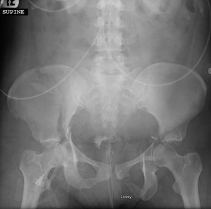
The patient was a 37-year-old female pedestrian struck by an automobile at an unknown speed, thrown into and struck by a second vehicle at an unknown speed, and subsequently brought to our Level 1 trauma center. The patient was initially unresponsive with declining mental status and required rapid sequence intubation in the trauma bay. She had obvious injuries on visual inspection including multiple superficial lacerations and abrasions, deformity to the right femur, shortened and externally rotated left leg, and a large open wound to the left foot. Initial standard trauma radiographs of the chest and pelvis were obtained. The single anterior-posterior (AP) chest film demonstrated several consecutive left segmental rib fractures with an associated pneumothorax. The AP pelvic radiograph showed a right iliac wing fracture, bilateral acetabulum fractures with left hip dislocation and femoral head medialization, right sacral fracture, and left inferior pubic ramus fracture (Figure 1). Physical examination demonstrated gross instability to right distal femur and left knee in coronal and sagittal planes, open and grossly unstable fracture to left midfoot. No pulses were palpable in the bilateral feet, but Doppler biphasic signals were audible at the right popliteal and posterior tibial arteries, as well as at the left popliteal and dorsalis pedis arteries.
The patient became hemodynamically unstable in the trauma bay requiring left chest tube placement, right femoral arterial line placement, and fluid resuscitation. Left hip closed reduction was performed as well as placement of bilateral distal femoral skeletal traction, and the left leg was irrigated of gross debris and splinted. The patient was transferred to the Intensive Care Unit for comprehensive multidisciplinary management and further resuscitation.
Computed tomography (CT) imaging of the head, neck, chest, abdomen, and pelvis, CT angiography of bilateral lower extremities (showing bilateral single vessel runoff), and extremity radiographs delineated the following injuries:
-
right iliac wing fracture
-
right both column acetabulum fracture
-
right zone 3 sacral fracture
-
pubic symphyseal dislocation
-
left iliosacral joint dislocation
-
left T-type and posterior wall acetabulum fracture
-
left hip dislocation with femoral head medialization
-
right distal femur fracture
-
left foot open 1st tarsometatarsal-fracture dislocation, 2nd metatarsal base fracture, 3rd metatarsal base fracture
-
left hand closed non-displaced 5th metacarpal base fracture
CT imaging of the pelvis was reconstructed with 3D imaging to assist with surgical planning (Figure 2, Figure 3).
On post-trauma day (PTD) 1, the patient was extubated and following commands. Tertiary survey was performed demonstrating complete absence of sensorimotor function in the tibial and peroneal distributions of the sciatic nerve in the left lower extremity. Sensation was intact over the medial aspect of left lower leg and anterior and medial thigh (obturator nerve). Quadriceps firing of left lower extremity was intact (femoral nerve). Neurovascular status of the bilateral upper extremities and right lower extremity were intact on tertiary exam.
The patient was taken to the operating room (OR) on PTD 1 to address the open fracture and stabilize her long bone fracture. She underwent left foot irrigation/debridement and operative fixation, right femur retrograde intramedullary nailing, and manipulation of bilateral knees demonstrating bilateral multiligamentous instability. On PTD 3, the patient returned to the OR for operative fixation of bilateral pelvis and acetabulum fractures. Intra-operatively a large left gluteal Morel-Lavallee lesion, traumatic lacerations to gluteal and short external rotator musculature, and complete left sciatic nerve traumatic laceration were found (Figure 4). In addition to fixation of the pelvic and acetabular fractures Figure 5, the sciatic nerve injury was primarily repaired with 6-0 nonabsorbable monofilament epineural suture and fibrin glue. The patient was placed into an ankle-foot-orthotic (AFO) to the left lower extremity. Hinged knee braces were applied for multiligamentous knee injuries. Post-operative CT scan of the pelvis demonstrated excellent reduction of the acetabulum fractures (Figure 6, Figure 7). Subsequent magnetic resonance imaging (MRI) of bilateral knees demonstrated bilateral multiligamentous injuries with an associated non-displaced left tibial plateau fracture. Finally, on PTD 29 the patient was taken to the OR for irrigation and debridement of her left necrotic gluteal wound and Morel-Lavallee lesion, with subsequent wound care and coverage performed by the plastic surgery service.
The patient was discharged to a rehabilitation facility on PTD 53 (50 days status post left sciatic nerve repair). At that time, the patient continued to have absence of motor function to the tibial and peroneal nerve distributions of the left lower extremity. Quadriceps firing remained intact. On sensory examination, she endorsed only a vague sensation of pressure upon deep palpation over the lateral lower leg, foot dorsum, and toes but no sensation upon testing fine touch, pain, or temperature in these distributions. Sensation remained intact over the medial aspect of the left lower leg and anterior and medial thigh.
The patient was lost to follow up until she returned to the ED approximately 6 months later for abdominal pain. On orthopaedic examination, there was no change in her left lower extremity sensorimotor status from time of discharge; however, she was able to ambulate with a hinged knee brace and AFO. Pelvic radiographs obtained at that time and showed hardware and fixation to be unchanged in alignment with interval bony healing (Figure 8). Following this evaluation, she was again lost to follow up until approximately 3 years postoperatively. She presented to an outside hospital due to left foot infection. The patient had developed chronic foot drop and a pressure ulcer on the lateral aspect of the foot with associated osteomyelitis. She underwent irrigation and debridement of the foot with subsequent intravenous antibiotic therapy. Her physical exam of the lower extremities was unchanged compared to prior exams. Radiographs of the pelvis were obtained and showed hardware to be unchanged in alignment and pelvic fractures to be healed (Figure 9). She remained ambulatory with an AFO and hinged knee brace.
CONCLUSION
Traumatic injury to the sciatic nerve during acetabular fracture is rare, and, when it does occur, the nerve is generally found to be in continuity (Fassler et al. 1993). In cases where the sciatic nerve is found to be in continuity, 75% of patients go on to develop recovery with good functional outcomes (Simske et al. 2019). Neurolysis in these cases is not routinely recommended due to the high incidence of transient neuropraxia and spontaneous recovery. Little data exists on the prognosis or management guidelines of complete sciatic nerve laceration due to its rare occurrence. One case series showed one of two patients with partial sciatic nerve recovery following sciatic nerve laceration when treated with sciatic nerve neurolysis and sural nerve autograft at the time of injury(Maripuu et al. 2014). Both of these cases were due to acute penetrating trauma with large segments of nerve defect and zone of nerve injury, so grafting was necessary.
The present case demonstrates complete sharp laceration of the sciatic nerve from fracture without a large zone of nerve injury. Direct, end-to-end epineural repair of peripheral nerve lacerations is the standard of care when feasible (in the absence of large nerve defects requiring grafts) (Griffin et al. 2013). Large peripheral nerves will heal at a rate of 2-5 mm/day, meaning a high sciatic nerve lesion could take several months to demonstrate any clinical evidence of recovery (Schmidt and Leach 2003). Unfortunately, lack of innervation to the motor-end plates (neuromuscular junction) beyond six months is unlikely to result in full motor recovery, even in the presence of successful axon regeneration (Sakuma et al. 2016). It would be unlikely for a complete sciatic nerve laceration at the level of the acetabulum to demonstrate recovery following direct repair; nonetheless, laceration of the nerve must be recognized and repair must be attempted in accordance with the medical standard of care in order to optimize clinical outcome for the patient. Long term management options for patients with high sciatic nerve lesions without clinical recovery include functional bracing, fusions, and tendon transfers (in the setting of partial recovery).
To our knowledge, this is the first reported case in the literature of complete traumatic sciatic nerve laceration secondary to acetabular fracture treated with direct end-to-end epineural repair. Although these presentations are rare, it is imperative for the traumatologist to understand the likelihood of such an injury, the management options, and be familiar with outcomes data in order to properly counsel patients and manage expectations.


__anterior_view.png)
__posterior_view.png)


__anterior_view.png)
__posterior_view.png)


__anterior_view.png)
__posterior_view.png)


__anterior_view.png)
__posterior_view.png)

