Loading [Contrib]/a11y/accessibility-menu.js
Skip to main content
null
J Orthopaedic Experience & Innovation
  • Menu
  • Articles
    • Brief Report
    • Case Report
    • Data Paper
    • Editorial
    • Hand
    • Meeting Reports/Abstracts
    • Methods Article
    • Product Review
    • Research Article
    • Review Article
    • Review Articles
    • Systematic Review
    • All
  • For Authors
  • Editorial Board
  • About
  • Issues
  • Blog
  • "Open Mic" Topic Sessions
  • Advertisers
  • Recorded Content
  • CME
  • JOEI KOL Connect
  • search

RSS Feed

Enter the URL below into your favorite RSS reader.

https://journaloei.scholasticahq.com/feed
Research Article
Vol. 1, Issue 2, 2020November 24, 2020 EDT

Incidence of Sacroiliac Screw Implant Pain and Results of Screw Removal

Taylor P Gurnea, Miguel A Gonzalez, Alexander R Turner, Alastair E Moody, Peter Althausen,
si screwtits screwhardware removal
Copyright Logoccby-nc-nd-4.0 • https://doi.org/10.60118/001c.17957
J Orthopaedic Experience & Innovation
Gurnea, Taylor P, Miguel A Gonzalez, Alexander R Turner, Alastair E Moody, and Peter Althausen. 2020. “Incidence of Sacroiliac Screw Implant Pain and Results of Screw Removal.” Journal of Orthopaedic Experience & Innovation 1 (2). https:/​/​doi.org/​10.60118/​001c.17957.
Save article as...▾
Download all (5)
  • Figure 1a
    Download
  • Figure 1b
    Download
  • Figure 2a
    Download
  • Figure 2b
    Download
  • Figure 3
    Download

Sorry, something went wrong. Please try again.

If this problem reoccurs, please contact Scholastica Support

Error message:

undefined

View more stats

Abstract

Introduction: Percutaneous sacroiliac(SI) screw fixation of pelvic fractures has become commonplace. Routine removal of SI screws has been recommended by some physicians while others remove only symptomatic implants citing risks associated with this procedure. Few publications exist documenting the incidence of SI screw related hardware pain, rates of SI screw removal, the results of implant removal and the risks and complications associated with this procedure. Our objective is to document the incidence and clinical impact of symptomatic sacroiliac drew removal at a community level II trauma centers over a 12-year period.

Methods: Operative records were queried to identify 250 patients with pelvic ring injuries treated with SI screw placement at our institution. Records were analyzed to identify fracture classification, implant removal, reason for implant removal, analogue pain scales before and after implant removal and complications after implant removal.

Results: During the 12 year study period from 2006-2018, 300 posterior pelvic ring screws were placed in 250 patients. 46/250 (18.4%) patients underwent implant removal, accounting for 66 screws. 43 patients cited painful hardware as the reason for removal and 3 patients cited infection. Average time to removal was 6.7 months. Mean analog pain scales were 4.91 and 0.51 before and after removal respectively. No intra-operative or post-operative complications were encountered. All patients reported improvement in pain with screw removal

Conclusions: Postoperative pain following SI screw placement attributed to retained implant occurs in 18% of patients. Despite the previously documented proximity of neuromuscular structures, no intra-operative or postoperative complications were observed in patients who underwent screw removal. All patients reported significant relief of symptoms following implant removal. In patients experiencing pain following SI screw placement, screw removal 6 months following initial fixation is a safe and effective procedure.

Introduction

Sacroiliac(SI) and transiliac-transsacral(TITS) screw fixation have become well accepted means of posterior ring fixation in pelvic fractures requiring operative fixation. Chronic pain, disability and sexual dysfunction are well documented complications of pelvic ring injury and fixation. Retained posterior implants have been cited as a possible source of pain due to decreased mobility, arthritis and chronic pain (Quade et al. 2019). There is disagreement among surgeons as to the true incidence of symptomatic implants, the benefit of implant removal and the safety of this procedure. Some suggest routine removal of implants (Culemann et al. 2004; Gänsslen, Hüfner, and Krettek 2006). Others recommend removal only in event of complications (Abumi et al. 2000; Chip Routt, Simonian, and Mills 1997). Our objective is to document the incidence of symptomatic posterior pelvic screw fixation, the complication rate of this procedure and the clinical outcome associated with screw removal.

Materials and Methods

Following investigational review board approval, the trauma database for our hospital’s four trauma fellowship trained orthopedic surgeons was queried from January 2006 to January 2018 to identify patients treated with sacroiliac (SI) or transiliac-transsacral (TITS) screw fixation.

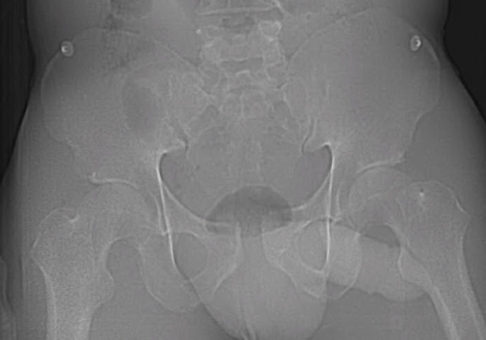
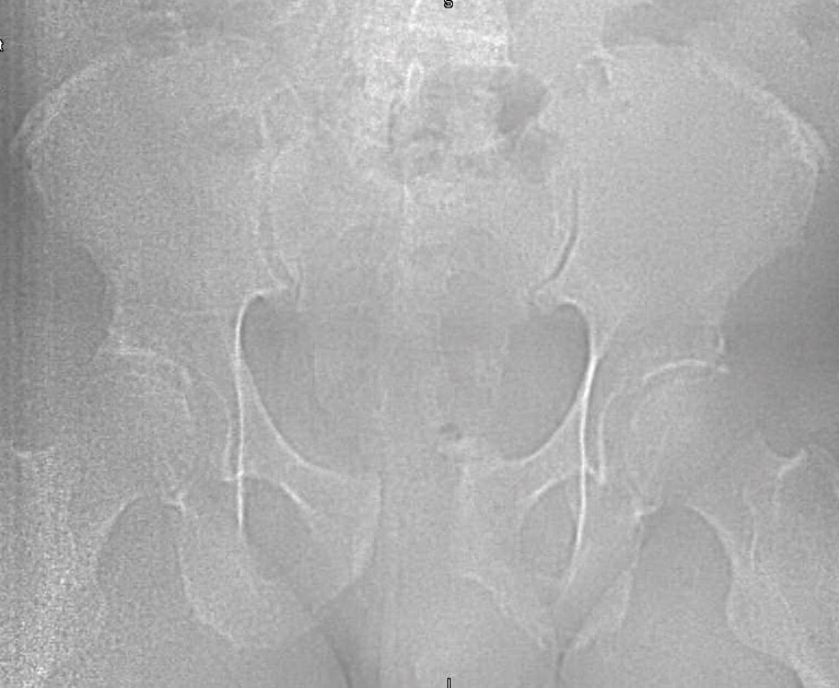
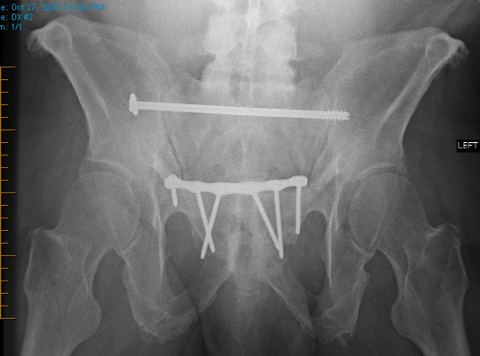
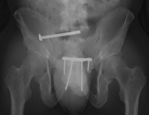
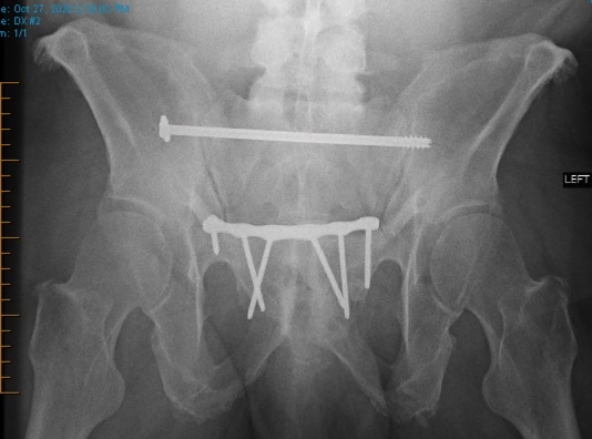
Chart review was undertaken to obtain basic demographic variables such as age, sex, smoking and diabetic history. Injury radiographs were reviewed to determine fracture type and Young-Burgess fracture classification. Preoperative and postoperative radiographs are demonstrated in figures 1A, 1B, 2A and 2B. Operative records were reviewed to determine which patients underwent screw removal and if any adverse intra-operative events occurred. Hospital charts and clinic charts were reviewed to document any postoperative complications and record pre and postoperative visual analog pain scales.

Figure 1a
Figure 1b
Figure 2a
Figure 2b

Results

Two hundred and fifty patients with 300 posterior pelvic ring screws were identified. 195 screws were SI and 105 screws were TITS. 46/250 (18.4%) patients underwent implant removal, accounting for 66 screws. Mean patient age was 42.5 years (+/-2.4). There were 28 males and 18 females. 43 patients cited pain as reason for removal and 3 patients cited infection. (Table 1) In patients requiring screw removal, fracture patterns by the Young and Burgess classification were 17 APC2, 5 APC3, 7 LC1, 13 LC2, and 4 LC3. (Table 2) Average time to implant removal was 6.7 months (+/- 1.8 months). 23.5 percent of SI screws and 19.7 percent of TITS screws were removed (p=0.61). No intra-operative complications or postoperative complications occurred. Some measure of pain relief occurred in all patients with mean preoperative and postoperative pain scales of 4.91 and 0.51 respectively (p<0.001). Mean follow up time for patients with implant removal was 34 months (+/- 3.1). (Table 3)

Discussion

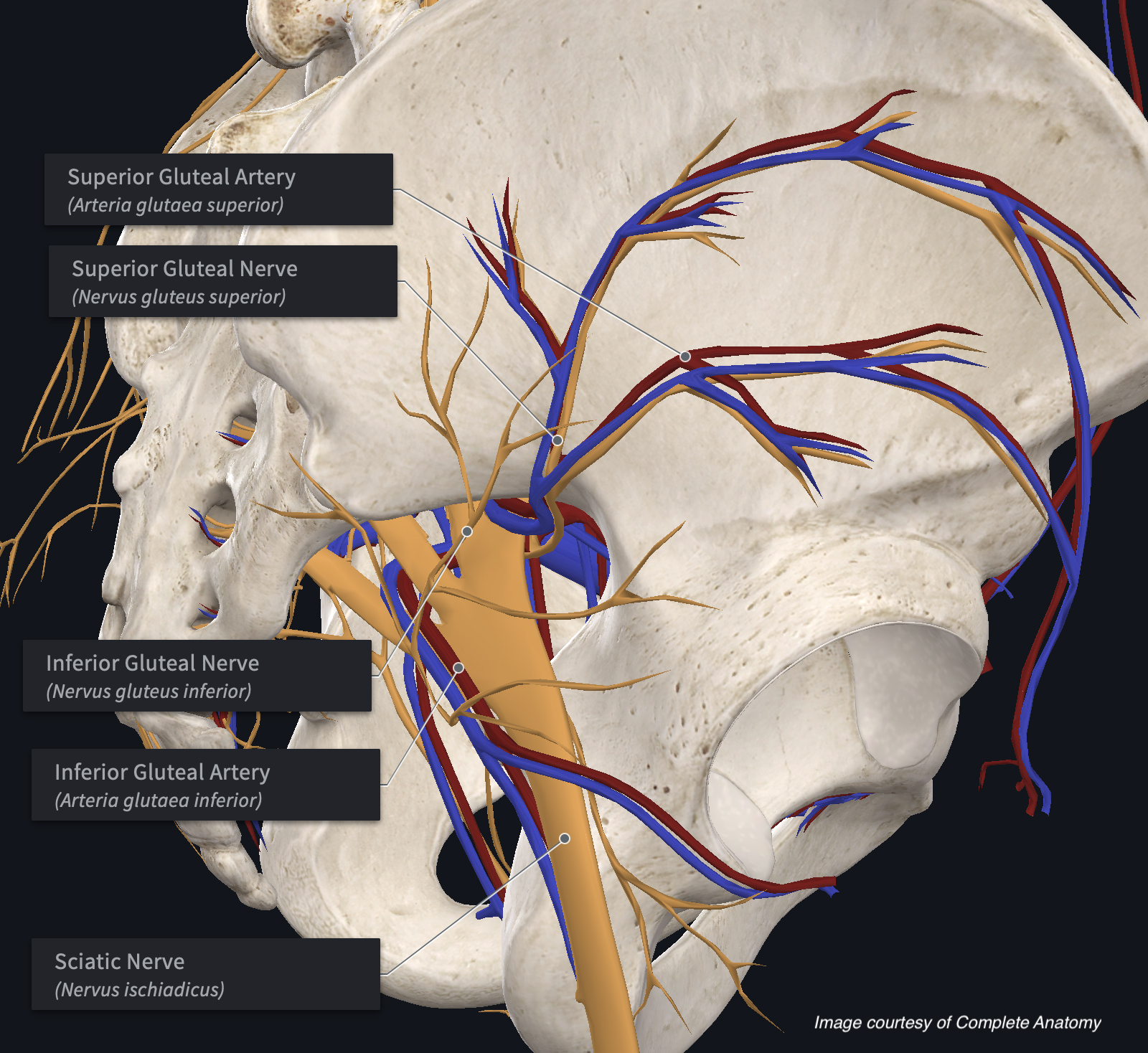
Posterior pelvic ring fixation with SI or TITS fixation is a well-accepted, successful means of fixation with good outcomes. Despite its widespread utilization, a paucity of publications exists on the indications and results of implant removal (Abumi et al. 2000; Culemann et al. 2004; Gänsslen, Hüfner, and Krettek 2006; Chip Routt, Simonian, and Mills 1997). Several case reports have demonstrated the proximity of neurovascular structures to these screws and demonstrated the possible risks associated with screw removal (Ebraheim et al. 1997; Mirkovic et al. 1991; Chip Routt, Simonian, and Mills 1997; Templeman et al. 1996). Figure 3 presents a diagram showing the proximity of neurovascular structures most likely to be damaged during placement or removal surgery.

Figure 3
Figure 3

In 2019, Quade et al found a 5.4% incidence of implant removal in patients treated with SI or TITS screw fixation. No intra-operative or postoperative complications occurred during implant removal and 83% of patients had improved symptoms with implant removal. Average time to removal was 10.7 months (Quade et al. 2019). Our study supports these findings. Our patients had a slightly higher incidence of implant pain and removal. In addition, patients did benefit from implant removal. In this study, average time to implant removal was 6.7 months, demonstrating that patients with painful implants can safely have their implants removed 4 months sooner than previously published.

This study is retrospective in nature and as a result is subject to certain bias. It is possible that some patients went elsewhere for screw removal after the data collection period was finished. However, our facility is geographically isolated and such a possibility is unlikely. Visual analog scale pain scales are subject to recall bias, but this should be similar across all patients. Pelvic ring fractures are complex injuries with many possible associated bone, muscle, ligament, nerve, and tendon injuries that could cause pain unrelated to implant position.

In conclusion, posterior pelvic ring fixation with SI or TITS screws is a well-accepted, successful means of posterior pelvic ring fracture fixation. Symptomatic implants are a reported complication occurring in 18% of patients. Implant removal is a safe procedure that is associated with improved subjective pain relief. Over 6 months following the index operation, implant removal appears to be a safe and effective procedure.

Submitted: September 18, 2020 EDT

Accepted: November 08, 2020 EDT

References

Abumi, Kuniyoshi, Michinori Saita, Takahiro Iida, and Kiyoshi Kaneda. 2000. “Reduction and Fixation of Sacroiliac Joint Dislocation by the Combined Use of S1 Pedicle Screws and the Galveston Technique.” Spine 25 (15): 1977–83. https:/​/​doi.org/​10.1097/​00007632-200008010-00018.
Google Scholar
Chip Routt, M. L. Jr, Peter T. Simonian, and William J. Mills. 1997. “Iliosacral Screw Fixation: Early Complications of the Percutaneous Technique.” Journal of Orthopaedic Trauma 11 (8): 584–89. https:/​/​doi.org/​10.1097/​00005131-199711000-00007.
Google Scholar
Culemann, U., G. Tosounidis, H. Reilmann, and T. Pohlemann. 2004. “Injury to the Pelvic Ring. Diagnosis and Current Possibilities for Treatment.” Unfallchirurg 107 (1169–81): 1169–81.
Google Scholar
Ebraheim, Nabil A., Rongming Xu, Ashok Biyani, and Matthew C. Nadaud. 1997. “Morphologic Considerations of the First Sacral Pedicle for Iliosacral Screw Placement.” Spine 22 (8): 841–46. https:/​/​doi.org/​10.1097/​00007632-199704150-00002.
Google Scholar
Gänsslen, Axel, Tobias Hüfner, and Christian Krettek. 2006. “Percutaneous Iliosacral Screw Fixation of Unstable Pelvic Injuries by Conventional Fluoroscopy.” Oper Orthop Traumatol 18:225–44.
Google Scholar
Mirkovic, S., J. J. Steinman, C. C. Edwards, M. Schaffler, J. Massie, and S. R. Garfin. 1991. “Anatomic Consideration for Sacral Screw Placement.” Spine 16 (Supplement): S286-294. https:/​/​doi.org/​10.1097/​00007632-199106001-00022.
Google Scholar
Quade, Jonathan, Gennadiy Busel, Michael Beebe, Darryl Auston, Anjan R. Shah, Anthony Infante, Benjamin Maxson, David Watson, Roy W. Sanders, and Hassan R. Mir. 2019. “Symptomatic Iliosacral Screw Removal After Pelvic Trauma-Incidence and Clinical Impact.” Journal of Orthopaedic Trauma 33 (7): 351–53. https:/​/​doi.org/​10.1097/​bot.0000000000001453.
Google Scholar
Templeman, David, Andrew Schmidt, J. Freese, and Irwin Weisman. 1996. “Proximity of Iliosacral Screws to Neurovascular Structures after Internal Fixation.” Clinical Orthopaedics & Related Research 329 (August):194–98. https:/​/​doi.org/​10.1097/​00003086-199608000-00023.
Google Scholar

This website uses cookies

We use cookies to enhance your experience and support COUNTER Metrics for transparent reporting of readership statistics. Cookie data is not sold to third parties or used for marketing purposes.

Powered by Scholastica, the modern academic journal management system