Loading [Contrib]/a11y/accessibility-menu.js

This website uses cookies

We use cookies to enhance your experience and support COUNTER Metrics for transparent reporting of readership statistics. Cookie data is not sold to third parties or used for marketing purposes.

Skip to main content
null
J Orthopaedic Experience & Innovation
  • Menu
  • Articles
    • Brief Report
    • Case Report
    • Case Series
    • Conference Proceedings
    • Data Paper
    • Editorial
    • Meeting Reports/Abstracts
    • Methods Article
    • Product Review
    • Research Article
    • Review Article
    • Review Articles
    • Systematic Review
    • All
  • For Authors
  • Editorial Board
  • About
  • Issues
  • Blog
  • "Open Mic" Topic Sessions
  • Advertisers
  • Recorded Content
  • CME
  • JOEI KOL Connect
  • Resident Research League
  • search
  • RSS feed (opens a modal with a link to feed)

RSS Feed

Enter the URL below into your favorite RSS reader.

https://journaloei.scholasticahq.com/feed
ISSN 2691-6541
Review Article
September 06, 2026 EDT

Diagnosis, Indications, Complications, and Technical Considerations for In-office Needle Arthroscopy: A Scoping Review

Antonio Henry, DO, MSPH, Nathaniel Mercer, MD, Vanessa Boggiano, D.O., Arianna Gianakos, DO,
needle arthroscopyin-office needle arthroscopydiagnostic arthroscopy
Copyright Logoccby-nc-nd-4.0 • https://doi.org/10.60118/001c.162493
J Orthopaedic Experience & Innovation
Henry, Antonio, Nathaniel Mercer, Vanessa Boggiano, and Arianna Gianakos. 2026. “Diagnosis, Indications, Complications, and Technical Considerations for In-Office Needle Arthroscopy: A Scoping Review.” Journal of Orthopaedic Experience & Innovation, September 6. https://doi.org/10.60118/001c.162493.
Save article as...▾
Download all (1)
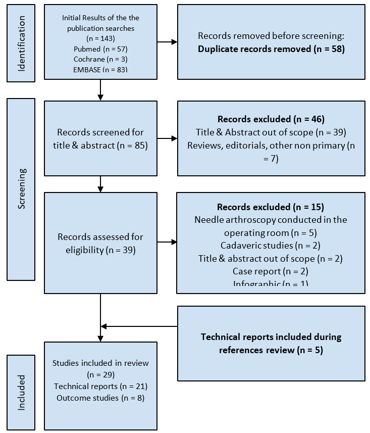
  • Figure 1. PRISMA Diagram
    Download

Error

Sorry, something went wrong. Please try again.

If this problem reoccurs, please contact Scholastica Support

Error message:

undefined

View more stats

Abstract

Purpose

Review the diagnostic and therapeutic indications, complications, and technical considerations for the use of In-office needle arthroscopy (IONA).

Methods

Authors conducted a scoping review using PubMed, Cochrane, and EMBASE evaluating IONA studies published between June 2013 and October 2024. Inclusion criteria were primary literature written in English assessing diagnostic capability, functional outcomes, or patient-reported outcomes. Qualitative analysis of technical reports assessed the pearls, pitfalls and step-by-step guides to performing IONA. Case reports, reviews, infographics, cost-effectiveness, cadaveric, animal, and operating room use of arthroscopy studies were excluded. Diagnostic capability, procedure length, clinical outcomes and technical considerations were evaluated.

Results

The search yielded 143 results, 29 of which (8 clinical outcome studies, 21 technical reports) met inclusion criteria, covering the shoulder, knee, foot/ankle joints. Level of evidence ranged from IV-V. IONA was used to diagnose intra-articular pathology pertaining to long head of the biceps tendinopathy, full thickness osteochondral defects of the femoral condyle, meniscus tears, Achilles tendinopathy, and anterior/posterior hindfoot pathologies. IONA procedures included: biceps tenotomy, medial meniscal repair, anterior talofibular ligament repair, ankle impingement debridement, first metatarsophalangeal joint cheilectomy, Achilles and peroneal tendoscopy, and intra-articular lavage. Multiple studies reported post-operative improvements in clinical outcome measures, including the visual analog scale, Foot and Ankle Outcome Score, Tegner Lysholm Knee Scoring Scale, and PROMIS pain and function domains. Several studies reported high patient satisfaction and return to activity following IONA.

Conclusion

The current literature on IONA consists predominantly of Level IV-V evidence, including small case series and technical reports. While available studies suggest IONA is technically feasible and may be associated with short-term improvements in patient reported pain and functional outcomes in select indications, the absence of comparative trials, standardized outcome reporting, and long term follow up limits definitive conclusions regarding safety, effectiveness, or superiority to conventional arthroscopy or advanced imaging.

Introduction

In-office needle arthroscopy (IONA) is an emerging minimally invasive technique that enables direct visualization and, in select cases, treatment of intra-articular pathology under local anesthesia in a clinic-based setting. Advances in optics, camera systems, and portal instrumentation have expanded the feasibility of performing diagnostic and therapeutic arthroscopy outside the operating room, thereby reducing reliance on advanced imaging modalities and, in some cases, general anesthesia.

IONA provides several potential advantages over traditional care models. Patients can receive immediate visualization of pathology and, in select cases, undergo definitive treatment in a single encounter. This streamlined care pathway minimizes delays associated with magnetic resonance imaging (MRI), preoperative evaluations, and operating room scheduling. Early studies suggest that IONA can reduce patient burden and resource utilization by accelerating diagnosis and eliminating the need for multiple visits (Voigt et al. 2014). Prior systematic reviews have reported superior diagnostic accuracy in the evaluation of certain knee pathologies, however the procedures included in these analyses were performed in the operating room rather than the office setting (Zhang et al. 2019). Additional studies have reported potential reductions in healthcare costs, increased patient satisfaction, and shorter recovery periods in select populations (Voigt et al. 2014; Gianakos and Kennedy 2024; McMillan, Schwartz, et al. 2017; Mercer, Samsonov, et al. 2022; Munn et al. 2023; Shah et al., n.d.).

Existing systematic reviews have also demonstrated the technical feasibility of needle arthroscopy across multiple joints, including the shoulder, knee, and ankle, with early reports describing its use in conditions such as long head of the biceps tendinopathy, meniscal pathology, and ankle impingement syndromes (Burt et al. 2023). Despite growing clinical interest, however, the available literature remains largely composed of technical reports and small case series, with limited high-quality evidence directly comparing in-office needle arthroscopy to conventional arthroscopy or advanced imaging modalities.

Importantly, prior reviews have focused primarily on diagnostic needle arthroscopy performed in operative settings and have emphasized cost-effectiveness or diagnostic accuracy relative to MRI (Amin et al. 2019; Gill et al. 2018; Wagner et al. 2021). These analyses have not specifically examined procedures performed in the office setting under local anesthesia, nor have they synthesized technical considerations and clinical outcomes across multiple joints. Accordingly, the present study aims to map the existing literature on in-office needle arthroscopy, including diagnostic indications, therapeutic applications, clinical outcomes, complications, and procedural techniques. This scoping review is intended to characterize current clinical experience and identify gaps in the evidence rather than to determine comparative effectiveness. To our knowledge, this study represents the first scoping review synthesizing technical reports and available clinical outcomes literature on the diagnostic and therapeutic indications, outcomes, and complications of IONA.

Methods

Study Eligibility

Articles written in English, published from June 2013 to October 2024, and which analyzed IONA were eligible for inclusion. June 2013 approximates the commercial introduction of small-bore needle arthroscopy systems and 10 years prior to the inception of the current study. These articles were primary literature that assessed diagnostic capability, functional, or patient outcomes or technical reports providing pearls, pitfalls and step-by-step guides to performing IONA. The exclusion criteria included any cadaver studies, animal studies, case reports, reviews, infographics, studies analyzing cost-effectiveness, and studies utilizing needle arthroscopy in the operating room.

Literature Search

A scoping review of PubMed, EMBASE, and Cochrane databases was performed based on the Preferred Reporting Items for Systematic Reviews and Meta-Analysis for Scoping Review (PRISMA-ScR) guidelines for articles published between June 2013 and October 2024. The following search term was used: (“needle arthroscopy” OR “arthroscopy”) AND (“in-office” or “in office”). Consistent with PRISMA-ScR methodology, this review was designed to map the breadth of available literature rather than perform quantitative synthesis or pooled outcome analysis. Given the heterogeneity of study designs, joints evaluated, procedural indications, and outcome measures, meta-analysis was not feasible and was not attempted. Search strings for each database are listed in Table 1. The search was intentionally broad to capture variations in terminology and indexing related to in-office needle arthroscopy. To mitigate the risk of missing relevant studies, reference lists of all included articles were independently reviewed after full-text screening.

Table 1.Database Search Strategy
PubMed ("needle arthroscopy" OR "arthroscopy") AND ("in-office" or "in office")
Cochrane ("needle arthroscopy" OR "arthroscopy") AND ("in-office" OR "in office")
Embase ('needle arthroscopy'/exp OR 'needle arthroscopy' OR 'arthroscopy'/exp OR 'arthroscopy') AND ('in-office' OR 'in office')

Study Selection and Data Abstraction

Two independent reviewers independently screened all titles, abstracts, full texts, and reference lists. Data extraction was also performed independently in duplicate using a predefined standardized template (Table 2), with disagreements resolved by a senior author. Formal inter-rater agreement statistics (e.g., kappa coefficients) were not calculated for title/abstract or full-text screening stages; this represents a methodological limitation of the present review. Consistent with PRISMA-ScR guidance, inter-rater reliability metrics are not mandated for scoping reviews; however, future scoping reviews in this area should incorporate these statistics to strengthen methodological transparency.

Table 2.Study Characteristics
Study no. Study Design Focus Diagnostic indication Therapeutic indication Sample Size Level of evidence Patient Demographics Key points Limitations / Complications
1 Andreozzi et al, 2022 outcomes Knee Inflammatory Arthritis Inflammatory Arthritis 12 4 9 male, 3 female; median Age = 57 (IQR = 8); median BMI = 24.6; race: 11 White, 1 Asian
  • IONA utility, safety, and accuracy in diagnosis during treatment of drug-resistant monoarticular inflammatory arthritis
  • Median operating time 12 minutes
  • Success rate of specimen collection 97%
  • Small sample size
  • Patient restrictions may not extrapolate to other inflammatory conditions
2 Annibaldi et al, 2022 outcomes Knee ACL Repair Healing - 15 4 11 males, 4 females; mean Age = 33.1 (21-55)
  • Tegner Lysholm Knee Scoring Scale (TLKSS), Knee Injury and Osteoarthritis Outcome Score (KOOS), and International Knee Documentation Committee (IKDC) wear used to assess ACL healing
  • The mean TLKSS was 97.86, the mean KOOS was 98.08 and the mean subjective IKDC was 96.71.
  • Small sample size
  • No control group of ACL repair and MRI
3 Bi et al, 2023 technique guide Knee Focal chondral and osteochondral lesions of the anterior femoral condyle Focal chondral and osteochondral lesions of the anterior femoral condyle 1 5 DNR
  • Describes IONA with chondroplasty and cartilage allograft extracellular matrix for the treatment of focal cartilage lesions of anterior femoral condyle
  • None listed
4 Bradsell et al, 2022 technique guide Knee Intra-articular knee pathology Diagnostic Modality 1 5 DNR
  • Describes needle arthroscopy use in standardized diagnostic approach of intra-articular knee pathology using Arthrex
  • No fluid irrigation hinders visualization in hemarthotic and synovitic patients
5 Butler et al, 2023 technique guide Foot/ankle ATFL pathology ATFL repair 1 5 male, age = 17
  • Describes NA use in assessing & treating anterior ankle sprains
6 Butler et al, 2024 outcomes Foot/ankle Healing of Chronic Achilles Tendinopathy Repair - 12 4 Mean Age = 50.9,
  • Measures diagnostic, functional, and patient satisfaction outcomes after use of IONA for Achilles tendoscopy
  • Two complications: (1) sural nerve neurapraxia that resolved 4 months postoperatively, (1) persistent Achilles pain and stiffness refractory to physical therapy and extracorporeal shockwave therapy
7 Chen et al, 2022 technique guide Foot/ankle Posterior Ankle Impingement Syndrome, FHL tendinopathy, osteochondral lesions f the posterior hindfoot Diagnostic Modality 1 5 DNR
  • Describes needle arthroscopy use in endoscopy for posterior hindfoot pathologies
  • Provides technical video with step-by-step guide
  • None listed
8 Colasanti et al, 2023 technique guide Shoulder LHB tendinopathy LHB tenotomy/tenodesis 1 5 DNR
  • Describes biceps tenodesis using IONA for treatment of long head of the biceps tendinopathy
  • None listed
9 Colasanti et al, 2022 technique guide Foot/ankle Anterior Ankle Impingement Diagnostic Modality 1 5 DNR
  • Describes NA use in correcting anterior ankle impingement
  • Cautions to avoid local neurovasculature
  • None listed
10 Colasanti et al, 2022 outcomes Foot/ankle - Anterior Ankle Impingement 31 4 18 male, 13 female; mean Age = 41.7 +/- 15.5; mean BMI = 27.3 +/- 5.7
  • Assessed mean post-op FAOS-reported symptoms, pain, daily activities, sports activities, & quality of life.
  • Small and heterogeneous sample
11 Daggett et al, 2020 technique guide Shoulder LHB tendinopathy Diagnostic Modality 1 5 DNR
  • Describes IONA use in standardized diagnostic approach of intra-articular shoulder pathology
  • Describes, patient comfort, bleeding, and infection considerations
  • No fluid irrigation hinders visualization in hemarthritic and synovitic patients
12 Dankert et al, 2022 technique guide Foot/ankle Tibialis Posterior tendinopathy Tibialis posterior tendon debridement 1 5 DNR
  • Describes use of IONA for tibialis posterior tendoscopy
  • None listed
13 DiBartola et al, 2021 outcomes Knee Healing of horizontal cleavage meniscal tears (medial & lateral) Diagnostic modality 8 4 Mean Age = 32; mean BMI = 25.4
  • Assessed healing of horizontal cleavage meniscal tears
  • No patients had recurrence of pre-op symptoms or evidence of retear
  • One patient demonstrated pre-op grade III changes, no patients demonstrated post-op grade III changes
  • Small sample size
14 Fariyike et al, 2024 technique Guide Shoulder SLAP tear Superior Labral tear debridement 1 5 DNR
  • Describes use of IONA for treatment of SLAP tears
  • High learning curve with use of 0 degree scope
15 Gauci et al, 2021 technique guide Shoulder - LHB tenotomy 1 5 DNR
  • Describes IONA feasibility in biceps isolated tenotomy
  • Provides a technique video with considerations for posterior/anterior approach, installation 7 arthroscopic scissors use
  • Conversion to classic biceps tenotomy in ⅙ patients due to uncontrolled intra-procedural pain
  • Visualization challenging
16 Gauci et al, 2023 outcomes Shoulder - LHB tenotomy 20 4 DNR
  • Measured subjective and objective outcomes after use of IONA for isolated long head of the biceps tenotomy
17 Kanakamedala et al, 2022 technique guide Foot/ankle Common peroneal tendinopathy Diagnostic modality 1 5 DNR
  • Describes use of IONA for tendoscopy of common peroneal tendon pathologies
  • None listed
18 Kaplan et al, 2022 technique guide Foot/ankle Hallux Rigidus Cheilectomy 1 5 DNR
  • Describes needle arthroscopy use in correcting hallux rigididus
  • Joint space patency maintained via externally applied traction
  • None listed
19 Kirschner et al, 2023 technique Guide Knee Meniscal tear Meniscal repair 1 5 DNR
  • Describes IONA with meniscectomy and/or meniscus repair for treatment of meniscal tears
  • None listed
20 Labib et al, 2015 technique guide Foot/ankle Intra-articular foot pathologya Diagnostic modality 1 5 DNR
  • Describes needle arthroscopy use in diagnosing intra-articular ankle pathology
  • Describes portal placement, indications, structures to visualize and complications
  • None listed
21 McMillan et al, 2017 technique guide Knee Intra-articular knee pathologyb Diagnostic modality 1 5 DNR
  • Describes standardized diagnostic approach for intra-articular pathology
  • 3 portal sites (1 medial, 2 lateral)
  • No fluid irrigation hinders visualization in hemarthitic and synovitic patients
22 McMillan et al, 2019 outcomes Knee/Shoulder (multiple) Intra-articular pathology Diagnostic modality 1419 4 Age: 14 to 78
  • Measures complication rates associated with IONA
23 Mercer et al, 2022 technique guide Foot/ankle Achilles tendinopathy Diagnostic modality 1 5 DNR
  • Describes NA use in achilles tendinopathy
  • Provides technical video with step-by-step guide
24 Mercer et al, 2022 outcomes Foot/ankle - Posterior Ankle Impingement 10 4 4 male, 6 female; meanAge = 41.9 +/- 15.5; meanBMI = 28.3 +/- 6.3
  • mean preoperative FAOS scores demonstrated improvement after IONA
  • 7 patients who participated in sports activities before the IONA procedure. Within this group, all patients returned to play at a median time of 4.1 weeks (range, 1-14 weeks). The median time to return to work was 3.4 ± 5.3 days.
  • Small sample size
  • Short follow-up time excludes later occurrence of posterior ankle impingement syndrome symptoms
25 Mercer et al, 2022 technique guide Foot/ankle ATFL tear ATFL repair with suture-tape augmentation 1 5 DNR
  • Describes needle arthroscopy use in correcting ATFL pathology
  • Describes use of 3 portals, including 1 accessory
  • None listed
26 Neal et al, 2023 technique Guide Knee Lateral parapatellar tightness Lateral parapatellar retinacular release 1 5 DNR
  • Describes lateral parapatellar retinacular release using IONA for treatment of lateral parapatellar tightness
  • None listed
27 Owusu-Sarpong et al, 2023 technique guide Shoulder Subacromial impingement Subacromial decompression with acromioplasty 1 5 DNR
  • Describes IONA of shoulder with acromioplasty fot treatment of subacromial impingement
  • None listed
28 Patel et al, 2018 technique guide Knee Medial meniscus pathology Diagnostic modality 1 5 DNR
  • Describes use of Mi-eye needle arhroscopy for diagnosis of intra-articular knee pathology
  • Uses transpatellar and accessory superior lateral/superior medial portals in addition to medial & lateral portals
  • Unfamiliarity with 0 degree scope
  • Diminished vision compared with operative arthroscopy
29 Trang et al, 2022 technique guide Knee Evaluation of osteochondral allograft transplant - 1 5 DNR
  • Describes needle arthroscopy use in postoperative evaluation of osteochondral allograft implant
  • Adequate visualization is challenging
  • Cost not much different than MRI

a structures visualized: Anterior talus and tibia plafond; Posterior talus and capsule including the PITFL is possible in loose ankles. Lateral gutter and fibula, AITFL, and syndesmotic synovial fold. Medial gutter and malleolus.
b ACL insertions, intercondylar notch. Lateral meniscus, lateral femoral condylar and tibial cartilage. Medial meniscus, midbody, medial femoral condylar and tibial cartilage.
c abbreviations: DNR = did not report, IQR = interquartile ratio, BMI = body mass index

Results

The initial search yielded 57 PubMed, 3 Cochrane, and 83 EMBASE articles. Further sample derivation is listed in Figure 1. Thirty-nine articles were included for full-text review, of which 10 were subsequently excluded. An additional 5 technical reports identified through reference list review were incorporated, yielding a final included sample of 29 studies: 21 technical reports and 8 outcome studies, all with level IV or V evidence categorization.
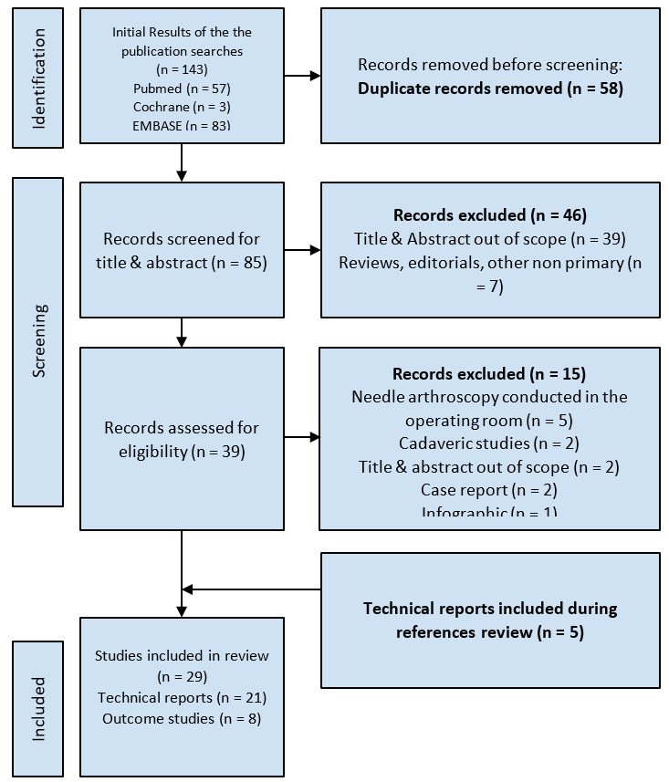
Figure 1
Figure 1.PRISMA Diagram

Study Characteristics

A total of 29 studies (8 outcome, 21 technical reports) were included in this scoping review. Six studies evaluated IONA in the glenohumeral joint (Colasanti et al. 2023; Daggett et al. 2020; Fariyike et al. 2024; Gauci et al. 2021; 2023; Owusu-Sarpong et al. 2023). Ten studies evaluated IONA in the knee (Andreozzi et al. 2022; Annibaldi et al. 2022; Bi et al. 2023; Bradsell et al. 2022; DiBartola et al. 2021; Kirschner et al. 2023; McMillan, Saini, et al. 2017; Neal et al. 2023; Patel et al. 2018; Trang et al. 2022). Twelve studies evaluated IONA in the foot and/or ankle (Butler et al. 2024; 2023; Chen et al. 2022; Colasanti, Kaplan, et al. 2022; Colasanti, Mercer, et al. 2022; Kanakamedala et al. 2022; Dankert et al. 2022; Kaplan et al. 2022; Labib and Slone 2015; Mercer, Gianakos, et al. 2022; Mercer, Azam, et al. 2022). One study evaluated multiple joints (McMillan et al. 2019).

Indications - Diagnosis

Twenty-two studies reported on the diagnostic indications of IONA. For the shoulder, IONA was used to diagnose subacromial impingement, superior labrum anterior to posterior (SLAP) lesions, and LHB tendinopathy (Colasanti et al. 2023; Daggett et al. 2020; Gauci et al. 2021; 2023; Owusu-Sarpong et al. 2023). Intra-articular knee pathology including lesions of the anterior cruciate ligament (ACL), medial and lateral meniscus were commonly diagnosed using IONA (Bradsell et al. 2022; DiBartola et al. 2021; Kirschner et al. 2023; McMillan, Saini, et al. 2017; Patel et al. 2018; McMillan et al. 2019). Additionally IONA in the knee was also used to assess healing of osteochondral allograft implants and ligamentous repair up to 12-24 months after surgery (Annibaldi et al. 2022; Trang et al. 2022). In the foot and ankle, IONA was used to diagnose impingement of the anterior and posterior ankle, and tendinopathy of the flexor hallucis longus (FHL), tibialis posterior, common peroneal muscles and Achilles, anterior talofibular ligament (ATFL) pathology, and hallux rigidus (Mercer, Samsonov, et al. 2022; Butler et al. 2024; 2023; Chen et al. 2022; Colasanti, Kaplan, et al. 2022; Kanakamedala et al. 2022; Dankert et al. 2022; Mercer, Gianakos, et al. 2022). Labib et al. reported IONA for diagnosis of a variety of intra articular foot and ankle pathologies such as osteochondral defects, instability, sinus (Kaplan et al. 2022) tarsi syndrome, impingement, tears, tendonosis, tenosynovitis, and sesamoid fractures (Labib and Slone 2015). Table 2 demonstrates the indications by body region described.

Indications - Treatment

Eleven studies described surgical procedures utilizing IONA (Fariyike et al. 2024; Gauci et al. 2021; Owusu-Sarpong et al. 2023; Andreozzi et al. 2022; Bi et al. 2023; Kirschner et al. 2023; Neal et al. 2023; Butler et al. 2023; Colasanti, Kaplan, et al. 2022; Colasanti, Mercer, et al. 2022; Kaplan et al. 2022; Garufi et al. 2023). Shoulder procedures included (1) SLAP lesion repair, (2) LHB tenodesis and/or tenotomy, and (3) subacromial decompression. Knee procedures using IONA included (1) lateral parapatellar retinacular release, (2) chondroplasty of focal cartilage lesions of the anterior femoral condyle, (3) medial meniscal repair, and (4) management of intra articular bacterial arthritis. Ankle procedures included (1) ATFL repair, (2) anterior ankle impingement debridement. Lastly, IONA was utilized for treatment for minimally invasive cheilectomy for hallux rigidus.

Clinical Evidence and Outcomes

One shoulder (Gauci et al. 2023), three knee (Andreozzi et al. 2022; Annibaldi et al. 2022; DiBartola et al. 2021) three foot and ankle (Mercer, Samsonov, et al. 2022; Butler et al. 2024; Colasanti, Mercer, et al. 2022) and one multiple-joint (McMillan et al. 2019) study evaluated outcomes following IONA. Five outcome studies compared pre- and postoperative outcomes using various validated instruments as reported in Table 3. These included: the Foot and Ankle Outcome Score (FAOS), Mean Tegner Lysholm Knee Scoring Scale (TLKSS) Mean Lysholm Score, Tegner Activity score, International Knee Documentation Committee Score (IKDS), and International Cartilage Repair Society Grades (ICRSG), Patient-Reported Outcomes Measurement Information System (PROMIS) (Mercer, Samsonov, et al. 2022; Gauci et al. 2023; Annibaldi et al. 2022; Butler et al. 2024; Colasanti, Mercer, et al. 2022).

Table 3.Outcome Studies
Study no. Study Group 1 (n) Group 2 (n) What was assessed? Result
SHOULDER 16 Gauci et al, 2023 pre-op post-op Mean duration of procedure = 24 min; VAS SSV; subjective shoulder constant VAS = 7 → 1 (p=.005); constant = 44 → 82 (p=.006), SSV = 53% → 93% (p=.005)
KNEE 1 Andreozzi et al, 2022 sample none Operating time, success rate of specimens achieved for pathologic analysis, iatrogenic chondral injuries, minor complications, postoperative pain, Likert scale, willingness to repeat biopsy, macroscopic synovitis, Krenn score Operating time = 12 min(median), success rate of specimens achieved for pathologic analysis = 97%, iatrogenic chondral injuries = 0, minor complications = 1, postoperative pain = 2, Likert scale = 4.4, willingness to repeat biopsy(yes) = 11(91.7%), macroscopic synovitis: low = 3, moderate = 5, severe = 4, Krenn score = 2 (median)
2 Annibaldi et al, 2022 2nd look arthroscopy 2-year follow-up Mean TLKSS KOOS, IKDC; ACL Healing Mean TLKSS= 97.5; KOOS = 98.1; IKDC = 96.7; ACL healing: graded A x11 patients, graded B x4 patients; KT-1OOO measurements maximum manual side-to-side difference <2mm x10 patients, 3mm x5 patients; synovial coverage graded “Good” x10 patients, fair x5 patients; Howell scale MRI assessment type I x10 patients type II x4 patients, type III x1 patient
13 DiBartola et al, 2021 pre-op post-op MRI Grade @ 12.4 months (mean) ⅞ patients demonstrated grade III changes pre-op, ⅝ demonstrated grade III changes post-op
FOOT/ANKLE 6 Butler et al, 2024 pre-op post-op Mean Follow-up time 26.3 months; mean Victorian Institute of Sport Assessment- Achilles and visual analog scores; patient satisfaction; mean return to play time; mean return to work time mean Victorian Institute of Sport Assessment- Achilles score = 35.6 → 83.6 (p<.001); visual analog scores = 6.6 -> 1.3 (p<.001); mean patient satisfaction = 4.5/5; mean return to play time = 5.9 weeks; mean return to work time = 4.2 days
10 Colasanti et al, 2022 pre-op post-op Mean FAOS (symptoms, pain, ADL, sports, QOL); mean PROMIS (pain interference, pain intensity) FAOS (symptoms: 64.9 +/- 13.6 → 79.4 +/- 11.9; pain 71.2 +/- 13.3 → 82.9 +/- 15.3; ADL: 69.6 +/- 14.4 → 83.5 +/- 15.4; sports: 60.8 =?- 14.8 → 71.9 +/- 18.5; QOL: 50.2 +/- 15.7 → 64.3 +/- 21.4); PROMIS (pain interference: 60.29 +/- 6.8 → 55.53 +/- 7.61; pain intensity: 50.89 +/- 8.4 → 44.52 +/- 6.9) all p<0.001
24 Mercer et al, 2022 pre-op post-op Mean follow-up time 13.3 month; FAOS (symptoms, pain, ADL, sports, QOL); PROMIS (pain interference, pain intensity) All mean pre-op FAOS scores demonstrated improvement after IONA
MULTIPLE 22 McMillan et al, 2019 sample none Major and minor complications Vasovagal events (1.6% in knees, 3% in shoulders), Persistent pain >24 hours = 4 knees n(0.3% of cases), post arthroscopy MRI needed in 1.4% of cases, 0 infections, no major complications

Abbreviations: VAS = visual analog score; SSV = subjective shoulder value; TLKSS = Tegner Lysholm Knee Scoring Scale KOOS = knee injury and osteoarthritis outcome score; IKDC = International Knee Documentation Committee; FAOS = Foot and ankle outcome score; QOL = quality of life; PROMIS = Patient-reported outcome measurement information system; adl = Activities of daily living

Shoulder

Gauci et al. performed an isolated long head of the biceps tenotomy on 20 participants for isolated biceps brachii pathology and reported significant improvements in the mean visual analog score (VAS) and subjective shoulder value and Constant (Gauci et al. 2023).

Knee

Andreozzi et al. evaluated the safety and efficacy of IONA in the diagnosis of drug-resistant monoarticular inflammatory arthritis in a study comparing pre and postoperative outcomes in 12 patients. The authors reported a mean surgical time of 12 minutes with a 97% specimen achievement rate. In addition there were no iatrogenic chondral injuries. Eleven of the twelve patients reported willingness to repeat the procedure (Andreozzi et al. 2022). Annibaldi et al. demonstrated IONA is effective in assessing ACL healing in 15 patients two years after repair reporting a excellent outcomes scores including TLKSS, KOOS, and IKDC scores of 97.5, 98.1, and 96.7, respectively (Annibaldi et al. 2022).

Ankle/Foot

Colasanti et al. evaluated outcomes following IONA for ankle debridement in 31 patients with anterior ankle impingement demonstrating a 22% improvement in symptoms, 16% improvement in pain, 20% improvement in ADLs, and 28% improvement in Quality of Life (QOL) for Foot and Ankle Outcome Score (FAOS) (Colasanti, Mercer, et al. 2022). Additionally there was a 9% decrease in pain interference and a 12% decrease in pain intensity for Patient-Reported Outcomes Measurement Information System (PROMIS). Mercer et al. similarly demonstrated significant improvements in all pre-op FAOS scores after IONA for treatment of posterior ankle impingement (Mercer, Samsonov, et al. 2022). Lastly, Butler et al. evaluated IONA for Achilles tendoscopy in 12 participants (Butler et al. 2024). Post operative visual analog scale scores for golf, running, walking, weight training and other performance sports all showed significant improvement and mean patient satisfaction score was 4.5/5.

Second Look Arthroscopy with IONA

DiBartola et al. assessed horizontal cleavage tears of the medial and lateral meniscus at 6 months follow up after IONA surgical repair. All tears demonstrated complete healing with IONA. Seven patients demonstrated grade III changes pre-operatively, and 5 patients demonstrated grade III changes post-operatively (DiBartola et al. 2021).

Annibaldi et al. assessed healing of ACL repairs 14 days after surgery in 15 patients. The mean TLKSS, KOOS, and IKDC were 97.86, 98.08, and 96.71, respectively (Annibaldi et al. 2022). IONA evaluated ACL healing, tension, and synovial coverage, finding a statistically significant correlation with MRI appearance, as measured by the Howell scale for signal intensity.

Complications

Eight of the 29 studies reported complications and limitations with use of IONA (Mercer, Samsonov, et al. 2022; Daggett et al. 2020; Fariyike et al. 2024; Andreozzi et al. 2022; Bradsell et al. 2022; McMillan, Saini, et al. 2017; Patel et al. 2018; Trang et al. 2022; Butler et al. 2024). Reported complication rates were low across included studies; however, meaningful aggregation is limited by heterogeneity in reporting and absence of standardized definitions. The largest series (n = 1,419) reported no major complications and minor complication rates below 5%, most commonly vasovagal events. Given the predominance of retrospective case series and technical reports, true complication incidence remains uncertain and may be underreported (McMillan et al. 2019).

Limitations of IONA

Four studies reported that lack of fluid irrigation, normally used with surgical arthroscopy, was a frequent hindrance of visualization of the joint space in hemarthritic and synovitic patients (Daggett et al. 2020; Bradsell et al. 2022; McMillan, Saini, et al. 2017; Trang et al. 2022). Additionally, the steep learning curve of using a zero degree scope presented a challenge to surgeons in two studies (Fariyike et al. 2024; Patel et al. 2018). Future research should describe how training pathways have addressed this learning curve. Additionally, it could investigate the effectiveness of equipment familiarization pathways, such as simulation-based or structured credentialing, compare them or others to one another, and determine their association with complications or patient reported outcomes.

Technical Feasibility and Procedural Considerations

The technical literature primarily describes portal placement, positioning, anesthetic technique, and procedural pearls. These reports are descriptive in nature and are not designed to evaluate safety or effectiveness.

i. Shoulder

Patient positioning and perioperative pharmaceutical use were highlighted as important aspects of IONA procedures. The patient was positioned with the back of the seat inclined at 70 to 80 degrees. The arm of the operative shoulder was placed on a well-padded Mayo stand, slightly flexed forward and abducted, to help facilitate easier access to the glenohumeral joint. One liter ratio of epinephrine to normal saline to simultaneously improve hemostasis and visualization. Portal management best practices included using posterior and anterosuperior portals with an additional lateral working portal for subacromial impingement, ensuring the viewing portal was opposite the aspect of the lesion and waiting a minimum of 10 minutes between intra-articular local anesthetic injection and portal incision to ensure optimal patient comfort.

ii. Knee

Considerations for diagnosis of intra-articular knee pathology include proper patient set-up determined by site of the lesion. For medial and lateral compartment evaluations, authors recommend keeping the patient supine with ability to flex the knee between 45 and 90 degrees. For patellofemoral joint inspection, the knee should be extended. Portals used include anteromedial and anterolateral, ensuring placement in the “soft spot” at the periphery of the patellar tendon approximately 1cm superior to the anterior tibial slope at the level of the inferior pole of the patella. Further specifics about portal placement in the “soft spot” are provided. Additionally, 3 cubic centimeter bursts of normal saline can be used to push away soft tissue and enhance visualization at the site of inspection.

iii. Foot and ankle

For anterior ankle pathology, the patient is placed in a supine position with a cushion placed under the leg just above the ankle, allowing for plantar flexion to facilitate joint distraction. For posterior pathology, the patient is positioned prone on pillows, ensuring comfort with proper head and leg support to minimize movement. Portal sites vary with the anatomical location of the pathology. Anterior ankle pathologies were addressed using anteromedial viewing and anterolateral portals, with the former placed lateral to the medial malleolus and medial to the tibialis anterior tendon and the latter placed medial to the lateral malleolus. Posterior hindfoot and Achilles tendon pathologies were addressed with posterolateral and posteromedial portals. The arthroscopy portal sites are located, cleaned with alcohol, and each portal is injected with 10 cc of 1% lidocaine. Using sterile technique, the extremity is prepped with a chlorhexidine gluconate and isopropyl alcohol solution, then draped. After five to 10 minutes, 20 cc of a 1:1 mixture of 1% lidocaine and 0.5% bupivacaine is injected through the portal sites into the joint. Portal stab incisions are made using an 11 blade, then the standard operating room procedure is performed with an inflow and outflow fluid irrigation system with a modifiable pressure of 35mmHg. Table 4 indicates technical considerations for all studies.

Table 4.Technique Guides
Study no. Study Pathology/injury Approach/portals Patient positioning Pearls/tips
SHOULDER 8 Colasanti et al, 2023 LHB tendinopathy Standard posterior arthroscopy portal, anterosuperior portal Beach-chair position, examination table positioned at 70 to 80 degrees
  • At least 10 minutes between portal and intra-articular local anesthetic injection and procedure
  • 5 cc of epinephrine in 1 L of moral saline for hemostasis and visualization
11 Daggett et al, 2020 diagnosis - LHB tendinopathy Standard posterior entry diagnostic portal seated
  • Ensure patients comfortably seated with positioning allowing easy access to shoulder
  • If visualization is impaired by bleeding, exchange of fluid can be performed to improve it, but if unsuccessful procedure should be terminated
14 Fariyike et al, 2024 SLAP tear Posterior arthroscopic portal, anterior Beach-chair position, examination table positioned at 70 to 80 degrees
  • At least 10 minutes between portal and intra-articular local anesthetic injection and procedure
  • 5 cc of epinephrine in 1 L of normal saline for hemostasis and visualization
15 Gauci et al, 2021 LHB tenotomy Anterior and posterior portal Supine Beach chair position (keep legs raised)
  • Shoulder must be in internal rotation, patient relaxed and warned at each step to keep them confident
  • Conventional arthroscopic scissors must be used to cut the biceps
  • Tenotomy must be performed by pushing the biceps at the same time with the elbow in flexion
27 Owusu-Sarpong et al, 2023 Subacromial impingement Posterior, anterior, lateral (working portal) Beach-chair position, examination table positioned at 70 to 80 degrees
  • At least 10 minutes between portal and intra-articular local anesthetic injection and procedure
  • 5 mL of epinephrine in 1 L of normal saline solution for hemostasis and visualization
KNEE 3 Bi et al, 2023 Chondral and osteochondral lesions of the knee Anterolateral, anteromedial Supine with operative leg hanging off examination table to allow opening of the joint space
  • Viewing portal opposite the lesions ideal for instrumentation/delivery, i.e., needle arthroscope in anterolateral portal for medial femoral condyle lesions
  • Thorough synovectomy around lesion, so after suctioning and transitioning to dry needle arthroscopy adequate visualization is achieved
4 Bradsell et al, 2022 diagnosis - intra-articular knee pathology Anterolateral; anteromedial Supine with 90 degrees Of knee flexion
  • Additional saline beyond initial insufflation may be used to improve visualization by serially injecting saline through nanoscope cannula
19 Kirschner et al, 2023 Meniscal tear Anterolateral, anteromedial Supine with operative knee hanging off table to allow opening of joint space
  • Minimum of 10 minutes between portal and intra-articular local anesthetic injection and procedure
  • Viewing portal opposite the lesion is ideal for instrumentation/delivery, i.e., needle arthroscopy in an anterolateral portal for medial meniscus lesion
21 McMillan et al, 2017 diagnosis - intra-articular knee pathology Superolateral; lateral infrapatellar; medial infrapatellar Supine with 30-45 degrees of Knee flexion
  • Flex the knee 45-90 degrees For medial and lateral compartment evaluations; extend the knee for patellofemoral joint inspection
  • Height of medial & lateral portals is approximately 1 cm superior to the anterior tibial slope, at the level of the inferior pole of the patella - the entry point is approximately 1 cm from the patellar tendon toward the “soft spot”
26 Neal et al, 2023 Lateral parapatellar tightness Anterolateral, anteromedial Supine with operative leg hanging off examination table to allow opening of the joint space
  • 10-minute window between initial local anesthetic (10 cc per portal and final joint load (20 cc) immediately before incision
  • Epinephrine in saline for hemostasis and visualization
28 Patel et al, 2017 diagnosis - intra-articular knee pathology Medial; lateral; transpatellar; superolateral; superomedial Supine with 90 degrees Of knee flexion (use bump)
  • Use a 10 or 20 ml syringe for better control of the prove
  • Use bursts (3 ml) of saline to push away soft tissue and allow for visualization at specific times during arthroscopy
29 Trang et al, 2022 Osteochondral allograft transplant Anterolateral; anteromedial Supine with knee supported in extension
  • None listed
FOOT/ANKLE 5 Butler et al, 2023 ATFL repair Anteromedial; anterolateral; accessory (1 cm anterior to tip of fibula) supine
  • Position the ankle in 30 degrees Of plantar flexion to prevent overtightening
  • Ensure adequate head and leg support to mitigate against lower extremity movement during procedure
7 Chen et al, 2022 Posterior hindfoot pathology lateral; medial Prone with foot and ankle hanging off foot of bed
  • Creation of portals with stab incisions through skin only followed by blunt dissection
  • Direction of instruments toward lateral border of third metatarsal during initial portal placement
  • Calcaneal distraction and ankle dorsiflexion to facilitate entry into the posterior tibiotalar joint
9 Colasanti et al, 2022 Anterior ankle impingement Anterolateral; anteromedial Supine with operated ankle hanging off edge of table
  • Gentle traction as necessary to open joint space and facilitate joint access
  • Adequate periosteal local anesthesia injection especially for patients with exotoses
12 Dankert et al, 2022 Tibialis posterior tendon dysfunction Proximal and distal tendoscopy portals Lateral decubitus position with operative ankle closest to table so medial aspect of operative ankle is exposed
  • “Walk” step-wise with the 18-gauge spinal needle posterior to the medial malleolus to identify the proper location for the inferior portal
  • Instruct patient to actively range their ankle to assess TPT gliding and identify any missed adhesions
  • Identifying the “glistening tendon” with right-angle forceps assists with correct cannula placement
17 Kanakamedala et al, 2022 Common peroneal tendon pathology Proximal and distal portals Lateral decubiitus position with lateral aspect of operative ankle exposed
  • Before making proximal portal, perform full diagnostic tendoscopy. Can place portal more proximal or distal depending on pathology (debriding accessory muscle or tendon tear proximally versus groove deepening distally)
  • If needed, can give additional anesthetic to facilitate conversion to open procedure
18 Kaplan et al, 2022 Cheilectomy for Hallux Rigidus Direct medial portal; dorsomedial; dorsolateral Supine with foot at edge of bed
  • Joint distraction is integral for intra articular anesthetic block
  • Careful placement of dorsolateral portal to obtain optimal positioning for bur;
  • Traction can be released after joint is accessed to loosen joint capsule and facilitate access to dorsal osteophyte
20 Labib et al, 2015 Diagnosis - intra-articular foot pathology Ankle anteromedial Supine with foot and ankle hanging off edge of bed
  • None listed
23 Mercer et al, 2022 diagnosis - Achilles tendonopathy Medial and lateral Prone with foot over edge of bed in neutral dorsiflexion-plantar flexion
  • Portal sites should allow for movement of the camera (medial or lateral to tendon). Central portal sites can cause difficulty positioning the camera as a large gastrocnemius complex may limit movement
  • Patient should be encouraged to actively range ankle to ensure removal of all adhesions
25 Mercer et al, 2022 ATFL pathology Anteromedial and anterolateral supine
  • Inject lidocaine at both sites and use a mixture with epinephrine and bupivacaine to limit bleeding
  • Help to prevent overtightening by having the patient plantarflex the ankle to approximately 30 degrees

Discussion

This scoping review summarizes the existing literature on the use of in-office needle arthroscopy (IONA) across multiple joints. The available evidence consists predominantly of technical reports and small, non-comparative case series that describe feasibility rather than comparative effectiveness. Although several outcome studies report short-term improvements in patient-reported measures and low rates of major complications, the predominance of low-level evidence and substantial heterogeneity preclude definitive conclusions regarding diagnostic accuracy, clinical effectiveness, or safety.

Shoulder

The shoulder represents one of the earliest and most frequently described applications of IONA, with five technical reports and a single study reporting quantitative clinical outcomes identified. Technical reports describe diagnostic visualization and treatment of select intra-articular pathologies, including long head of the biceps (LHB) tendinopathy, subacromial impingement, and superior labral pathology managed with limited debridement or tenotomy. Gauci et al. reported improvements in visual analog scale (VAS) and Subjective Shoulder Value (SSV) following in-office procedures, suggesting that patients may tolerate the intervention in this setting (Gauci et al. 2023). However, the absence of comparator groups, blinded assessments, or standardized reference standards limits interpretation of these findings beyond feasibility and short-term symptom change.

Knee

The knee literature includes seven technical reports and three outcome studies. Technical descriptions focus on diagnostic assessment of intra-articular pathology and procedural feasibility for chondral, ligamentous, and tendinous conditions, including cartilage debridement, evaluation of horizontal cleavage tears, and postoperative assessment following anterior cruciate ligament repair. Outcome studies did not consistently demonstrate clinical benefit for therapeutic intervention but did suggest potential utility for postoperative healing assessment and second-look evaluation. Notably, available data do not establish whether IONA provides incremental diagnostic or clinical value over routine physical examination and standard postoperative follow-up, particularly during active rehabilitation. Future comparative studies are required to clarify whether in-office visualization alters clinical decision-making or patient outcomes in this context.

Second-Look Arthroscopy and Synovial Biopsy

Emerging evidence suggests a role for IONA in second-look arthroscopy and synovial biopsy, particularly in inflammatory and postoperative conditions. In one study evaluating monoarticular arthritis, most patients expressed willingness to undergo repeat in-office procedures (11 of 12 patients), with high rates of successful specimen collection and short procedural duration (Andreozzi et al. 2022). While real-time intra-articular visualization may offer theoretical advantages over arthrocentesis alone, these findings remain preliminary. Interpretation is limited by verification bias, as patients undergoing IONA were already selected for invasive evaluation, and by the absence of blinded comparisons to established reference standards. To minimize such bias, blinded diagnostic accuracy designs should be included in future studies. Additionally, comparative studies evaluating diagnostic yield, clinical impact, and cost-effectiveness relative to arthrocentesis and advanced imaging are needed.

Foot and Ankle

Nine technical reports and three outcome studies describe the use of IONA in the foot and ankle. Reported diagnostic and therapeutic indications include anterior ankle impingement, ATFL pathology, cheilectomy for hallux rigidus, posterior hindfoot pathology, and tendinopathies involving the Achilles, tibialis posterior, and common peroneal tendons. Technical reports primarily describe procedural feasibility, portal placement, and visualization of intra-articular and peri-tendinous pathology.

Outcome studies report short-term improvements in patient-reported measures following IONA for anterior and posterior ankle impingement, including improvements in pain and functional domains. One outcome study evaluating IONA for healing assessment following chronic Achilles tendinopathy reported favorable functional outcomes and high patient satisfaction, with 11 of 12 patients indicating willingness to undergo the procedure again (Butler et al. 2024). In this cohort, one patient experienced a sural nerve neurapraxia that resolved within four months, and one patient reported persistent Achilles pain and stiffness refractory to physical therapy. These findings underscore the importance of careful patient selection, procedural counseling, and consideration of potential neurologic risk.

As with other joints, interpretation of these findings is limited by small sample sizes, lack of comparator groups, and short follow-up duration. No study directly compared IONA-based assessment or intervention with physical examination, rehabilitation-based evaluation, or operative arthroscopy. Accordingly, while these reports suggest technical feasibility and short-term symptom improvement in select indications, further prospective, comparative studies are needed to determine whether the potential benefits of direct in-office visualization outweigh procedural risks and whether IONA meaningfully alters clinical decision-making or outcomes in foot and ankle pathology.

Safety Considerations

Although complication rates cannot be generalized across heterogeneous procedures and indications, reported adverse events in the existing literature are infrequent and typically minor. No major complications were identified, with minor events including vasovagal episodes, transient neuropraxia, and short-term postoperative discomfort (Mercer, Azam, et al. 2022; McMillan et al. 2019; Garufi et al. 2023; Szachnowski et al. 1995; Kim et al. 2005). A larger retrospective analysis of 335 office-based knee arthroscopies, which reported major complication rates of 1.2 percent and minor complications in 12.8 percent of cases, without any long-term morbidity or mortality (Szachnowski et al. 1995). Similarly, prospective trials and systematic reviews involving the knee, ankle, and wrist have reported no device-related complications or serious adverse events (Burt et al. 2023; Gill et al. 2018). Given the majority of literature in the present study are technical reports, true complication incidence may likely be underreported. Perceived safety of this modality should be interpreted cautiously and within the context of selective patient populations and limited follow-up. Although patient-specific susceptibility may contribute, mitigation strategies such as preprocedural counseling and the use of physical counterpressure maneuvers like leg-crossing and muscle tensing may improve patient tolerance and reduce the likelihood of an event (Kim et al. 2005; Peterson and Cigrang 2003).

Other intraoperative considerations have not been discussed in the literature. In addition to vasovagal events and unlike OR-arthroscopy, IONA introduces risks specific to an awake patient: patient movement, pain response, electrocautery tolerance, bleeding management, and protocols for procedural termination or conversion must be further qualified and quantified in future literature. More robust description of patient selection criteria, pre-procedural counseling protocols, and contingency planning when patients cannot tolerate therapeutic maneuvers intraoperatively are needed as well.

Limitations

This review is limited by the nature and quality of the available evidence. Most included studies were technical reports or small, non-comparative case series, with only eight studies reporting clinical outcomes using validated measures. As such, the literature primarily reflects technical feasibility and early clinical experience rather than comparative effectiveness. Outcome reporting was highly heterogeneous, with inconsistent documentation of follow-up duration, clinical significance, and complications, precluding quantitative synthesis or meta-analysis.

Interpretation of diagnostic performance is further constrained by verification bias and the absence of blinded diagnostic accuracy studies or standardized reference standards. Additionally, demographic reporting was inconsistent across studies. Only one technical report included patient demographic data, and while most outcome studies reported age and body mass index, reporting formats varied and other potentially relevant characteristics were infrequently described (Patel et al. 2018). More consistent reporting of patient demographics and baseline characteristics would improve interpretation and generalizability. Generalizability is also limited by the characteristics of the populations studied. In-office needle arthroscopy is not universally covered by insurance, which may select for patients with higher health literacy and socioeconomic status. Further demographic correlations with undergoing elective procedures such as IONA exist (e.g, race and ethnicity) which systematically excludes portions of the broader population. Patient characteristics should be included in future research, especially for a modality that has potential to reduce time to diagnosis and treatment, a factor that has been correlated with healthcare access. Reported outcome studies also included relatively few patients with obesity, potentially limiting applicability to broader clinical populations where visualization and procedural complexity may differ. Future studies should also implement multi-surgeon and multi-center design to minimize observer bias and optimize generalizability.

Finally, outcome measures varied widely across studies, even within the same joint, with inconsistent reporting of functional outcomes, patient-reported measures, procedural metrics, and complications. As in-office needle arthroscopy continues to evolve, future investigations should prioritize prospective, comparative study designs with standardized reporting of outcomes, complications, and health system impacts to better define its clinical role.

Conclusion

The current literature on in-office needle arthroscopy consists predominantly of technical reports and small case series. Available studies suggest that IONA is technically feasible and may be associated with short-term improvements in patient-reported outcomes in select indications; however, the predominance of low-level evidence and absence of comparative studies preclude definitive conclusions regarding effectiveness, safety, or superiority over conventional arthroscopy or advanced imaging. Further prospective, comparative investigations are required before broader clinical adoption can be recommended.

Submitted: March 29, 2026 EDT

Accepted: May 23, 2026 EDT

References

Amin, N., L. McIntyre, T. Carter, J. Xerogeanes, and J. Voigt. 2019. “Cost-Effectiveness Analysis of Needle Arthroscopy versus Magnetic Resonance Imaging in the Diagnosis and Treatment of Meniscal Tears of the Knee.” Arthroscopy: The Journal of Arthroscopic & Related Surgery 35 (2): 554-562.e513. https:/​/​doi.org/​10.1016/​j.arthro.2018.09.030.
Google Scholar
Andreozzi, V., E. Monaco, C. Garufi, et al. 2022. “In-Office Needle Arthroscopic Synovial Biopsy Is an Effective Diagnostic Tool in Patients With Inflammatory Arthritis.” Arthroscopy, Sports Medicine, and Rehabilitation 4 (6): e2099–106. https:/​/​doi.org/​10.1016/​j.asmr.2022.10.003.
Google Scholar
Annibaldi, A., E. Monaco, M. Daggett, et al. 2022. “In-Office Needle Arthroscopic Assessment after Primary ACL Repair: Short-Term Results in 15 Patients.” Journal of Experimental Orthopaedics 9 (1): 89. https:/​/​doi.org/​10.1186/​s40634-022-00528-1.
Google Scholar
Bi, A. S., C. A. Colasanti, N. Kirschner, et al. 2023. “In-Office Needle Arthroscopy With Cartilage Allograft Extracellular Matrix Application for Cartilage Lesions of the Knee.” Arthroscopy Techniques 12 (9): e1507–13. https:/​/​doi.org/​10.1016/​j.eats.2023.04.023.
Google Scholar
Bradsell, H., A. Lencioni, K. Shinsako, and R. M. Frank. 2022. “In-Office Diagnostic Needle Arthroscopy Using the NanoScopeTM Arthroscopy System.” Arthroscopy Techniques 11 (11): e1923–27. https:/​/​doi.org/​10.1016/​j.eats.2022.07.006.
Google Scholar
Burt, J., V. Smith, C. W. Gee, J. V. Clarke, and A. J. Hall. 2023. “The Role of Outpatient Needle Arthroscopy in the Diagnosis and Management of Musculoskeletal Complaints: A Systematic Review of the Arthrex NanoScope.” Knee 42: 246–57. https:/​/​doi.org/​10.1016/​j.knee.2023.04.003.
Google Scholar
Butler, J. J., A. I. Brash, M. T. Azam, B. DeClouette, and J. G. Kennedy. 2023. “The Role of Needle Arthroscopy in the Assessment and Treatment of Ankle Sprains.” Foot and Ankle Clinics 28 (2): 345–54. https:/​/​doi.org/​10.1016/​j.fcl.2023.01.005.
Google Scholar
Butler, J. J., G. W. Randall, L. Schoof, et al. 2024. “Excellent Clinical Outcomes and Rapid Return to Activity Following In-Office Needle Tendoscopy for Chronic Achilles Tendinopathy.” Arthroscopy, Sports Medicine, and Rehabilitation, 100937. https:/​/​doi.org/​10.1016/​j.asmr.2024.100937.
Google Scholar
Chen, J. S., D. J. Kaplan, C. A. Colasanti, et al. 2022. “Posterior Hindfoot Needle Endoscopy in the Office Setting.” Arthroscopy Techniques 11 (3): e273–78. https:/​/​doi.org/​10.1016/​j.eats.2021.10.018.
Google Scholar
Colasanti, C. A., M. T. Azam, A. S. Bi, et al. 2023. “Reproducible and Effective Biceps Tenodesis Method Utilizing In-Office Nano-Arthroscopy.” Arthroscopy Techniques 12 (10): e1797–802. https:/​/​doi.org/​10.1016/​j.eats.2023.06.009.
Google Scholar
Colasanti, C. A., D. J. Kaplan, J. S. Chen, et al. 2022. “In-Office Needle Arthroscopy for Anterior Ankle Impingement.” Arthroscopy Techniques 11 (3): e327–31. https:/​/​doi.org/​10.1016/​j.eats.2021.10.025.
Google Scholar
Colasanti, C. A., N. P. Mercer, J. V. Garcia, G. M. Kerkhoffs, and J. G. Kennedy. 2022. “In-Office Needle Arthroscopy for the Treatment of Anterior Ankle Impingement Yields High Patient Satisfaction with High Rates of Return to Work and Sport.” Arthroscopy: The Journal of Arthroscopic & Related Surgery 38 (4): 1302–11. https:/​/​doi.org/​10.1016/​j.arthro.2021.09.016.
Google Scholar
Daggett, M. C., B. Stepanovich, B. Geraghty, A. Meyers, J. Whetstone, and A. Saithna. 2020. “Office-Based Needle Arthroscopy: A Standardized Diagnostic Approach to the Shoulder.” Arthroscopy Techniques 9 (4): e521–25. https:/​/​doi.org/​10.1016/​j.eats.2019.12.003.
Google ScholarPubMed CentralPubMed
Dankert, J. F., N. P. Mercer, D. J. Kaplan, et al. 2022. “In-Office Needle Tendoscopy of the Tibialis Posterior Tendon with Concomitant Intervention.” Arthroscopy Techniques 11 (3): e339–45. https:/​/​doi.org/​10.1016/​j.eats.2021.10.027.
Google Scholar
DiBartola, A.C., A. Rogers, P. Kurzweil, M.V. Knopp, and D.C. Flanigan. 2021. “In-Office Needle Arthroscopy Can Evaluate Meniscus Tear Repair Healing as an Alternative to Magnetic Resonance Imaging.” Arthroscopy, Sports Medicine, and Rehabilitation 3 (6): e1755–60. https:/​/​doi.org/​10.1016/​j.asmr.2021.08.003.
Google Scholar
Fariyike, B., W. H. Neal, A. S. Bi, et al. 2024. “In-Office Needle Arthroscopy for Superior Labral Tear Debridement.” Arthroscopy Techniques 13 (5): 102956. https:/​/​doi.org/​10.1016/​j.eats.2024.102956.
Google Scholar
Garufi, C., F. Spinelli, M. Leopizzi, et al. 2023. AB0087 THE SYNOVIAL EXPRESSION OF PSTAT1-3-5 IN PATIENTS WITH KNEE TREATMENT-RESISTANT MONOARTHRITIS IN THE COURSE OF RHEUMATOID AND PSORIATIC ARTHRITIS. https:/​/​doi.org/​10.1136/​annrheumdis-2023-eular.6343.
Google Scholar
Gauci, M.-O., B. Monin, A. Rudel, B. Bige, N. Bronsard, and J.-F. Gonzalez. 2023. “Results Of In-Office Biceps Isolated Tenotomy In A Daily Practice. A Prospective Study.” Journal of Shoulder and Elbow Surgery 32 (5): e246–47. https:/​/​doi.org/​10.1016/​j.jse.2023.02.029.
Google Scholar
Gauci, M.-O., B. Monin, A. Rudel, L. Blasco, B. Bige, and P. Boileau. 2021. “In-Office Biceps Tenotomy with Needle Arthroscopy: A Feasibility Study.” Arthroscopy Techniques 10 (5): e1263–68. https:/​/​doi.org/​10.1016/​j.eats.2021.01.022.
Google Scholar
Gianakos, A. L., and J. G. Kennedy. 2024. “In-Office Needle Arthroscopy: Indications, Surgical Techniques, Tips, and Tricks.” JAAOS Global Research & Reviews 8 (2): e23. https:/​/​doi.org/​10.5435/​JAAOSGlobal-D-23-00124.
Google Scholar
Gill, T. J., M. Safran, B. Mandelbaum, B. Huber, R. Gambardella, and J. Xerogeanes. 2018. “A Prospective, Blinded, Multicenter Clinical Trial to Compare the Efficacy, Accuracy, and Safety of In-Office Diagnostic Arthroscopy With Magnetic Resonance Imaging and Surgical Diagnostic Arthroscopy.” Arthroscopy 34 (8): 2429–35. https:/​/​doi.org/​10.1016/​j.arthro.2018.03.010.
Google Scholar
Kanakamedala, A., J. S. Chen, D. J. Kaplan, et al. 2022. “In-Office Needle Tendoscopy of the Peroneal Tendons.” Arthroscopy Techniques 11 (3): e365–71. https:/​/​doi.org/​10.1016/​j.eats.2021.11.002.
Google Scholar
Kaplan, D. J., J. S. Chen, C. A. Colasanti, et al. 2022. “Needle Arthroscopy Cheilectomy for Hallux Rigidus in the Office Setting.” Arthroscopy Techniques 11 (3): e385–90. https:/​/​doi.org/​10.1016/​j.eats.2021.11.005.
Google Scholar
Kim, K. H., J. G. Cho, K. O. Lee, et al. 2005. “Usefulness of Physical Maneuvers for Prevention of Vasovagal Syncope.” Circ J 69 (9): 1084–88. https:/​/​doi.org/​10.1253/​circj.69.1084.
Google Scholar
Kirschner, N., S. Owusu-Sarpong, W. H. Neal, et al. 2023. “In-Office Needle Arthroscopy with Meniscal Repair for Meniscal Lesions of the Knee.” Arthroscopy Techniques 12 (10): e1821–26. https:/​/​doi.org/​10.1016/​j.eats.2023.06.013.
Google Scholar
Labib, S. A., and H. S. Slone. 2015. “Office-Based Needle Arthroscopy for the Foot and Ankle.” Techniques in Foot & Ankle Surgery 14 (1): 9–11. https:/​/​doi.org/​10.1097/​BTF.0000000000000073.
Google Scholar
McMillan, S., A. Chhabra, J. D. Hassebrock, E. Ford, and N. H. Amin. 2019. “Risks and Complications Associated with Intra-Articular Arthroscopy of the Knee and Shoulder in an Office Setting.” Orthopaedic Journal of Sports Medicine 7 (9): 2325967119869846. https:/​/​doi.org/​10.1177/​2325967119869846.
Google Scholar
McMillan, S., S. Saini, E. Alyea, and E. Ford. 2017. “Office-Based Needle Arthroscopy: A Standardized Diagnostic Approach to the Knee.” Arthroscopy Techniques 6 (4): e1119–24. https:/​/​doi.org/​10.1016/​j.eats.2017.03.031.
Google Scholar
McMillan, S., M. Schwartz, B. Jennings, S. Faucett, T. Owens, and E. Ford. 2017. “In-Office Diagnostic Needle Arthroscopy: Understanding the Potential Value for the US Healthcare System.” Am J Orthop 46 (5): 252–56.
Google Scholar
Mercer, N. P., M. T. Azam, N. Davalos, et al. 2022. “Anterior Talofibular Ligament Augmentation with Internal Brace in the Office Setting.” Arthroscopy Techniques 11 (4): e545–50. https:/​/​doi.org/​10.1016/​j.eats.2021.12.005.
Google Scholar
Mercer, N. P., A. L. Gianakos, D. J. Kaplan, et al. 2022. “Achilles Paratenon Needle Tendoscopy in the Office Setting.” Arthroscopy Techniques 11 (3): e315–20. https:/​/​doi.org/​10.1016/​j.eats.2021.10.024.
Google Scholar
Mercer, N. P., A. P. Samsonov, J. F. Dankert, et al. 2022. “Improved Clinical Outcomes and Patient Satisfaction of In-Office Needle Arthroscopy for the Treatment of Posterior Ankle Impingement.” Arthroscopy, Sports Medicine, and Rehabilitation 4 (2): e629–38. https:/​/​doi.org/​10.1016/​j.asmr.2021.12.004.
Google Scholar
Munn, D., J. Burt, C. Gee, C. Mclaren, J. Clarke, and A. Hall. 2023. “Moving Orthopaedic Procedures out of the Operating Theatre: Outpatient Needle Arthroscopy Can Reduce Cost & Waste, and Increase Inpatient Capacity Compared to Conventional Knee Arthroscopy.” The Knee 42: 143–52. https:/​/​doi.org/​10.1016/​j.knee.2023.03.009.
Google Scholar
Neal, W. H. E., N. Kirschner, S. Owusu-Sarpong, et al. 2023. “In-Office Needle Arthroscopy of the Knee With Lateral Parapatellar Retinacular Release.” Arthrosc Tech 12 (10): e1809–13. https:/​/​doi.org/​10.1016/​j.eats.2023.06.011.
Google Scholar
Owusu-Sarpong, S., B. Fariyike, C. A. Colasanti, et al. 2023. “In-Office Nano-Arthroscopy of the Shoulder with Acromioplasty.” Arthroscopy Techniques 12 (8): e1423–28. https:/​/​doi.org/​10.1016/​j.eats.2023.04.012.
Google Scholar
Patel, K. A., D. E. Hartigan, J. L. Makovicka, D. L. Dulle 3rd, and A. Chhabra. 2018. “Diagnostic Evaluation of the Knee in the Office Setting Using Small-Bore Needle Arthroscopy.” Arthrosc Tech 7 (1): e17–21. https:/​/​doi.org/​10.1016/​j.eats.2017.08.044.
Google Scholar
Peterson, A. L., and J. A. Cigrang. 2003. “Management of Vasovagal Syncope: Controlling or Aborting Faints by Leg Crossing and Muscle Tensing.” Circulation 107 (20): e198. https:/​/​doi.org/​10.1161/​01.CIR.0000074257.16387.40.
Google Scholar
Shah, A., A. Abbas, J. Lex, T. Hauer, J. Abouali, and J. Toor. n.d. “IN-OFFICE NEEDLE ARTHROSCOPY (IONA) IS A COST-EFFECTIVE ALTERNATIVE FOR OPERATING ROOM DIVERSION IN MEDIAL MENISCECTOMY.” Paper. Orthopaedic Proceedings 2022. https:/​/​doi.org/​10.1186/​s13018-023-03866-7.
PubMed CentralPubMed
Szachnowski, P., N. Wei, W. J. Arnold, and L. M. Cohen. 1995. “Complications of Office Based Arthroscopy of the Knee.” J Rheumatol 22 (9): 1722–25.
Google Scholar
Trang, G., S. R. Del Sol, S. Jenkins, et al. 2022. “Evaluation of Osteochondral Allograft Transplant Using In-Office Needle Arthroscopy.” Arthrosc Tech 11 (12): e2243–48. https:/​/​doi.org/​10.1016/​j.eats.2022.08.032.
Google Scholar
Voigt, J. D., M. Mosier, and B. Huber. 2014. “Diagnostic Needle Arthroscopy and the Economics of Improved Diagnostic Accuracy: A Cost Analysis.” Applied Health Economics and Health Policy 12: 523–35. https:/​/​doi.org/​10.1007/​s40258-014-0109-6.
Google Scholar
Wagner, E. R., J. M. Woodmass, Z. R. Zimmer, et al. 2021. “Needle Diagnostic Arthroscopy and Magnetic Resonance Imaging of the Shoulder Have Comparable Accuracy with Surgical Arthroscopy: A Prospective Clinical Trial.” Arthroscopy: The Journal of Arthroscopic & Related Surgery 37 (7): 2090–98. https:/​/​doi.org/​10.1016/​j.arthro.2021.03.006.
Google Scholar
Zhang, K., R. J. Crum, K. Samuelsson, E. Cadet, O. R. Ayeni, and D. de Sa. 2019. “In-Office Needle Arthroscopy: A Systematic Review of Indications and Clinical Utility.” Arthroscopy 35 (9): 2709–21. https:/​/​doi.org/​10.1016/​j.arthro.2019.03.045.
Google Scholar

Attachments

Powered by Scholastica, the modern academic journal management system