Loading [Contrib]/a11y/accessibility-menu.js

This website uses cookies

We use cookies to enhance your experience and support COUNTER Metrics for transparent reporting of readership statistics. Cookie data is not sold to third parties or used for marketing purposes.

Skip to main content
null
J Orthopaedic Experience & Innovation
  • Menu
  • Articles
    • Brief Report
    • Case Report
    • Case Series
    • Conference Proceedings
    • Data Paper
    • Editorial
    • Meeting Reports/Abstracts
    • Methods Article
    • Product Review
    • Research Article
    • Review Article
    • Review Articles
    • Systematic Review
    • All
  • For Authors
  • Editorial Board
  • About
  • Issues
  • Blog
  • "Open Mic" Topic Sessions
  • Advertisers
  • Recorded Content
  • CME
  • JOEI KOL Connect
  • search
  • RSS feed (opens a modal with a link to feed)

RSS Feed

Enter the URL below into your favorite RSS reader.

https://journaloei.scholasticahq.com/feed
ISSN 2691-6541
Research Article
April 02, 2026 EDT

From Our Perspective…Why Total Hip Arthroplasties Dislocate After Spinal Fusion: Functional Mechanics Uniting Hip and Spine Surgery

Russell Bodner, MD, Richard Lyon, DC,
spinopelvic relationshipsTHA biomechanicsTHA and spine fusion planningTHA dislocation
Copyright Logoccby-nc-nd-4.0 • https://doi.org/10.60118/001c.153896
J Orthopaedic Experience & Innovation
Bodner, Russell, and Richard Lyon. 2026. “From Our Perspective…Why Total Hip Arthroplasties Dislocate After Spinal Fusion: Functional Mechanics Uniting Hip and Spine Surgery.” Journal of Orthopaedic Experience & Innovation, April. https://doi.org/10.60118/001c.153896.
Download all (9)
  • Figure 1a. The geometric pelvic-acetabular construct with central triangle integrating acetabular angles Anteinclination (AI), Sacroacetabular Angle (SAA), Pelvic Acetabular Angle (PAA) to pelvic angles Pelvic Incidence (PI), Sacral Slope (SS), Pelvic Tilt (PT). The hip position is measured through the Pelvic Femoral Angle (PFA). The functional safe zone measure, Combined Sagittal Index, CSI, is defined as CSI=AI+PFA. Pelvic tilt (red line) change affects the spatial position of the center of the S1 endplate, mobility the length of the SS triangle side (blue line), each of which independently alter the AI cup angle (green line).
    Download
  • Figure 1b. Sitting clinical postoperative lateral radiograph illustrating the complete construct angles described in Figure 1. The highlighted 62° critical angle (yellow) is the cup Anteinclination, AI (sitting range 55°+/- 10°). PI= 45.2, (90-PI)=44.7, SS=9.0, PT=36.6, SAA=71.2, PAA=64.0, PFA=127.1. SAA+PAA+(90-PI) the 3 corners of the triangle always equal 180°, SS+PT always equal PI, PFA+AI= CSI, the validated functional safe zone parameter replacing the Lewinnek zone, here 127.1+62.2=189.3, well above the published limit of 151 and mean value of 180°.
    Download
  • Figure 2. Hip joint specific spinopelvic-acetabular angles. The cup AI, the planning angles PT, PAA and the combined hip flexion/extension angle PFA. PT and PI share the anatomically fixed line (red) connecting the centers of the hip axis and the S1 endplate, PAA describes the cup’s relationship to the PT through its complementary angle, PAA=(90-AI)+PT, alternatively, 90-(AI-PT), utilizing the fundamental quotient bridging the cup AI angle to that of the moving pelvis. AI is the sagittal cup inclination angle composed of operative plane anteversion with an elliptical inclination contribution. An optimal stand/sit pairing for PI=60°, pelvic mobility 25° is shown with the 30° standing cup AI increasing to 55° sitting identically with the pelvic movement.
    Download
  • Figure 3. The four unique applications of the triangular construct considering solely the distinct hip-based ratios of angular change. Ratios are determined by parameters being fixed/mobile under the specific condition being studied.
    Download
  • Table 1. Normative architectural values for all sagittal plane parameters based on a functional normative pelvic parameters relationship defined as SS=0.67PI+5, PT=0.33PI-5, a 3:2:1 ratio of angular change for PI:SS:PT. This creates an equal relationship between cup and the SS/PT values forming an isosceles triangle solving for both the unknown SAA/PAA angles as (90+PI)/2 and AI as SAA-SS or 90-(PAA-PT). Whole number parameter ratios across the table rows are 6:4:2:​-1:3:-3 for PI:SS:PT:AI:PAA:(AI-PT).
    Download
  • Figure 4. Case example of cup planning method and calculating effects of spinal intervention.
    Download
  • Table 2. Tilt Adjusted normative Standing AI cup values by Pelvic Incidence at 25° Pelvic Mobility, illustrating the Spinopelvic rule of 4s. PT expresses a 4°:1° direct relationship to optimal AI cup orientation (columns), PI 4°:1° inversely at constant PT (rows).
    Download
  • Table 3. Mobility adjusted Standing AI cup orientation at normalized pelvic tilt angles by PI. Mobility creates an arc of excursion with a 2° change altering both the standing and sitting cup angles 1° in opposing directions. Thus, as mobility increases the standing AI decreases (columns), PI:SS:PT:AI exhibiting ratios of change as 6:4:2:-1 (rows).
    Download
  • Table 4. Normative tilt adjusted values for the bridging quotient (AI-PT) over the PI range. The quotient changes in an inverse 3:4 ratio to the PT change. SAA and PAA change 3° reciprocally as PT changes 4°. It is the combination of the 4°:1° PT:AI tilt ratio with the 2:1 mobility effect that quantitatively determines functional cup adjustments from normative values.
    Download

Error

Sorry, something went wrong. Please try again.

If this problem reoccurs, please contact Scholastica Support

Error message:

undefined

View more stats

Abstract

Background

Spinopelvic alignment influences spinal fusion and hip replacement planning yet integrative biomechanical principles remain quantitatively elusive. This study models an acetabular component functionally within the postural system, determining individualized cup orientations in a novel fashion utilizing spinal pelvic tilt (PT) parameters, and quantitatively defines acetabular positional changes following spinal surgery.

Methods

Intrinsic spinopelvic mechanics of an implanted hemispherical component was studied using applications of a triangular construct consisting of pelvic and acetabular parameters. Utilizing Pelvic Incidence (PI), spinal Pelvic Tilt (PT), Pelvic Acetabular Angle (PAA), Pelvic Femoral Angles (PFA), in standing and sitting, normative sagittal cup Anteinclination (AI) angles were geometrically calculated. Corrections keeping cup AI and PAA optimally oriented for changes in PT and pelvic mobility were determined. Acetabular component angle changes resulting from corrective spinal PT and mobility alterations were derived.

Results

Cup position tilt adjustment of PT:AI occurs by ratio of 4°:1°, a 4° PI increase for any given PT angle also decreases AI 1°, termed “The Spinopelvic Rule of 4s”. The quotient (AI-PT) bridges the tilt and mobility sensitive cup relationships to the moving pelvis, through the equation, PAA=90-(AI-PT). These relationships revert to a new fixed anatomic PT:AI ratio of 1°:1° following component implantation, subsequent PT change by spinal procedures retrovert the cup AI 0.75° per 1° PT decrease from the new postsurgical optimal orientation. Additionally, in sitting 0.5° retroversion also occurs for each 1° of pelvic mobility lost.

Conclusion

Hip and spine surgery are biomechanically interwoven and quantifiable through sagittal plane angular functional relationships. Through understanding these conditional ratios of angular change between the hip angles PT, AI, and PAA, changes above and below the pelvis can be fully integrated. This supports common risk planning algorithms to proactively address high-risk patients with dual disease from post-spinal fusion dislocations.

Clinical Relevance

Longer PT altering lumbar fusions, particularly crossing the sacrum, should be performed prior to THA due to the substantial alterations in pelvic tilt and mobility. When an acetabular component is present prior to multilevel spine fusions, its orientation should be analyzed during planning to assess risk and minimize postoperative posterior hip dislocations.

Introduction

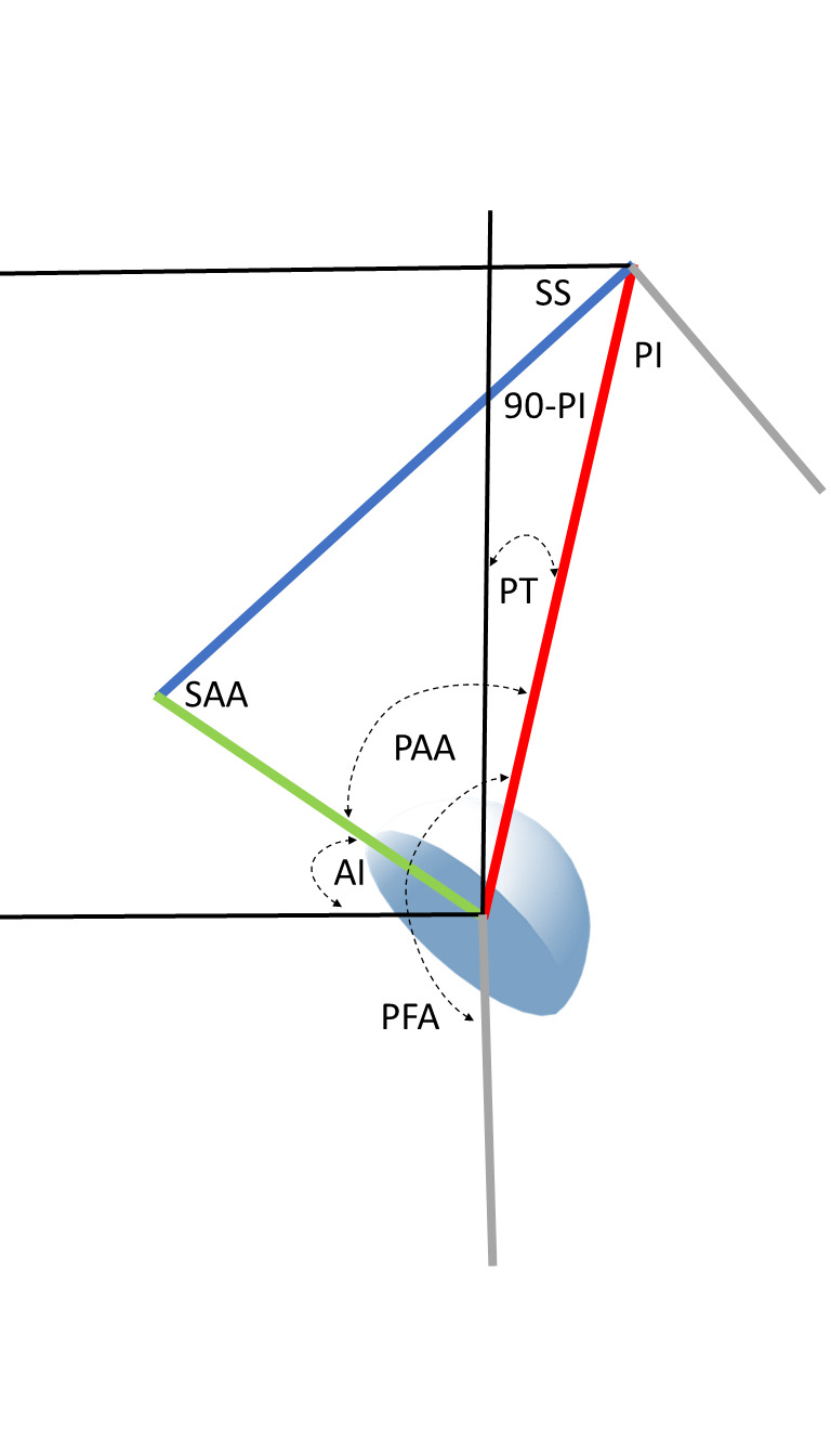
It is important for both hip and spine surgeons to understand the determinants of safe acetabular cup position as procedures in both specialties may substantially alter the orientation and stability of hip replacement components (Lazennec, Brusson, and Rousseau 2011; Esposito et al. 2015; Buckland et al. 2015; Esposito et al. 2018; DelSole et al. 2017; Heckmann et al. 2018; Buckland et al. 2017; Grammatopoulos et al. 2019; Bernstein et al. 2019; Innmann et al. 2022). Research has shown the functional behavior of the hip joint is accomplished through integration between the hip, including the femur and acetabulum, and the lumbar spine as together they coordinate balanced posture in different positions (Heckmann et al. 2018; Ferrero, Liabaud, Challier, Lafage, et al. 2016; Lazennec et al. 2004; Heckmann et al. 2021). The total hip planning paradigm was fashioned upon coronal anatomic reconstruction and until recently did not address the dynamic orientation of the components. Spinopelvic studies have correlated cup orientation with dislocation by measuring sagittal parameters associated with standing pelvic imbalance and mobility abnormalities to a sitting pose (Heckmann et al. 2018, 2021; Grammatopoulos et al. 2022). The biomechanical relationship of sagittal functional cup orientation to pelvic parameter measurements was described by Lazennec (Lazennec et al. 2004), the Anteinclination (AI) cup angle clinically developed by Dorr (Stefl et al. 2017), and recently geometrically modelled (Bodner 2022). The primary relationship employs two horizontally referenced functional angles, the Sacral Slope of S1, (SS), and the sagittal AI cup angle (Kanawade, Dorr, and Wan 2014), to algebraically define an optimizable integrated hip-spine angle, the Sacroacetabular Angle, (SAA) (Lazennec et al. 2004) (Figure 1). However, the hip joint and cup also share an intrinsic functional relationship to the vertically referenced spinal Pelvic Tilt, (PT), defined as the Pelvic Acetabular Angle, (PAA) (Bodner 2022).

Figure 1a
Figure 1a.The geometric pelvic-acetabular construct with central triangle integrating acetabular angles Anteinclination (AI), Sacroacetabular Angle (SAA), Pelvic Acetabular Angle (PAA) to pelvic angles Pelvic Incidence (PI), Sacral Slope (SS), Pelvic Tilt (PT). The hip position is measured through the Pelvic Femoral Angle (PFA). The functional safe zone measure, Combined Sagittal Index, CSI, is defined as CSI=AI+PFA. Pelvic tilt (red line) change affects the spatial position of the center of the S1 endplate, mobility the length of the SS triangle side (blue line), each of which independently alter the AI cup angle (green line).
Figure 1b
Figure 1b.Sitting clinical postoperative lateral radiograph illustrating the complete construct angles described in Figure 1. The highlighted 62° critical angle (yellow) is the cup Anteinclination, AI (sitting range 55°+/- 10°). PI= 45.2, (90-PI)=44.7, SS=9.0, PT=36.6, SAA=71.2, PAA=64.0, PFA=127.1. SAA+PAA+(90-PI) the 3 corners of the triangle always equal 180°, SS+PT always equal PI, PFA+AI= CSI, the validated functional safe zone parameter replacing the Lewinnek zone, here 127.1+62.2=189.3, well above the published limit of 151 and mean value of 180°.

Spinal procedures addressing degeneration and deformity may alter both standing PT and functional pelvic mobility when correcting for sagittal imbalance from loss of lordosis in the lumbar spine, thus there needs to be a protocol to analyze if spinal surgery correction will place a hip replacement at risk for instability. This addresses the decision tree as to the proper order of operation in hip-spine patients, which has been based simply on clinical symptomatology or outcomes and not inclusive of understanding the biomechanical effects of one operation on the other. The purpose of this investigation is to expand upon the existing sagittal cup planning methodology employing Sacral Slope sided calculations to define pelvic-cup mechanics through the spinal PT and solely hip-based angles that integrate with spinal surgery planning principles, utilize these parameters in a novel fashion to determine a biomechanically optimum AI cup position, and provide quantitative risk analysis for the potentially deleterious effects of spinal fusion surgeries on established THA cup positions. Secondarily, we attempt to provide a common biomechanical language for clinically relevant planning discussion between the two specialties (Chevarria, Douleh, and York 2021). Finally, we demonstrate the power of a geometrical sagittal hip-spine construct as a biomechanical tool and illustrate 4 applications addressing surgical spinopelvic related questions solvable with mathematical precision.

Methods

This is a descriptive mathematical study of the sagittal plane functional mechanics of the spinopelvic postural system including a hemispherical acetabular component. The cup may be incorporated into the spinopelvic system using a triangular parameter construct created by combining 2 known angular equations. Firstly, the pelvic parameters are related by the equation defining Pelvic Incidence, PI, a morphologic and functional descriptor of pelvic construction (Legaye et al. 1998). Algebraically, the constant PI equals the sum of the reciprocal functional parameters SS and PT as PI=SS+PT. The acetabular parameters are related by the equation defining the Sacroacetabular Angle of Lazennec, (SAA), as SAA=SS+AI (Lazennec et al. 2004). Superimposing these 2 equations on the sagittal plane of the pelvis reveals the central triangular structure, (Figure 1) connecting three points in space, the anatomic hip and S1 endplate centers and the SAA angle vertex at the intersection of the cup AI tangent line and the SS line (Bodner 2022). The 3 angles created inside this triangle are the anatomically fixed apex angle of (90°-PI), the SAA angle and at the hip joint itself, the Pelvic Acetabular Angle, (PAA), relating the AI cup angle to the PT angle in a similar fashion as the SAA relates the cup to the SS angle. In any individual, SAA and PAA behave reciprocally just as their underlying constituents, SS and PT do. The hip joint combined flexion/extension parameter, Pelvic Femoral Angle, PFA, is measured from S1 center to hip center then continuing parallel to the anterior femoral cortex towards the knee center (Ike et al. 2018).

Applications of the Triangular Model

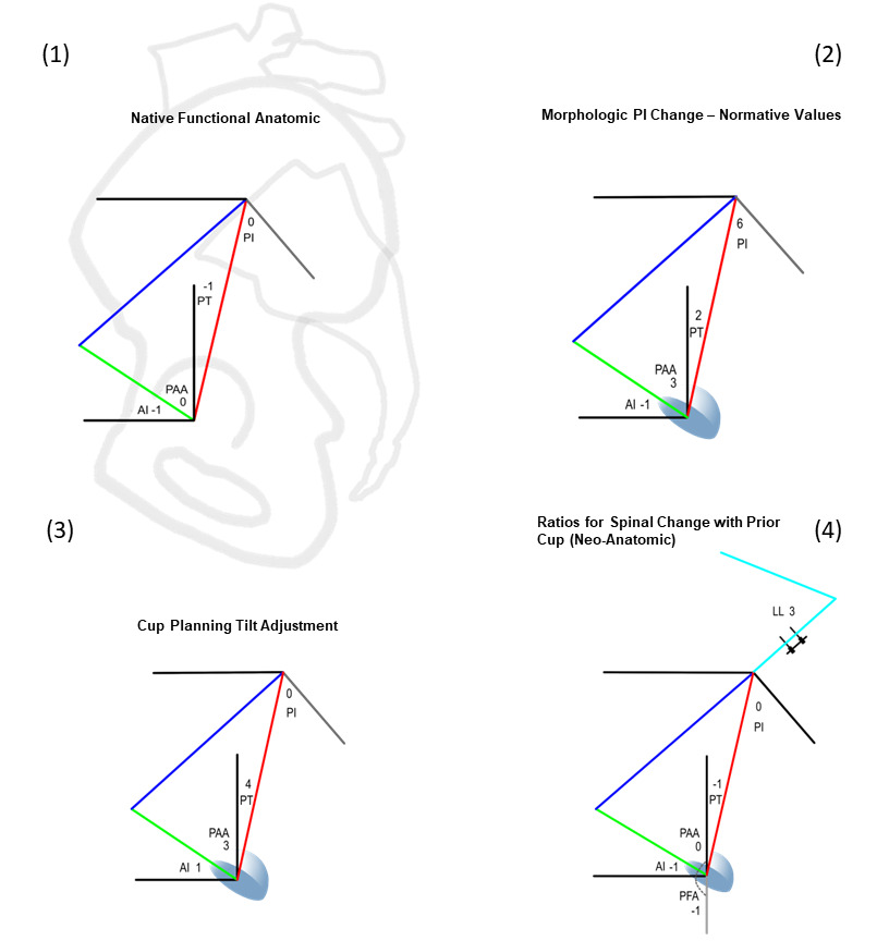
The integration of the pelvic parameters used in spinal surgery, PI, SS, and PT with the sagittal cup angle AI and the angular relationships of the cup AI to the SS (SAA) and to the PT (PAA) function as a tool to answer different spinopelvic questions. This is accomplished by inserting the angular relationships between the parameters found in any physical situation, such as in the native pelvis where all the mobile parameters are fixed within the pelvic bone, thus SS, PT, AI, even the extraneous APP all move in a 1:1 fashion and PI is a constant. 4 different applications are shown in Figure 3 as the relationships in native anatomy, the normative relationships for any PI value, the relationships for calculating a tilt adjusted AI cup position, and the relationships when spinal surgery repositions the pelvis with a preexisting cup already in place. For simplicity, only the angular ratios for parameters crossing the hip joint associated with the PT side of the pelvis are shown, novel development of PT based parameters is the focus of this work.

Figure 2
Figure 2.Hip joint specific spinopelvic-acetabular angles. The cup AI, the planning angles PT, PAA and the combined hip flexion/extension angle PFA. PT and PI share the anatomically fixed line (red) connecting the centers of the hip axis and the S1 endplate, PAA describes the cup’s relationship to the PT through its complementary angle, PAA=(90-AI)+PT, alternatively, 90-(AI-PT), utilizing the fundamental quotient bridging the cup AI angle to that of the moving pelvis. AI is the sagittal cup inclination angle composed of operative plane anteversion with an elliptical inclination contribution. An optimal stand/sit pairing for PI=60°, pelvic mobility 25° is shown with the 30° standing cup AI increasing to 55° sitting identically with the pelvic movement.
Figure 3
Figure 3.The four unique applications of the triangular construct considering solely the distinct hip-based ratios of angular change. Ratios are determined by parameters being fixed/mobile under the specific condition being studied.

(1) In the native pelvis, all relationships are anatomically fixed and positional parameters PT, AI move 1°:1°.

(2). Determining normative parameter values for changing PI angles requires an angular ratio describing the relationship of the pelvic parameters PI:PT, here 3:1, if PI increases by 6°, PT raises 2°. When SAA and PAA share an equal relationship to the cup AI angle this creates an isosceles condition and AI becomes discoverable algebraically.

(3). For Individual cup tilt adjustment, PT and SS, PAA and SAA each become reciprocal, the cup AI passively altered +4°PT:+1°AI, the PAA and (AI-PT) quotient +4° PT:+3° PAA/-3° (AI-PT).

(4). Once impacted, the cup forms a new anatomic relationship with the pelvis. Any changes to pelvic tilt from spinal surgery alters PT, and AI in a 1°:1° fashion. As in native anatomy, PI, PAA, and (AI-PT) remain fixed constant angles creating discrepancy between optimal orientation pre and post spinal surgery. The lumbar lordosis conversion ratio is taken from Buckland et al (Buckland et al. 2015).

Normative values for pelvic and acetabular parameters

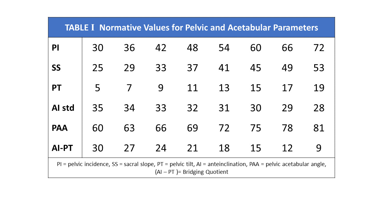
AI, SAA and PAA normative parameters have been adopted using linear equations describing normalized SS and PT relationships for any given PI (Bodner 2022). These equations, SS=0.67PI+5 and PT=0.33PI-5 define that for every 10° change in PI, a normal SS changes 6.67° and PT 3.33°, describing angular ratios of change as 3:2:1 for PI:SS:PT.. Normative values are defined when the cup shares an identical angular relationship to both SS and PT. At this orientation, the triangle’s acetabular angles SAA and PAA become equal at (90+PI)/2 each, this balanced hip-pelvis state creating an isosceles form for the triangle. This value is valid for a static standing position and remains unchanged only when the pelvic mobility between standing and upright seated postures is 25°. The resultant AI cup angle, the sole remaining unknown in the system becomes discoverable as a simple algebraic calculation using either SAA or PAA (Table 1).

Table 1
Table 1.Normative architectural values for all sagittal plane parameters based on a functional normative pelvic parameters relationship defined as SS=0.67PI+5, PT=0.33PI-5, a 3:2:1 ratio of angular change for PI:SS:PT. This creates an equal relationship between cup and the SS/PT values forming an isosceles triangle solving for both the unknown SAA/PAA angles as (90+PI)/2 and AI as SAA-SS or 90-(PAA-PT). Whole number parameter ratios across the table rows are 6:4:2:​-1:3:-3 for PI:SS:PT:AI:PAA:(AI-PT).

The Sagittal Parameters of the Hip Joint

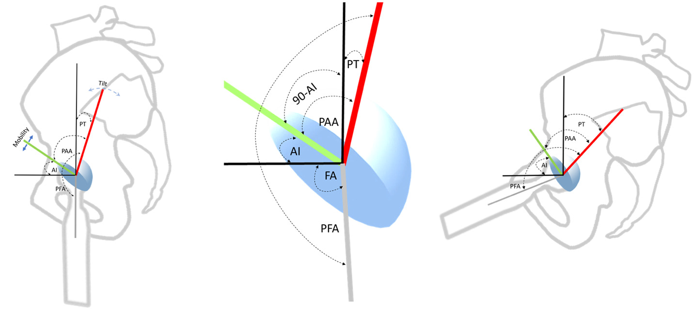
There are 5 angles with their apices at the hip joint that may be analyzed for individualized cup planning or understanding alterations caused by spinal realignment (Figure 2). From superior to inferior, they are PT, (90°-AI), PAA, AI, and PFA. Involuntarily modulating PT represents the body’s main mechanism to balance the trunk and head as they incline forward with aging and deterioration (Barrey et al. 2013; Diebo et al. 2015). PFA represents the collective orientations of the PT angle above and the femoral shaft angle below the hip center, it is intimately correlated to the truncal inclination parameter, the T1 Pelvic Angle, (TPA), thus PFA reactively counterbalances the sagittal spinal imbalance above the hip through flexion/extension of the joint itself (Hasagawa et al. 2016). Adding the calculated AI to the PFA in standing and sitting gives the validated functional safe zone parameter for both acute and chronic dislocation, Combined Sagittal Index, CSI (Heckmann et al. 2018; Grammatopoulos et al. 2022). PAA is algebraically the sum of (90-AI) + PT, the complementary angle to AI is necessary as a consequence of PT being vertically referenced.

Transition from SS based to PT calculation

Functional cup positions have been determined by the spatial position of the pelvis in standing and upright sitting using SS angles measured at the S1 endplate (Kanawade, Dorr, and Wan 2014). Calculations are geometrically simple using the SS as it shares the horizontal reference with AI, (SAA=AI+SS). Determination using the PT requires employing complementary angles including (90-AI), a constituent of the PAA, as again PAA=(90-AI)+PT. Rewriting the equation, PAA=90-(AI-PT) uses the very same bridging cup to pelvis quotient as found in the SAA equation, SAA=PI+(AI-PT).

The quotient (AI-PT) represents a mathematical bridge between the surgically created cup AI angle and the measured PT angle, linking a patient’s intrinsic pelvic morphology and postural compensations to a potentially ideal orientation for the cup. This model has shown a 1° change to PT alters an optimally planned AI target 0.25° to keep it optimal, a ratio of 1:4, an increased PT gives an increased AI (Figure 3). Applying this ratio to the PAA equation solves that for +1° PT change, the PAA angle will increase 0.75°, as PAA reflects the cup’s relationship to the increased PT. SAA decreases reciprocally to preserve the integrity of the central 180° triangle.

Employing PT, cup tilt adjustment is calculated for every 1° the measured PT diverges from the normalized position; in similar fashion to coronal tilt adjustment performed for divergence from a neutral APP pelvic position. The ideal standing AI becomes discoverable by Standing AI= 90-(PAA-PT). Finally, 90-(AI-PT) will confirm the individualized tilt corrected PAA angle and its reciprocal SAA angle.

Functional Mobility Effects on Acetabular Parameters

Functional analysis utilizing the standing and sitting positions requires optimizing the cup’s orientation to an individual’s arc of pelvic excursion as measured by the delta pelvic mobility. Traditionally measured by the Sacral Slope change between standing and sitting, this excursion is identical using PT change. Mobility has a direct effect on the AI, SAA, and PAA angles (Figure 4), the effect increases or decreases the cup’s arc of excursion in similar fashion to opening and closing a hand fan, a 1° change alters each end of the fan by 0.5° with stiffness raising the standing AI and decreasing the sitting AI. Geometrically, mobility of 25° creates an isosceles triangle, the length of the modifiable apex-SS side matching the anatomically fixed S1-hip center side. Stiffness shortens the SS side of the triangle from the equal isosceles length in both postures. Each 1° loss in mobility increases both the SAA and the standing AI angles 0.5° and decreasing the sitting AI 0.5°. Increased mobility has the opposite effect. Mobility effects are constant across all spinopelvic applications, whether calculating for normative values or tilt adjusted cup positions using either SS or PT.

Figure 4
Figure 4.Case example of cup planning method and calculating effects of spinal intervention.

a. THA planning algorithm: measurements on presentation, PI 60°, PT stand/sit 27/42°, PFA 196/129°, mobility-delta 15°. The variance from normative PI 60 values is 12° increased PT tilt and 10° decreased mobility. Calculating using Application 3 ratios- standing AI increases 3° (12 x 0.25) for tilt adjustment, +5° for mobility (10 x 0.5) to 38°, sitting becomes 53°, (AI-PT) becomes 38-27=11° from the normative 15° (30-15°) and PAA becomes 90-11=79°.

b. Effect of spine surgery with 5° PT correction: PAA, (AI-PT) are fixed in a neo-anatomic fashion at 79° and 11° (Application 4), mobile parameters now change 1°:1°, 1° tilt correction retroverts AI 0.75° from the ideal 0.25° used in optimal cup planning. AI is lowered to 33/48° from 38/53° with the optimal cup orientation recalculated using Application 3 as 36.75/51.75°, (AI-PT)=14.75° and PAA=90-14.75=75.25°. The surgical change retroverts the cup AI 3.75° from its new optimum, coincidently, -5° PT also retroverts the cup 3.75° in absolute radiographic plane anteversion.

c. Additional effect of spine surgery decreasing stand/sit mobility from 15° to 9°: PT stand/sit becomes 22/31°, AI 33/42°. Recalculated optimal AI adds +3° to standing, -3° to sitting becoming 39.75/48.75°, the actual AI position is 6.75° retroverted to its new optimal (5° radiographic retroversion) In sitting, the cumulative AI effect of spine surgery is additive, a 53° AI pre-fusion position altered to 42°, an 11° AI change (8° radiographic coronal retroversion) without consideration of further risk enhancement by compensatory femoral hyperflexion.

Results

PT based cup calculations- Rule of 4s

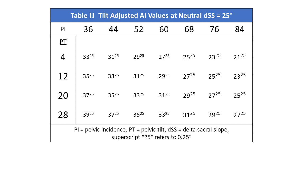
Tilt adjusted AI cup positions based on preoperatively measured PI and PT values are demonstrated in Table 2. The ratio of angular change between the cup AI and PT is identical to using SS, 1° to 4°, the correction from normative values may be calculated either using the variance of measured PT from the PI normal values or the PI variance from the PI for which that measured PT would be normal. This Rule of 4s defines when the PT value is 4° greater than the normative PT value for the measured PI, the ideal cup AI orientation will be 1° greater than the normative AI value, and as the PI for any measured PT increases 4°, the ideal cup AI decreases 1°.

Table 2
Table 2.Tilt Adjusted normative Standing AI cup values by Pelvic Incidence at 25° Pelvic Mobility, illustrating the Spinopelvic rule of 4s. PT expresses a 4°:1° direct relationship to optimal AI cup orientation (columns), PI 4°:1° inversely at constant PT (rows).

Effect of Mobility on Calculated AI cup angles

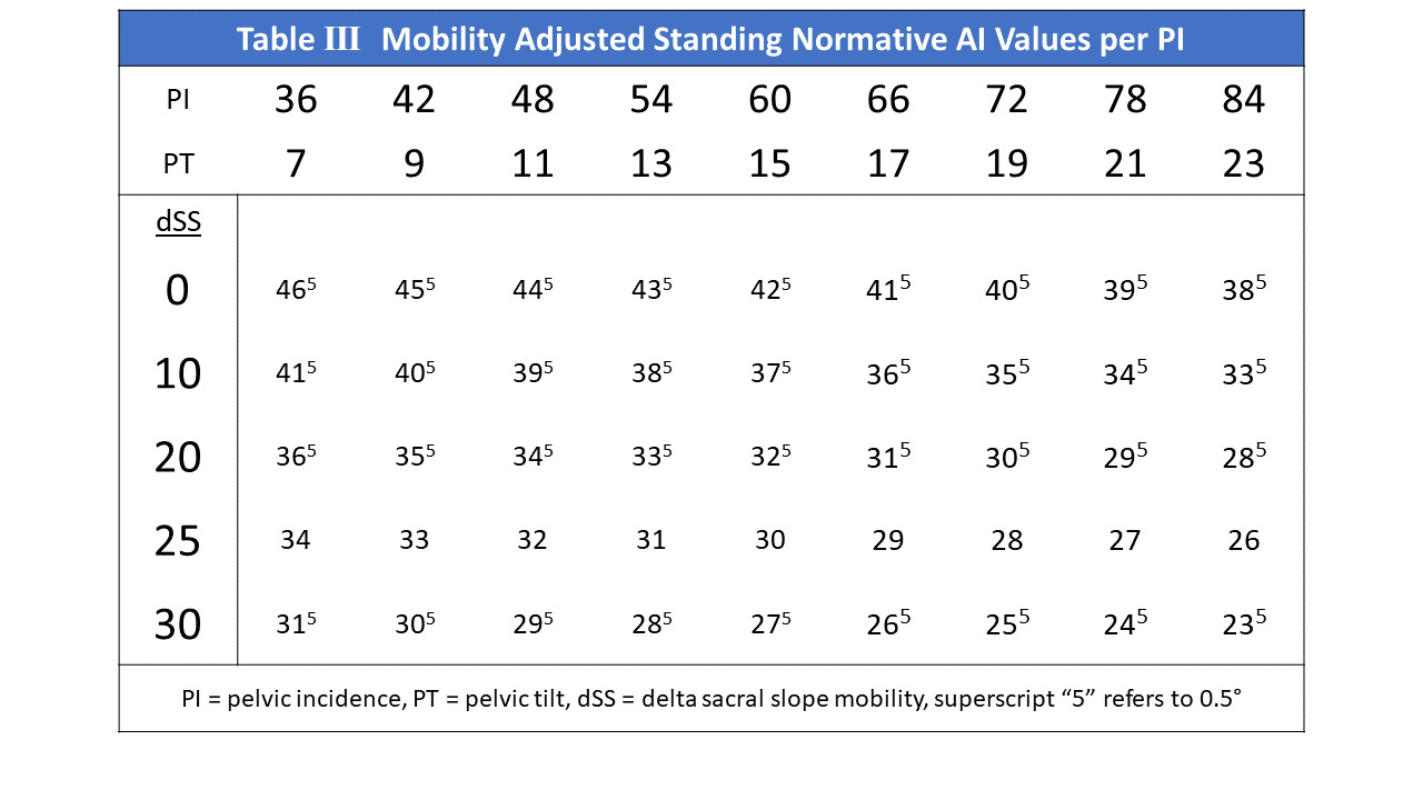
Table 3 demonstrates the mobility adjusted AI cup angles for normative PT values for any PI. The normative values creating the isosceles triangle condition are those in the 25° mobility row. As explained, a 1° increase in mobility will decrease the standing AI 0.5° for any PI and PT, a 6° increase in PI and 2° increase in PT will decrease the optimal AI 1° in standing. Mobility has the same effect on the AI value in the (AI-PT) quotient; for a given PT, a 1° increase in mobility lowers the AI 0.5° lowering this quotient by 0.5°. This results in increasing the PAA 0.5°, decreasing SAA 0.5° reciprocally.

Table 3
Table 3.Mobility adjusted Standing AI cup orientation at normalized pelvic tilt angles by PI. Mobility creates an arc of excursion with a 2° change altering both the standing and sitting cup angles 1° in opposing directions. Thus, as mobility increases the standing AI decreases (columns), PI:SS:PT:AI exhibiting ratios of change as 6:4:2:-1 (rows).

Solution for tilt adjustment of the (AI-PT) quotient

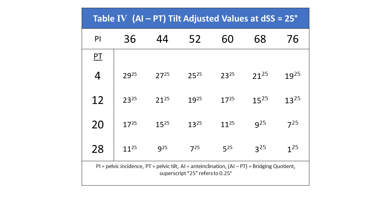
The relationship between an ideal cup orientation and the changing pelvic tilt creates a quotient of (0.25-1) = -0.75° for each 1° increase in PT (Table 4). A 1° PI increase for any given PT value decreases (AI-PT) by -0.25°. This adjusted quotient may be subtracted from 90° to produce the PAA angle, or added to the PI angle to produce the tilt adjusted SAA angle.

Table 4
Table 4.Normative tilt adjusted values for the bridging quotient (AI-PT) over the PI range. The quotient changes in an inverse 3:4 ratio to the PT change. SAA and PAA change 3° reciprocally as PT changes 4°. It is the combination of the 4°:1° PT:AI tilt ratio with the 2:1 mobility effect that quantitatively determines functional cup adjustments from normative values.

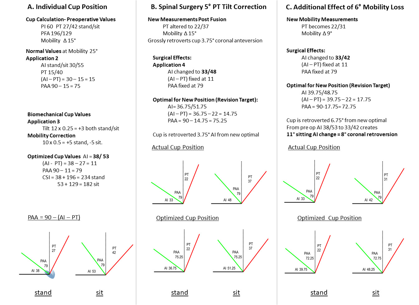
The full sequence of determining biomechanically optimal acetabular parameters, including cup AI, (AI-PT), PAA, and the CSI using PI/PT/PFA preoperative measurements is illustrated in Figure 4, column A.

Effect of Spinal Reconstruction on Cup Orientation

After cup implantation, the SAA and PAA angles become immutably fixed, creating a neo-anatomic state, the fourth spinopelvic construct application (Figure 3(4)). All changes in PT tilt and mobility angles alter AI in a 1:1° fashion instead of the tilt adjustment 4:1° ratio employed in the optimal planning application. For an optimally implanted cup, a 1° decrease in PT then retroverts the cup AI 0.75° (~0.55° radiographic anteversion at inclination of 45°) in both the standing and sitting postures from the postoperative orientation to keep it optimal (Figure 4, column B). Likewise (AI-PT) remains fixed, remaining 0.75° greater than ideal per 1° PT surgical decrease. Additional retroverting effect occurs sitting for each degree of stand/sit pelvic mobility lost from spinal fusions, magnifying this position’s risk. Each 1° loss in mobility retroverts the AI sitting position 0.5° from the ideal orientation, the combined effects of tilt and mobility are additive in sitting while tilt and mobility effects oppose in standing (Figure 4, column C). Thus, a 1° PT correction combined with a 1° loss of mobility retroverts AI 1.25° beyond the new sitting ideal, (~ 0.9° radiographic anteversion loss).

Case Example- Step by step case employing this methodology (Figure 4).

Column A- Calculation of Biomechanically Optimized Cup Orientation

  1. Measure patient PI, PT, PFA stand/ sitting: PI 60, PT 27/42, delta SS= 15°, PFA 196/129

  2. Compare PT standing to normative PT for PI 60: 27 vs 15, a +12° PT variance

  3. Apply 4:1 Tilt correction:

    • +12° PT correction= +3° standing AI correction (Rule of 4s) from normative AI of 30°= 33° AI standing

    • (AI-PT) standing correction = -0.75 x 12= 9° Normative for PI 60= (30-15) =15° 15-9= 6°

  4. Mobility correction:

    • 10° stiffness correction from neutral 25° to measured 15° dSS applies +5° to standing AI, -5° to sitting AI, +5° to (AI-PT). Therefore, the targeted AI becomes 38/53° stand/sit, (AI-PT) becomes 11° making SAA 60+11=71° and PAA 90-11=79°. Sitting (AI-PT) confirms (53-42)=11°.
  5. CSI calculation: CSI=AI+PFA stand/sit. Standing limit <244° for posterior impingement, >151° for anterior impingement

Stand- 38+196=234°, Sit- 53+129=182°, this cup is safe within limits.

Column B- Effect of lowering PT 5° through spinal surgery:

  1. A. PT lowered to 22° without mobility change. The cup AI lowers directly to 33/48° stand/sit. The optimal cup orientation for 22° PT should be 36.75/51.75°, AI delta 3.75° in both postures. The net effective retroversion of the cup is 0.75° lower AI than ideal for every 1° PT is surgically decreased, (0.55° coronal plane anteversion), the cup has been retroverted 3.75° surgically and is now 2° retroverted to its coronal ideal. Once implanted, (AI-PT) remains constant as cup and pelvis now move 1:1.

Column C- Effect of Additional 6° loss of pelvic mobility

  1. The cup AI becomes 33/42° stand/sit. The optimal orientation should be 39.75/48.75. In toto, cup AI has been altered from initial AI 38/53 ° to 33/42 by the combined spinal surgery effects, 5 and 11° of AI retroversion change, a coronal sitting anteversion loss of approximately 8°, and 5° of coronal retroversion from its new ideal cup orientation.

Discussion

Balanced postures are governed by control of the body’s line of gravity in the sagittal plane, requiring coordinated movements between the spine-pelvis-hip complex (Lazennec, Brusson, and Rousseau 2011; Dubousset 1994; Schwab et al. 2006). Positional changes to sit or bend require considerable mobility between these regions, accomplished through pelvic rotation about the hip joint, spinal shape change, and horizontal translations called shifts (Stefl et al. 2017; Diebo et al. 2016; Ferrero, Liabaud, Challier, Lefage, et al. 2016; Lazennec et al. 2020). As functional awareness grows in both hip and spine surgery, it becomes crucial to develop a common language in which procedures on one side of the pelvis can be understood in terms of effects on the other. This is difficult with the sagittal standing priority in spinal planning and the supine coronal plane paradigm in THA (Masquefa et al. 2015; Liu et al. 2019) APP-derived tilt adjustment in THA planning has been an accepted method of coordinating cup position to that of the pelvis. Intrinsically however, these relationships are quantitatively defined by the equations for the SAA and PAA angles, determining a functionally constant optimal angular relationship of a hemispheric cup to measured or predicted SS and PT values respectively. Postural control has been shown to be modified through PT and PFA, the latter highly correlated with the spinal T1 Pelvic Angle of truncal inclination, (TPA), and PT is an important parameter within corrective spinal planning (Lafage et al. 2009; Protopsaltis et al. 2014). The quotient, (AI-PT) represents a bridge between the tilt and mobility sensitive cup orientation and the functional morphology and orientation of the pelvis, adding (AI-PT) to the PI produces SAA or subtracting it from 90° produces PAA. The key biomechanical concept is the angular ratio of the cup change to pelvic position change differs when planning to place a component freely into the pelvis than when it moves within the pelvis after it is implanted. Acetabular implantation creates new fixed anatomic relationships, functioning as the adjacent segment to the lumbar spine and sensitive to subsequent spinal surgical changes altering pelvic tilt and/or mobility. In a sense, spinal surgery can create an unwitting THA revision of cup orientation and function.

This triangular model allows individualized AI cup calculation through either the SS or PT value, alterations from normative values derived by the tilt adjustment rule of 4s, 4° of SS or PT change will alter an ideally placed cup 1° in both standing and sitting, with mobility’s 2:1 effect of 2° alteration changing standing and sitting AI each 1° in opposing directions. For determining optimal cup position, working through the SS value is simpler geometrically as AI and SS are both measured from parallel horizontal reference lines. But it is the PT value that integrates the hip to the gravity management system, which can be directly used to determine effects on cup position of spinal procedures.

The hip joint is the pivot point for postural pelvic compensatory rotation, the pelvis rotating back adjusting for truncal inclination after which femur and knee flexion can be recruited (Barrey et al. 2011; Lazennec, Brusson, and Rousseau 2013). Anterior pelvic rotation is typically caused by hip flexion contractures. The sum of these compensations may be measured through the PFA hip joint angle as it includes both the PT above and the femoral angle below. A high standing PFA indicates a hyperextended hip, a low sitting indicates a hyperflexed hip, the spinopelvic risk factors for THA impingement leading to instability (Heckmann et al. 2018; Grammatopoulos et al. 2019). The CSI, (AI+PFA), describes how safe optimal cup orientation may be determined through hip-based angles, avoiding impingements from improper cup orientation while including the non-modifiable spinopelvic state (Tezuka et al. 2019). This analysis supports prior work recommending sagittal plane workups in any patient with history of back pain or surgery (Vigdorchik et al. 2019; Grammatopoulos et al. 2023), provides a method of personalized cup orientation based on a patient’s individual biomechanics and provides a basis for development of predictive models for spinal fusion risk as this could provide improved indications for pre-fusion higher constraint acetabular components in at risk primary THA patients.

Discussion regarding sequencing the proper order of operation in patients requiring hip and spine procedures has been framed by the presence of neurological symptoms, flexion contracture, and primary location of pain, there has not been a biomechanical basis to fully interpret the effects or warn of postoperative hip instability following spinal procedures (Malkani et al. 2019; Parilla et al. 2019). Using AI and the SAA/PAA, the effects of lordosis restoring PT reduction on AI cup orientation become quantitative. Spine surgery decreases PT and reduces the mobility of the lumbosacral articulation, both retroverting an implanted cup and increasing the sitting position impingement risk. This PT tilt adjustment retroverts a cup AI 0.75° from ideal for every degree the pelvis is repositioned compounded by any mobility loss leaving the sitting cup AI another 0.5° retroverted for every lost 1° of mobility. However, repositioning an overly anteverted cup in standing may actually improve the construct orientation.

These findings have significant clinical ramification, particularly for spine surgeons planning longer segment lordosis producing spinal fusions (changing spinal tilt) and those crossing the lumbosacral junction (producing pelvic stiffness) in patients with preexisting hip replacements. The sitting position retroversion produced may destabilize a hip that has lower preexisting anteversion, this can be analyzed preoperatively through a sitting lateral view of the pelvis. AI cup angles near the established sitting lower limit of 45° (Tezuka et al. 2019) can stimulate a discussion with the patient or referral to a hip surgeon to discuss risk associated with THA instability. Additionally, if a significant change in PT or multilevel fusion planned, this should be performed prior to a hip replacement and then the hip planned based on the restored pelvic position and residual mobility measures. Integrated planning software automating the risk analysis is the possible future application of this analysis.

The limitations of this analytic model lie in its assumptions and theoretical nature. The adopted equation for the pelvic parameters produces SS/PT values within 1° of published equations derived in spinal literature for the standing position (Bodner 2022). The 25° mobility neutral point defies explanation mathematically but the AI cup positions produced mirror those published by multiple authors (Tezuka et al. 2019; Grammatopoulos et al. 2023). Clinical studies specifically validating this planning algorithm are needed, however due to its sub-1° accuracy, assistive technology would be essential to achieve this type of precision.

In conclusion, spinopelvic relationships linking spine and hip planning parameters may be integrated through geometric representation of the pelvic and acetabular angles. Biomechanically optimal cup orientation may be determined using either the SS or PT angles, and geometry allows understanding the differences between planning for cup position and the effects of changing the orientation of a fixed cup’s position through spinal surgery. Though cup planning is simpler using SS values, it is through the PT side and quotient (AI-PT) that a direct relationship between the functional influences of the two specialties intersect.

Submitted: October 29, 2025 EDT

Accepted: December 06, 2025 EDT

References

Barrey, C., P. Roussouly, J. C. Le Huec, G. D’Acunzi, and G. Perrin. 2013. “Compensatory Mechanisms Contributing to Keep the Sagittal Balance of the Spine.” Eur Spine J 22 (suppl 6): 834–41. https:/​/​doi.org/​10.1007/​s00586-013-3030-z.
Google Scholar
Barrey, C., P. Roussouly, G. Perrin, and J.C. Le Huec. 2011. “Sagittal Balance Disorders in Severe Degenerative Spine. Can We Identify the Compensatory Mechanisms?” Eur Spine J 20 (Suppl 5): S626-33. https:/​/​doi.org/​10.1007/​s00586-011-1930-3.
Google Scholar
Bernstein, J., R. Charette, M. Sloan, and G.C. Lee. 2019. “Spinal Fusion Is Associated with Changes in Acetabular Orientation and Reductions in Pelvic Mobility.” Clin Orthop Relat Res 474 (8): 1788–97. https:/​/​doi.org/​10.1097/​CORR.0000000000000390.
Google Scholar
Bodner, R. J. 2022. “The Functional Mechanics of the Acetabular Component in Total Hip Arthroplasty.” J Arthroplasty 37 (11): 2199–2207. https:/​/​doi.org/​10.1016/​j.arth.2022.05.017.
Google Scholar
Buckland, A. J., V. Puvanesarajah, J. Vigdorchik, R. Schwarzkopf, A. Jain, E. O. Klineberg, R. A. Hart, J. J. Callaghan, and H. Hassanzadeh. 2017. “Dislocation of a Primary Total Hip Arthroplasty Is More Common in Patients with a Spinal Fusion.” Bone Joint J 99-B (5): 585–91. https:/​/​doi.org/​10.1302/​0301-620X.99B5.BJJ-2016-0657.R1.
Google Scholar
Buckland, A. J., J. M. Vigdorchik, F. J. Schwab, T. J. Errico, R. Lafage, C. Ames, S. Bess, J. Smith, G. M. Mundis, and V. Lafage. 2015. “Acetabular Anteversion Changes Due to Spinal Deformity Correction: Bridging the Gap between Hip and Spine Surgeons.” J Bone Joint Surg Am 97 (23): 1913–20. https:/​/​doi.org/​10.2106/​JBJS.O.00276.
Google Scholar
Chevarria, J. C., D. G. Douleh, and P. J. York. 2021. “The Hip-Spine Challenge.” J Bone Joint Surg Am 103 (19): 1852–60. https:/​/​doi.org/​10.2106/​JBJS.20.01728.
Google Scholar
DelSole, E. M., J. M. Vigdorchik, R. Schwarzkopf, T. J. Errico, and A. J. Buckland. 2017. “Total Hip Arthroplasty in the Spinal Deformity Population: Does Degree of Sagittal Deformity Affect Rates of Safe Zone Placement, Instability, or Revision?” J Arthroplasty 32 (6): 1910–17. https:/​/​doi.org/​10.1016/​j.arth.2016.12.039.
Google Scholar
Diebo, B. G., E. Ferrero, R. Lafage, V. Challier, B. Liabaud, S. Liu, J. M. Vital, T. J. Errico, F. J. Schwab, and V. Lafage. 2015. “Recruitment of Compensatory Mechanisms in Sagittal Spinal Malalignment Is Age and Regional Deformity Dependent: A Full-Standing Axis Analysis of Key Radiographical Parameters.” Spine (Phila Pa 1976) 40 (9): 642–49. https:/​/​doi.org/​10.1097/​BRS.0000000000000844.
Google Scholar
Diebo, B. G., J. H. Oren, V. Challier, R. Lafage, E. Ferrero, S. Liu, S. Vira, et al. 2016. “Global Sagittal Axis: A Step toward Full-Body Assessment of Sagittal Plane Deformity in the Human Body.” J Neurosurg Spine 25 (4): 494–99. https:/​/​doi.org/​10.3171/​2016.2.SPINE151311.
Google Scholar
Dubousset, J. 1994. “Three-Dimensional Analysis of the Scoliotic Deformity.” In Pediatric Spine: Principles and Practice, edited by S. L. Weinstein, 479–83. New York, NY: Raven Press.
Google Scholar
Esposito, C. I., K. M. Carroll, P. K. Sculco, D. E. Padgett, S. A. Jerabek, and D. J. Mayman. 2018. “Total Hip Arthroplasty Patients with Fixed Spinopelvic Alignment Are at Higher Risk of Hip Dislocation.” J Arthroplasty 33 (5): 1449–54. https:/​/​doi.org/​10.1016/​j.arth.2017.12.005.
Google Scholar
Esposito, C. I., B. P. Gladnick, Y. Y. Lee, S. Lyman, T. M. Wright, D. J. Mayman, and D. E. Padgett. 2015. “Cup Position Alone Does Not Predict Risk of Dislocation after Hip Arthroplasty.” J Arthroplasty 30 (1): 109–13. https:/​/​doi.org/​10.1016/​j.arth.2014.07.009.
Google Scholar
Ferrero, E., B. Liabaud, V. Challier, R. Lafage, B. G. Diebo, S. Vira, S. Liu, et al. 2016. “Role of Pelvic Translation and Lower-Extremity Compensation to Maintain Gravity Line Position in Spinal Deformity.” J Neurosurg Spine 24 (3): 436–46. https:/​/​doi.org/​10.3171/​2015.5.SPINE14989.
Google Scholar
Ferrero, E., B. Liabaud, V. Challier, R. Lefage, B.G. Diebo, S. Vira, S. Liu, et al. 2016. “Role of Pelvic Translation and Lower-Extremity Compensation to Maintain Gravity Line Position in Spinal Deformity.” J Neurosurg Spine 24 (3): 436–46. https:/​/​doi.org/​10.3171/​2015.5.SPINE14989.
Google Scholar
Grammatopoulos, G., A. Falsetto, E. Sanders, J. Weishorn, H. S. Gill, P. Beaule, M. M. Innmann, and C. Merle. 2022. “Integrating the Combined Sagittal Index Reduces the Risk of Dislocation Following Total Hip Replacement.” J Bone Joint Surg Am 104 (5): 397–411. https:/​/​doi.org/​10.2106/​JBJS.21.00432.
Google Scholar
Grammatopoulos, G., W. Gofton, Z. Jibri, M. Coyle, J. Dobransky, C. Kreviazuk, P.R. Kim, and P.E. Beaule. 2019. “2018 Frank Stinchfield Award: Spinopelvic Hypermobility Is Associated with an Inferior Outcome after THA: Examining the Effect of Spinal Arthrodesis.” Clin Orthop Relat Res 477 (2): 310–21. https:/​/​doi.org/​10.1097/​CORR.0000000000000367.
Google Scholar
Grammatopoulos, G., M. Innmann, P. Phan, R. Bodner, and G. Meermans. 2023. “Spinopelvic Challenges in Primary Total Hip Arthroplasty.” EFFORT Open Rev 8 (5): 298–312. https:/​/​doi.org/​10.1530/​EOR-23-0049.
Google Scholar
Hasagawa, K., M. Okamoto, S. Hatsushikano, H. Shimoda, M. Ono, and K. Watanabe. 2016. “Normative Values of Spino-Pelvic Sagittal Alignment, Balance, Age, and Health-Related Quality of Life in a Cohort of Healthy Adult Subjects.” Eur Spine J 25:3675–86. https:/​/​doi.org/​10.1007/​s00586-016-4702-2.
Google Scholar
Heckmann, N., B. McKnight, M. Stefl, N. A. Trasolini, H. Ike, and L. D. Dorr. 2018. “Late Dislocation Following Total Hip Arthroplasty: Spinopelvic Imbalance as a Causative Factor.” J Bone Joint Surg Am 100 (21): 1845–53. https:/​/​doi.org/​10.2106/​JBJS.18.00078.
Google Scholar
Heckmann, N., T. Tezuka, R. J. Bodner, and L. D. Dorr. 2021. “Functional Anatomy of the Hip Joint.” J Arthroplasty 36 (1): 374–78. https:/​/​doi.org/​10.1016/​j.arth.2020.07.065.
Google Scholar
Ike, H., L. D. Dorr, N. Trasolini, M. Stefl, B. McKnight, and N. Heckmann. 2018. “Spine-Pelvis-Hip Relationship in the Functioning of a Total Hip Replacement.” J Bone Joint Surg Am 100:1606–15. https:/​/​doi.org/​10.2106/​JBJS.17.00403.
Google Scholar
Innmann, M. M., J. C. F. Verhaegen, F. Reichel, B. Schaper, C. Merle, and G. Grammatopoulos. 2022. “Spinopelvic Characteristics Normalize 1 Year after Total Hip Arthroplasty: A Prospective Longitudinal, Case-Controlled Study.” J Bone Joint Surg Am 104 (8): 675–83. https:/​/​doi.org/​10.2106/​JBJS.21.01127.
Google Scholar
Kanawade, V., L. D. Dorr, and Z. Wan. 2014. “Predictability of Acetabular Component Angular Change with Postural Shift from Standing to Sitting Position.” J Bone Joint Surg Am 96 (12): 978–86. https:/​/​doi.org/​10.2106/​JBJS.M.00765.
Google Scholar
Lafage, V., F. Schwab, A. Patel, N. Hawkinson, and J. P. Farcy. 2009. “Pelvic Tilt and Truncal Inclination: Two Key Radiographical Parameters in the Setting of Adults with Spinal Deformity.” Spine (Phila Pa 1976) 34:E599-606. https:/​/​doi.org/​10.1097/​BRS.0b013e3181aad219.
Google Scholar
Lazennec, J. Y., A. Brusson, and M. A. Rousseau. 2011. “Hip-Spine Relations and Sagittal Balance Clinical Consequences.” Eur Spine J 20 (5(Suppl 5)): 686–98. https:/​/​doi.org/​10.1007/​s00586-011-1937-9.
Google Scholar
———. 2013. “Lumbar-Pelvic-Femoral Balance on Sitting and Standing Lateral Radiographs.” Orthop Traumatol Surg Res 99 (1 Suppl): S87-103. https:/​/​doi.org/​10.1016/​j.otsr.2012.12.003.
Google Scholar
Lazennec, J. Y., N. Charlot, M. Gorin, B. Roger, N. Arafati, A. Bissery, and G. Saillant. 2004. “Hip-Spine Relationship: A Radio-Anatomical Study for Optimization in Acetabular Cup Positioning.” Surg Radiol Anat 26 (2): 136–44. https:/​/​doi.org/​10.1007/​s00276-003-0195-x.
Google Scholar
Lazennec, J. Y., Y. Kim, D. Folinais, and A. E. Pour. 2020. “Sagittal Spinopelvic Translation Is Combined with Pelvic Tilt during Standing to Sitting Position: Pelvic Incidence Is a Key Factor in Patients Who Underwent Tha.” Arthroplast Today 6 (4): 672–81. https:/​/​doi.org/​10.1016/​j.artd.2020.07.002.
Google Scholar
Legaye, J., G. Duval-Beaupere, J. Hecquet, and C. Marty. 1998. “Pelvic Incidence: A Fundamental Pelvic Parameter for Three Dimensional Regulation of Spinal Sagittal Curves.” Eur Spine J 7 (2): 99–103. https:/​/​doi.org/​10.1007/​s005860050038.
Google Scholar
Liu, N., S.B. Goodman, P.F. Lachiewicz, and K.B. Wood. 2019. “Hip or Spine First? A Survey of Treatment Order for Patients with Concurrent Degenerative Hip and Spinal Disorders.” Bone Joint J 101-B (6 Supple B): 37–44. https:/​/​doi.org/​10.1302/​0301-620X.101B6.BJJ-2018-1073.R1.
Google Scholar
Malkani, A. L., K. J. Himschoot, K. L. Ong, E. C. Lau, D. Baykal, J. R. Dimar, S. D. Glassman, and D. J. Berry. 2019. “Does Timing of Primary Total Hip Arthroplasty Prior to or after Lumbar Spine Fusion Have an Effect on Dislocation and Revision Rates.” J Arthroplasty 34 (5): 907–11. https:/​/​doi.org/​10.1016/​j.arth.2019.01.009.
Google Scholar
Masquefa, T., N. Verdier, O. Gille, L. Boissiere, I. Obeid, C. Maillot, C. Tournier, and T. Fabre. 2015. “Change in Acetabular Version after Lumbar Pedicle Subtraction Osteotomy to Correct Post-Operative Flat Back: EOS Measurements of 38 Acetabula.” Orthop Traumatol Surg Res 101 (6): 655–59. https:/​/​doi.org/​10.1016/​j.otsr.2015.07.013.
Google Scholar
Parilla, F. W., R. R. Shah, A. C. Gordon, S. M. Mardjetko, N. E. Cipparrone, W. M. Goldstein, and J. M. Goldstein. 2019. “Does It Matter: Total Hip Arthroplasty of Lumbar Spinal Fusion First? Preoperative Sagittal Spinopelvic Measurements Guide Patient-Specific Surgical Strategies in Patients Requiring Both.” J Arthroplasty 34 (11): 2652–62. https:/​/​doi.org/​10.1016/​j.arth.2019.05.053.
Google Scholar
Protopsaltis, T., F. J. Schwab, N. Bronsard, J. S. Smith, E. Klineberg, G. Mundis, D. J. Ryan, et al. 2014. “The T1 Pelvic Angle, a Novel Radiographic Measure of Global Sagittal Deformity, Accounts for Both Spinal Inclination and Pelvic Tilt and Correlates with Health-Related Quality of Life.” J Bone Joint Surg Am 96:1631–40. https:/​/​doi.org/​10.2106/​JBJS.M.01459.
Google Scholar
Schwab, F., V. Lafage, R. Boyce, W. Skalli, and J.P. Farcy. 2006. “Gravity Line Analysis in Adult Volunteers: Age-Related Correlation with Spinal Parameters, Pelvic Parameters, and Foot Position.” Spine (Phila Pa 1976) 31 (25): E959-67. https:/​/​doi.org/​10.1097/​01.brs.0000248126.96737.0f.
Google Scholar
Stefl, M., W. Lunergan, N. Heckmann, B. McKnight, H. Ike, R. Murgai, and L. D. Dorr. 2017. “Spinopelvic Mobility and Acetabular Component Position for Total Hip Arthroplasty.” Bone Joint J 99-B (1(Supple A)): 37–45. https:/​/​doi.org/​10.1302/​0301-620X.99B1.BJJ-2016-0415.R1.
Google Scholar
Tezuka, T., N. D. Heckmann, R. J. Bodner, and L. D. Dorr. 2019. “Functional Safe Zone Is Superior to the Lewinnek Safe Zone for Total Hip Arthroplasty: Why the Lewinnek Safe Zone Is Not Always Predictive of Stability.” J Arthroplasty 34 (1): 3–8. https:/​/​doi.org/​10.1016/​j.arth.2018.10.034.
Google Scholar
Vigdorchik, J. M., N. Eftekhary, A. Elbuluk, M. P. Abdel, A. J. Buckland, R. Schwarzkopf, S. Jerabek, and D. J. Mayman. 2019. “Evaluation of the Spine Is Critical in the Workup of Recurrent Instability after Total Hip Arthroplasty.” Bone Joint J 101-B (7): 817–23. https:/​/​doi.org/​10.1302/​0301-620X.101B7.BJJ-2018-1502.R1.
Google Scholar

Attachments

Powered by Scholastica, the modern academic journal management system