Loading [Contrib]/a11y/accessibility-menu.js
Skip to main content
null
J Orthopaedic Experience & Innovation
  • Menu
  • Articles
    • Brief Report
    • Case Report
    • Data Paper
    • Editorial
    • Hand
    • Meeting Reports/Abstracts
    • Methods Article
    • Product Review
    • Research Article
    • Review Article
    • Review Articles
    • Systematic Review
    • All
  • For Authors
  • Editorial Board
  • About
  • Issues
  • Blog
  • "Open Mic" Topic Sessions
  • Advertisers
  • Recorded Content
  • CME
  • JOEI KOL Connect
  • search

RSS Feed

Enter the URL below into your favorite RSS reader.

https://journaloei.scholasticahq.com/feed
Case Report
Vol. 1, Issue 1, 2020April 28, 2020 EDT

Topical Hydroquinone use leading to Endogenous Ochronosis: A Case Report

Charles Orth, James D. Phillips Jr.,
orthopaedicorthopedicochronosistotal knee arthroplasty
Copyright Logoccby-nc-nd-4.0 • https://doi.org/10.60118/001c.12662
J Orthopaedic Experience & Innovation
Orth, Charles, and James D. Phillips Jr. 2020. “Topical Hydroquinone Use Leading to Endogenous Ochronosis: A Case Report.” Journal of Orthopaedic Experience & Innovation 1 (1). https:/​/​doi.org/​10.60118/​001c.12662.
Save article as...▾
Download all (11)
  • Figure 1:  This Figure shows the preoperative AP radiograph of the affected knee with associated osteophytic lipping, joint sclerosis, and narrowing.
    Download
  • Figure 2: This Figure shows the preoperative lateral radiograph of the affected knee with associated subchondral sclerosis. There is significant joint space narrowing, sclerosis, and cystic changes of the patellofemoral joint. There is an incidental fabella noted as well.
    Download
  • Figure 3:  This is a postoperative lateral radiograph of the affected knee status post total knee arthroplasty.
    Download
  • Figure 4:  This is a postoperative AP radiograph of the affected knee status post total knee arthroplasty.
    Download
  • Figure 5: This is a representative coronal T1 MRI slice demonstrating flattening of the femoral condyles and chondral thinning.
    Download
  • Figure 6: This is an additional coronal T1 MRI slice that demonstrates lateral osteophytic changes, chondral fissuring, and femoral condyle flattening.
    Download
  • Figure 7: This image is a T2 MRI sagittal slice that demonstrates increased signal within the lateral meniscus, subchondral edema of the trochlea, and chondral thinning.
    Download
  • Figure 8:  This is a representative T2 MRI sagittal slice that demonstrates chondral thinning, increased signal within the meniscus, and a joint effusion.
    Download
  • Figure 9: This image is a T1 MRI sagittal slice with subchondral changes to the femoral condyle, with flattening anteriorly. There are signal changes within the cartilage and blunting of the posterior meniscus.
    Download
  • Figure 10: This T1 MRI sagittal slice demonstrates subchondral edema of the trochlea. There is partial thickness cartilage fissure of the weight-bearing femoral condyle with surface irregularity of the tibial plateau.
    Download
  • Figure 11: Cartilage and meniscal Figures intraoperative
    Download

Sorry, something went wrong. Please try again.

If this problem reoccurs, please contact Scholastica Support

Error message:

undefined

View more stats

Abstract

Ochronosis is a rare disease characterized by skin pigment changes and degenerative arthritis caused by deposition of homogentisic acid in joint cartilage. There are exogenous and endogenous forms of the disease with the endogenous form typically caused by alkaptonuria, or a defect in homogentisic acid oxidase enzyme. In this report we present a previously undocumented case of endogenous ochronosis caused by topical hydroquinone use in a 69-year-old Caucasian male without alkaptonuria.

Introduction

Ochronosis is a rare disease characterized by skin pigment changes and degenerative arthritis caused by deposition of homogentisic acid in joint cartilage. There are exogenous and endogenous forms of the disease with the endogenous form typically caused by alkaptonuria, or a defect in homogentisic acid oxidase enzyme. The orthopedic manifestations are typically present as spondylitis. This is thought to cause calcification and degeneration of joints. In this report we present a previously undocumented case of endogenous ochronosis caused by topical hydroquinone use in a 69-year-old Caucasian male without alkaptonuria.

Case Report

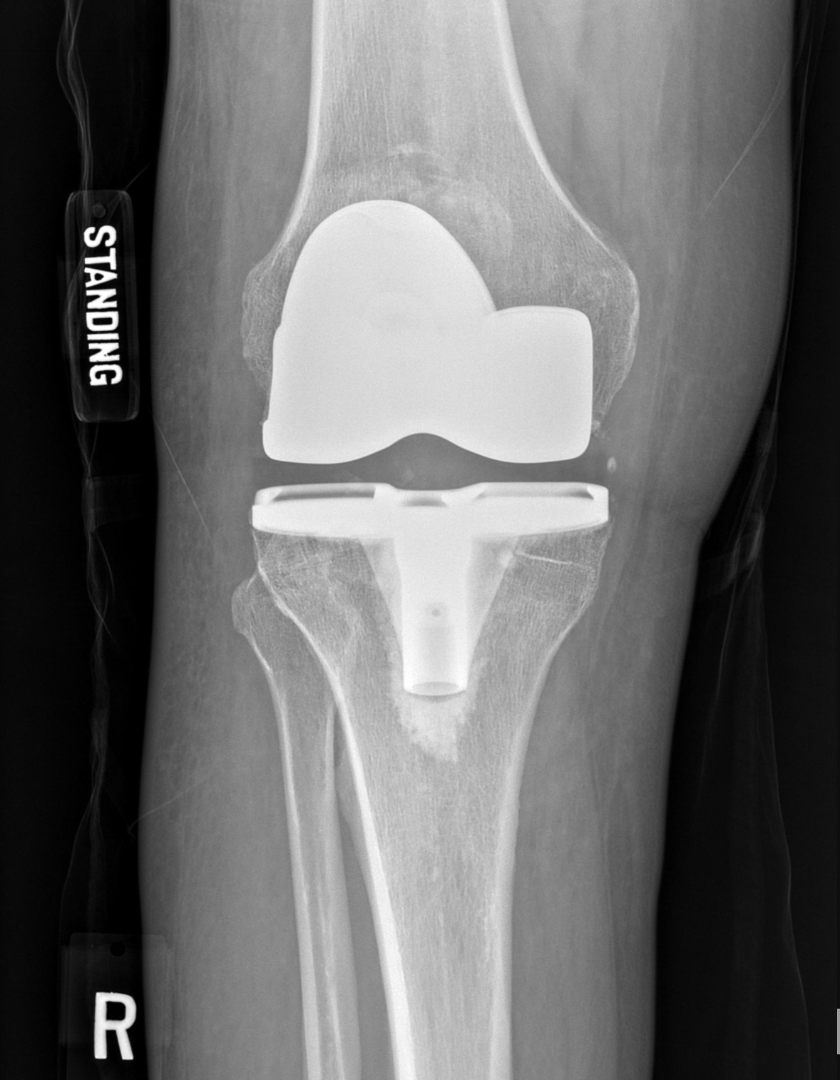
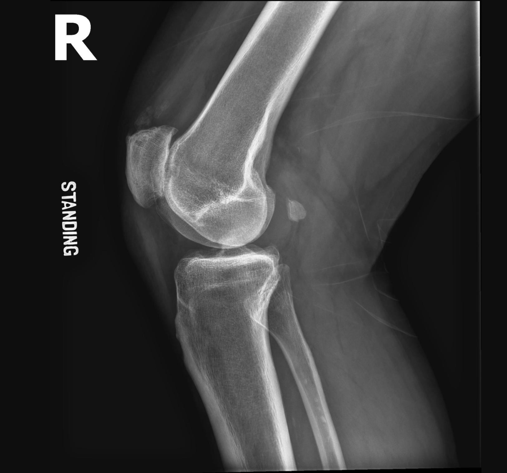
A 69-year-old male presented to the orthopedic clinic with right knee pain and x-ray evidence of osteoarthritis. He attempted conservative treatment including NSAIDs, activity modification, strengthening exercises, and intra-articular cortisone injections. He reported Losartan, Atorvastatin, ASA 81mg, Tamsulosin, Pantoprazole, Etodolac, and topical hydroquinone. He denied any history of alkaptonuria, black or discolored urine. He does report medical history of GERD, hypercholesterolemia, hypertension, and neuropathy. His surgical history includes bunion correction and previous right knee arthroscopy. There was no evidence of endogenous Ochronosis at the time of his previous knee arthroscopy.

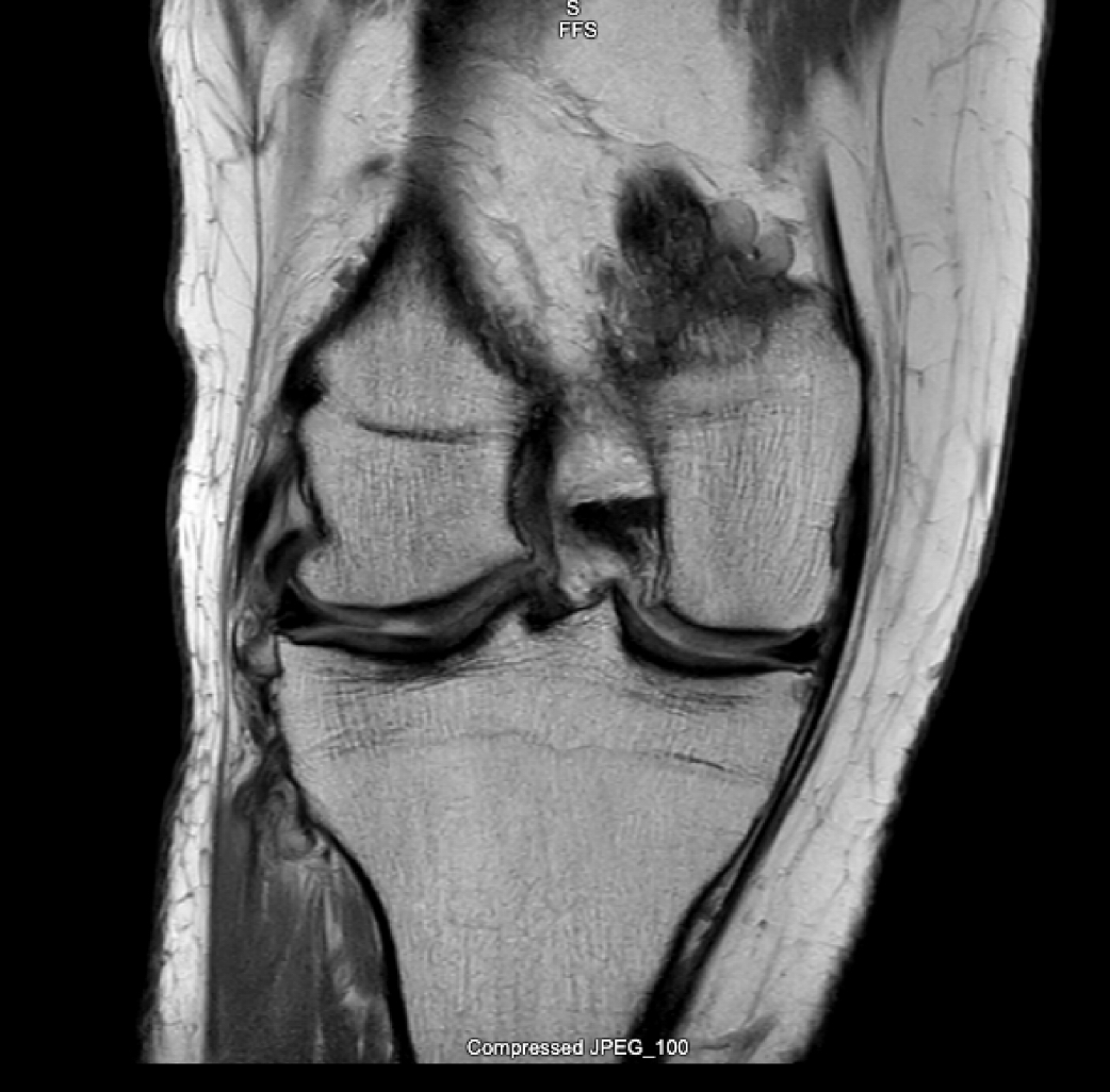
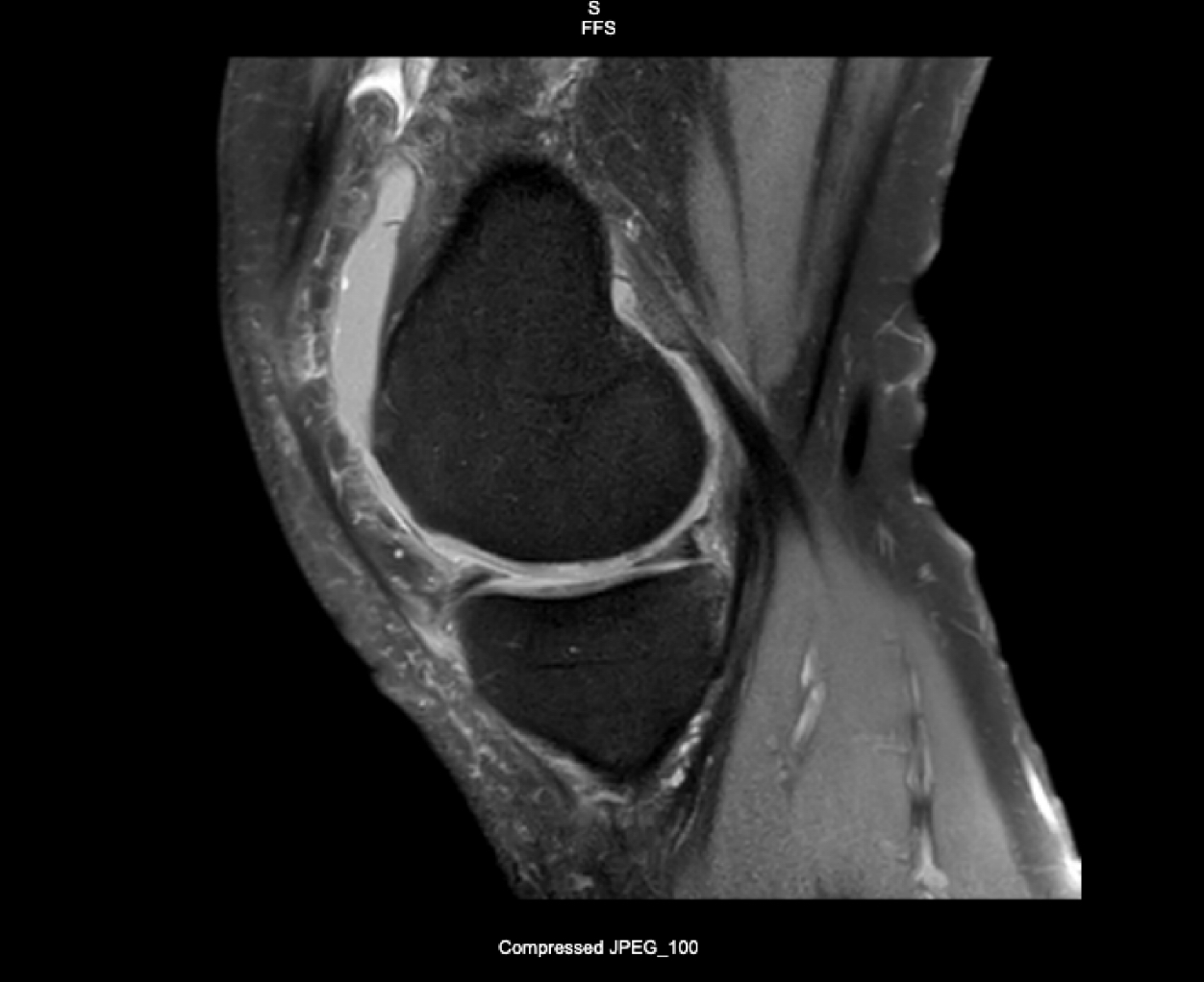
Physical exam demonstrates 0-110 degree range of motion of the right knee, tenderness and a small effusion of the right knee without warmth or erythema. No obvious skin pigment changes or brown-colored skin changes symmetrically on the cheeks typically seen with exogenous Ochronosis. He is 6 feet tall and 250 pounds. Preoperative x-rays and MRI are shown in Figures 1-10.

During a total knee arthroscopy intraoperative findings included gross blue/black discoloration of the cartilage surfaces of the distal femur, proximal tibia, and medial/lateral menisci [Figure 11]. There was eburnated bone in all three knee compartments. There was also blue/black hyperpigmentation minimally visualized in the synovial tissue. Postoperative pathology report demonstrated the same gross findings and hyperpigmentation of the cartilage. Postoperative work-up included culture/gram stain and urine organic acid testing. These tests all returned negative.

His postoperative course progressed as expected. He developed postoperative edema and had a negative doppler ultrasound. His physical therapist’s final report demonstrated 0/10 pain, 0-120 degree range of motion, and all goals met by > 90%.

Discussion

Ochronosis is a rare disease and can present in either exogenous or endogenous forms. Endogenous Ochronosis, or alkaptonuria is an autosomal recessive disease caused by an inborn defect of homogentisic acid oxidase, which results in the accumulation of homogentisic acid, a hydroquinone metabolite of tyrosine. Collagen of connective tissue, tendons, and cartilage is bound irreversibly by homogentisic acid and causes skin hyperpigmentation and/or arthropathy. The effect on cartilage is the most serious consequence of endogenous Ochronosis. The cartilage becomes brittle and fibrillated, leading to inflammation, progressive osteoarthritis, and denudation of subchondral bone (Kumar V,. Abbas A, Fausto N, Astor J. 2010). The small joints of the hands and feet are typically spared while the intervertebral disks, knees, hips, and shoulders are affected. Treatment of alkaptonuria is symptomatic and ultimately joint replacement if indicated (Ferreira et al. 2014).

Exogenous Ochronosis is evident by hyperpigmentation usually of the ear lobes, sclera, cheeks, and nose. Exogenous Ochronosis was first described by Findlay et al. and described the paradoxical hyperpigmentation caused by topical hydroquinone use for skin bleaching (Findlay, Morrison, and Simson 1975). The microscopic changes include banana-shaped yellow-pigmented inclusions and occasionally histiocytes (Engasser and Maibach 1984).

There have been multiple cases of topical hydroquinone use leading to exogenous Ochronosis (Gandhi, Verma, and Naik 2012; Merola et al. 2008). We believe this case represents the first instance of topical hydroquinone use leading to endogenous Ochronosis. Our patient had used topical hydroquinone for multiple years as skin bleaching treatment, and although he manifested no dermal side effects, the progression of his osteoarthritis and cartilaginous changes are consistent with those seen in endogenous Ochronosis.

Conclusion

Although this is the first report of topical hydroquinone use leading to evidence of endogenous Ochronosis in the absence of alkaptonuria to our knowledge, this should further provide credence that topical hydroquinone use is not without side effects. There have been overtures made to the FDA to evaluate the use of these topical agents for skin bleaching (2006) and perhaps further evaluation is warranted (Levitt 2007). A careful history, including medications viewed as noncontributory, for instance cosmetic and topical agents, should not be overlooked by orthopedic surgeons in the presurgical evaluation of osteoarthritis.

Figure 1
Figure 1:  This Figure shows the preoperative AP radiograph of the affected knee with associated osteophytic lipping, joint sclerosis, and narrowing.
Figure 2
Figure 2:This Figure shows the preoperative lateral radiograph of the affected knee with associated subchondral sclerosis. There is significant joint space narrowing, sclerosis, and cystic changes of the patellofemoral joint. There is an incidental fabella noted as well.
Figure 3
Figure 3:  This is a postoperative lateral radiograph of the affected knee status post total knee arthroplasty.
Figure 4
Figure 4:  This is a postoperative AP radiograph of the affected knee status post total knee arthroplasty.
Figure 5
Figure 5:This is a representative coronal T1 MRI slice demonstrating flattening of the femoral condyles and chondral thinning.
Figure 6
Figure 6:This is an additional coronal T1 MRI slice that demonstrates lateral osteophytic changes, chondral fissuring, and femoral condyle flattening.
Figure 7
Figure 7:This image is a T2 MRI sagittal slice that demonstrates increased signal within the lateral meniscus, subchondral edema of the trochlea, and chondral thinning.
Figure 8
Figure 8:  This is a representative T2 MRI sagittal slice that demonstrates chondral thinning, increased signal within the meniscus, and a joint effusion.
Figure 9
Figure 9:This image is a T1 MRI sagittal slice with subchondral changes to the femoral condyle, with flattening anteriorly. There are signal changes within the cartilage and blunting of the posterior meniscus.
Figure 10
Figure 10:This T1 MRI sagittal slice demonstrates subchondral edema of the trochlea. There is partial thickness cartilage fissure of the weight-bearing femoral condyle with surface irregularity of the tibial plateau.
Figure 11
Figure 11:Cartilage and meniscal Figures intraoperative

References

Engasser, P.G., and H.I. Maibach. 1984. “Ochronosis Caused by Bleaching Creams.” J Am Acad Dermatol 10:1072–73.
Google Scholar
EVANGELISTA, Priscila, José Sávio Parente, Romano Brasileiro, José Gerardo Paiva, Max Victor Freitas, Aline Moreira, Ana Paula Alves, and Igor Costa. 2018. “Secondary Osteoarthritis to Ochronotic Arthropathy - a Diagnostic Challenge.” Revista Da Associação Médica Brasileira 64 (7): 583–85. https:/​/​doi.org/​10.1590/​1806-9282.64.07.583.
Google Scholar
Ferreira, A.M.S.M., F.L. Santos, A.M.C. Costa, B.M.P. Barbosa, R.M.R. Rocha, and J.F. Fontes Lebre. 2014. “Knee Osteoarthrosis Secondary to Ochronosis: Clinical Case.” Rev Bras Ortop 49 (6): 675–80.
Google Scholar
Findlay, G.H., J.G.L. Morrison, and I.W. Simson. 1975. “Exogenous Ochronosis and Pigmented Colloid Milium from Hydroquinone Bleaching Creams.” Br J Dermatol 93:613–22.
Google Scholar
Gandhi, V., P. Verma, and G. Naik. 2012. “Exogenous Ochronosis After Prolonged Use of Topical Hydroquinone (2%) in a 50-Year-Old Indian Female.” Indian J Dermatol 57 (5): 394–95.
Google Scholar
Kumar V,. Abbas A, Fausto N, Astor J. 2010. Robbins and Cotran Pathologic Basis of Disease. 8th ed. Philadelphia: Elsevier/Saunders.
Google Scholar
Levitt, Jacob. 2007. “The Safety of Hydroquinone: A Dermatologist’s Response to the 2006 Federal Register.” Journal of the American Academy of Dermatology 57 (5): 854–72. https:/​/​doi.org/​10.1016/​j.jaad.2007.02.020.
Google Scholar
Merola, Joseph F., Shane Meehan, Ruth F. Walters, and Lance Brown. 2008. “Exogenous Ochronosis.” Dermatol Online J 15;14(10):6 (October).
Google Scholar

This website uses cookies

We use cookies to enhance your experience and support COUNTER Metrics for transparent reporting of readership statistics. Cookie data is not sold to third parties or used for marketing purposes.

Powered by Scholastica, the modern academic journal management system