Loading [Contrib]/a11y/accessibility-menu.js
Skip to main content
null
J Orthopaedic Experience & Innovation
  • Menu
  • Articles
    • Brief Report
    • Case Report
    • Data Paper
    • Editorial
    • Hand
    • Meeting Reports/Abstracts
    • Methods Article
    • Product Review
    • Research Article
    • Review Article
    • Review Articles
    • Systematic Review
    • All
  • For Authors
  • Editorial Board
  • About
  • Issues
  • Blog
  • "Open Mic" Topic Sessions
  • Advertisers
  • Recorded Content
  • CME
  • JOEI KOL Connect
  • search

RSS Feed

Enter the URL below into your favorite RSS reader.

https://journaloei.scholasticahq.com/feed
Case Report
Vol. 6, Issue 1, 2025May 21, 2025 EDT

Atraumatic myositis ossificans of the clavicle mimicking malignancy in a paediatric patient: a rare case report & review of literature.

Darren Hong Wen Lim, Arjandas Mahadev, Mohammad Ashik Bin Zainuddin, Kenneth Pak Leung Wong,
Myositis ossificansClavicleMalignancyIdiopathicCase report
Copyright Logoccby-nc-nd-4.0 • https://doi.org/10.60118/001c.126157
J Orthopaedic Experience & Innovation
Lim, Darren Hong Wen, Arjandas Mahadev, Mohammad Ashik Bin Zainuddin, and Kenneth Pak Leung Wong. 2025. “Atraumatic Myositis Ossificans of the Clavicle Mimicking Malignancy in a Paediatric Patient: A Rare Case Report & Review of Literature.” Journal of Orthopaedic Experience & Innovation 6 (1). https:/​/​doi.org/​10.60118/​001c.126157.
Save article as...▾
Download all (6)
  • Click here : https://joeipub.com/learning
    Download
  • Figure 1: Axial radiography of the left clavicle showing a calcified mass.
    Download
  • Figure 2: Axial (a) and Coronal (b) T1 weighted magnetic resonance images of a 3.1cm x 2.8cm x 2.3cm lesion in the middle-distal third of the left clavicle
    Download
  • Figure 3: Sagittal T1 weighted MRI with contrast (a) and T2 fblade MRI (b) showing that the lesion is inseparable from the adjacent periosteum in some areas.
    Download
  • Figure 4: PET Scan showing a markedly FDG-avid bone forming lesion in the left clavicle.
    Download
  • Figure 5: Radiography (axial view) of the left shoulder taken 2 months (a), 3 months (b) and 6 months (c) after the patient initially presented. The continual radiography showed the spontaneous resolution of the mass.
    Download

Sorry, something went wrong. Please try again.

If this problem reoccurs, please contact Scholastica Support

Error message:

undefined

View more stats

Abstract

Myositis ossificans (MO) is commonly characterised as a benign, self-limiting ossifying lesion in extraskeletal soft tissues. We present a case of MO of the clavicle in a 13 year old boy who did not have any history of preceding trauma. Multiple clinical investigations were required to reach the diagnosis of MO. Initial radiographic and imaging findings were inconclusive. Diagnosis was only achieved after biopsy and molecular study (COL1A1::USP6 gene fusion). Atraumatic MO presents a complex diagnostic challenge due to its close clinical similarity to bone and soft tissue tumours like osteosarcoma, requiring the correlation of histopathological and molecular findings with plain radiography and clinical observations. This was an exceedingly rare case of MO due to the location of the mass, the absence of a history of trauma and the patient’s age. To the best of our knowledge, there have been no previously published case reports of atraumatic MO of the clavicle in a paediatric patient.

Click here : https://joeipub.com/learning

INTRODUCTION

Myositis ossifican (MO) is commonly characterised as a benign, self-limiting ossifying lesion in extraskeletal soft tissues (Cherry et al. 2023). It is considered a rare type of heterotrophic ossificans that involves bone formation in soft tissue sites, including skeletal muscle, subcutaneous fat, tendon and nerve (W. T. Li, Horng, and Chien 2016). There are 3 main sub categories of myositis ossificans: fibrodysplasia ossificans progressiva (severe systemic form of heterotrophic ossificans), pseudomalignant/atraumatic MO (without a history of trauma) and circumscribed MO (with a background of trauma) (Rehman et al., n.d.). Clinically, MO can be initially misdiagnosed as malignancy (commonly osteosarcoma) due to its inherent similarities in the patient’s physical and radiographic findings.

We present a case of MO of the clavicle in a 13 year old boy who did not have any significant history of preceding trauma. Although MO can present in any ages, it is most commonly found in those aged 20 to 30 and is known to be rare in the paediatric population (Saad et al. 2021). MO also commonly presents in the extremities instead of the head and neck region, making our case exceedingly rare (Cherry et al. 2023). To the best of our efforts in reviewing existing literature, there have been no previously published case reports of atraumatic MO of the clavicle in a paediatric patient.

CASE REPORT

The 13-year-old school boy came with complains of a fast growing, painful mass over the medial end of the left shoulder over the last 2 months. He had difficulty achieving a full range-of-motion of the shoulder (mainly abduction) due to pain. He also complained of night pain which occasionally wakes him up at night. He had no history of fever or sudden loss of weight and appetite. He had also no known history of trauma, although we cannot exclude the possibility of any inciting event (ie secondary to a heavy school bag, physical activities in school). He had no significant past medical history.

CLINICAL FINDINGS

Upon inspection, there was an 8x7cm diffuse swelling over the medial half of the left clavicle. Skin over the clavicle was warm to touch and tender, with mild discoloration. Mild erythema was also noted. There was no palpable cervical or axillary lymph nodes and abdominal examination was unremarkable.

APTT 44.6 sec
Prothrombin Time 13.3 sec
Routine
Haemoglobin 13.0 g/dL
WBC Count 8.63 x10^9/L
Platelet Count 400 x10^9/L
RBC Count 5.86 x10^12/L
MCV 67.7 fL
MCH 22.2 pg
MCHC 32.7 g/dL
RBC Distribution Width 13.1 %
Mean Platelet Volume 10.3 fL
Lymphocyte Absolute 3.35 x10^9/L
ESR 9 mm/hr
Neutrophil 52.6 %
Lymphocyte 38.8 %
Monocyte 6.4 %
Eosinophil 2.0 %
Basophil 0.2 %
Neutrophil Absolute 4.54 x10^9/L
Monocyte Absolute 0.55 x10^9/L
Eosinophil Absolute 0.17 x10^9/L
Basophil Absolute 0.02 x10^9/L
Haematocrit 39.7 %

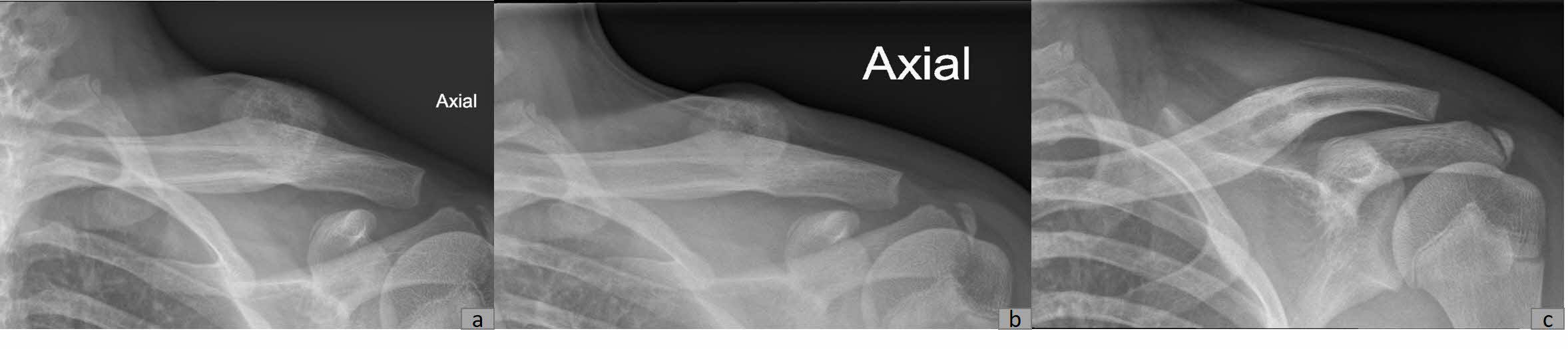
DIAGNOSTIC ASSESSMENT

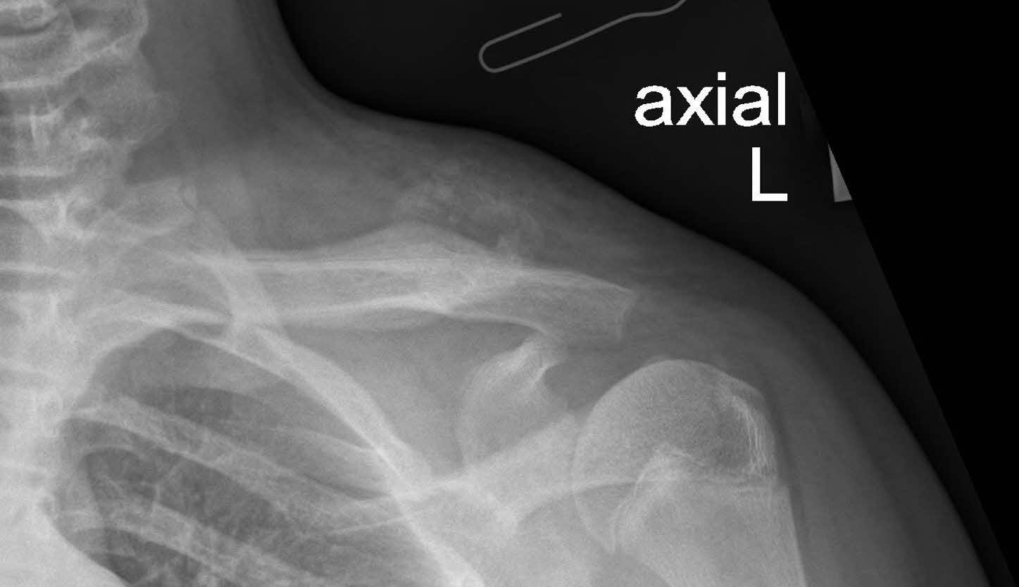
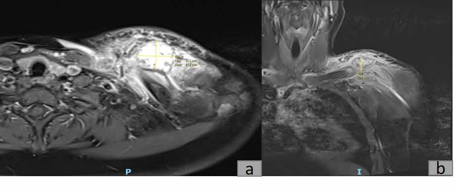
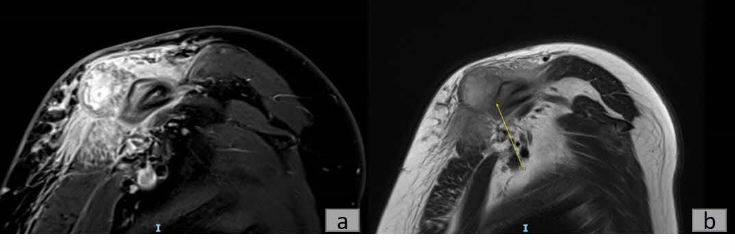
Plain radiography revealed a calcified mass with associated periosteal reaction along the left clavicular shaft (Figure 1). An MRI scan was also done and reported an avidly-enhancing T1w-isointense and T2w-hyperintense lesion in the middle-distal third of the left clavicle, measuring approximately 3.1cm x 2.8cm x 2.3cm (Figure 2). In certain areas, the lesion was inseparable from the adjacent periosteum (Figure 3), which itself was significantly thickened and enhancing. There was also increased T2W marrow edema with enhancement and some adjacent soft tissue swelling. No associated fracture was noted. The MRI report was suggestive of a malignant lesion such as soft tissue sarcoma.

X-ray of a shoulder joint AI-generated content may be incorrect.
Figure 1:Axial radiography of the left clavicle showing a calcified mass.
A close-up of a scan of a human body AI-generated content may be incorrect.
Figure 2:Axial (a) and Coronal (b) T1 weighted magnetic resonance images of a 3.1cm x 2.8cm x 2.3cm lesion in the middle-distal third of the left clavicle
A close-up of a person's head AI-generated content may be incorrect.
Figure 3:Sagittal T1 weighted MRI with contrast (a) and T2 fblade MRI (b) showing that the lesion is inseparable from the adjacent periosteum in some areas.

Incisional biopsy and molecular gene testing were performed. Microscopic analysis of the biopsy revealed multiple fragments with variable cellularity, with some showing features of maturation. Some fragments showed plump stromal cells with bland nuclear features and a background of fibrocollagenous to hyalinised stroma with some osseous debris. There were trabeculae of woven bone bordered by plump osteoblasts with occasional osteoclasts and a fibrinous exudate. No significant cytological atypia was noted. In addition, molecular testing identified a COL1A1::USP6 gene fusion (via Archer FusionPlex Anchored multiplex PCR). Interphase fluorescence in situ hybridization (FISH) for MDM2 (12q15) was also performed and this test was negative for MDM2 gene amplification.

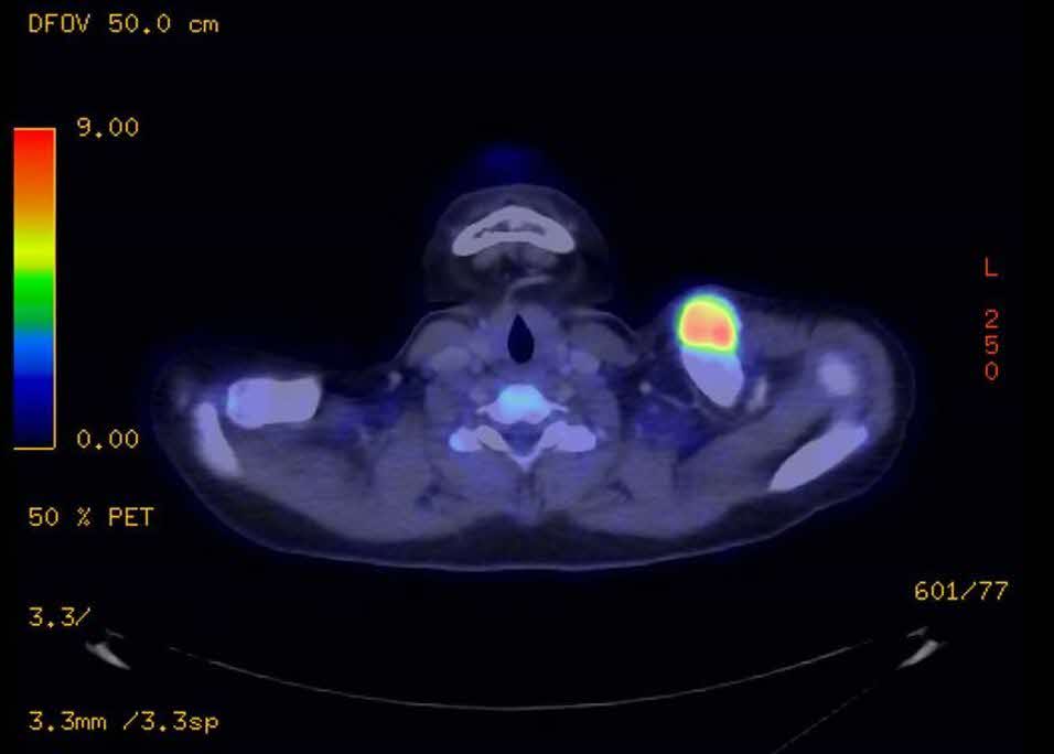
While awaiting the histopathological report, a PET/CT scan was also performed to look for potential metastasis. Report from the PET/CT scan (Figure 4) showed a markedly FDG-avid bone forming lesion (SUV MAX value: 9.7) in the left clavicle, with no other FDG-avid lesion detected in the body.

A close-up of a x-ray AI-generated content may be incorrect.
Figure 4:PET Scan showing a markedly FDG-avid bone forming lesion in the left clavicle.

THERAPEUTIC INTERVENTION AND FOLLOW UP

Subsequently, the patient was referred to musculoskeletal oncology given the high suspicion of malignancy. Conservative treatment was adopted for this patient, including a 3 months prescription of Indomethacin and continual radiographic monitoring. Spontaneous resolution of the mass was observed over the next six months, as evidenced by plain radiography (Figure 5). At the fifth month of follow-up, the patient was asymptomatic and exhibited normal function with a full range of motion in the left shoulder. Consequently, the patient was given an open dated review.

A x-ray of a human shoulder AI-generated content may be incorrect.
Figure 5:Radiography (axial view) of the left shoulder taken 2 months (a), 3 months (b) and 6 months (c) after the patient initially presented. The continual radiography showed the spontaneous resolution of the mass.

DISCUSSION

The exact pathogenesis of MO is not well understood in literature. In cases of circumscribed MO preceded by a history of trauma, it is hypothesized that trauma caused an inappropriate differentiation of fibroblasts into osteogenic cells (Walczak, Johnson, and Howe 2015). Some studies have suggested that Endothelial-mesenchymal transition could contribute to the development of MO. Inflammatory cytokines (bone morphogenetic protein-2 & -4) cause endothelial-derived mesenchymal stem cells to differentiate into chondrocytes or osteoblasts, resulting in bone formation in soft tissue (Walczak, Johnson, and Howe 2015).

However, pathogenesis of atraumatic MO remains unclear. Atraumatic MO is significantly rarer than circumscribed MO (Rehman et al., n.d.). Clinically, atraumatic MO can mimic bone and soft tissue malignancy, and plain radiography is insufficient to diagnose MO.

The process of MO can be grouped into 3 stages: early (first 4 weeks), intermediate (4- 8 weeks), and mature (months later). Calcification typically occurs during the intermediate stage, while mature bone formation develops in the mature stage. Bone tumours, like osteosarcoma, can also exhibit calcification and bone formation. Diagnosis of MO by plain radiography is generally possible during the mature stage, when the characteristic features, such as bone formation with central lucency, become apparent.

However, in the early stage of MO, radiography and MRI cannot fully confirm the diagnosis of MO and exclude the possibility of malignancy. Delaying diagnosis by waiting for the tumour to mature is not advisable, as such delays in treatment – particularly if the tumour turns out to be osteosarcoma – can have severe consequences and poor prognosis. In this case, the patient presented to us 2 months after first noticing the growing mass in his clavicle. Given the duration, the tumour was likely not yet mature, rendering MRI and radiography inconclusive. Consequently, biopsy and molecular testing were therefore indicated in this case with diagnostic uncertainty.

The main differential diagnoses being considered were Parosteal osteosarcoma and Synovial sarcoma.

Parosteal osteosarcoma (PO) is a well-differentiated low-grade malignant bony tumour (Kumar, Barwar, and Khan 2014). It is the most common subtype of surface osteosarcoma. PO is characterized as a slow growing bony mass with a “cauliflower-like” appearance. On radiography, PO usually presents as a radiodense lesion arising from the metaphyseal region of long bones (Prabowo et al. 2020), and may present with a “string sign”: a thin, radiolucent line between the tumour and the bone cortex (Idrees, Zarrar, and Mujtaba 2023). In most findings, the lesion is poorly demarcated with calcifications. Studies have observed that in osteosarcoma, calcification begins at the center and spreads towards the periphery, unlike in MO where it begins at the periphery and progresses inwards (Man et al. 2011). In our case, peripheral calcification can be seen on radiography and MRI. Moreover, amplification of the MDM2 gene, an oncogene commonly associated with low-grade osteosarcoma and recognized as an effective diagnostic marker for this osteosarcoma (Dujardin et al. 2011) (sensitivity = 81.3%, specificity = 100%) (L. Li et al. 2024), was also absent in our patient. Both findings made a diagnosis of osteosarcoma very unlikely.

Synovial sarcoma is a malignant slow-growing soft tissue tumour that can arise from anywhere in the body (Saad et al. 2021). Most SS patients present with a prolonged history of a painless slow-growing mass, with an average duration of 2-4 years before seeking medical assistance. Conversely, Patients with MO usually seek clinical evaluation a few months after the occurrence, presenting with pain due to the inflammatory process during the early stage as mentioned above. On radiography, approximately 30% of SS cases present with “fine, stippled” calcification (Wilkerson et al. 2012). Calcifications in SS are predominantly situated at the periphery, resembling the pattern observed in MO. Bony erosion in synovial sarcoma can also occur when the tumour is located in close proximity to bone structures, though it is observed in only about 22% of cases (Luczyńska et al. 2014). On the other hand, MO never presents with bony erosion (Luczyńska et al. 2014). In our patient, the lesion was closely situated near the left clavicle yet no bony erosion was observed. Given that our patient had no bony erosion and presented with a fast-growing painful mass, a diagnosis of SS was unlikely. Granted, more gene expression studies (Transducin-Like Enhancer 1 (TLE1)) can be done to definitively exclude SS (El Beaino et al. 2020).

In our case, histopathological and molecular findings, correlated with imagery findings and clinical history, were used to arrive at a diagnosis of MO. There were immature woven bone formations, coupled with no signs of significant cytological atypia. Ideally, the presence of progressive maturation of the lesion from the centre outwards, known as “zoning” phenomenon, should be used as a diagnostic feature (Nishio et al. 2010). However, due to the nature of the biopsy (incisional), we were unable to observe this. Despite this, the detection of a COL1A1::USP6 gene fusion in molecular testing provides substantial evidence supporting a diagnosis of myositis ossificans (MO), given its established association with MO in several case series. In one study, COL1A1-USP6 rearrangement was identified in 5 out of 7 cases of MO (Švajdler et al. 2019), while another series reported USP6 rearrangements in 8 of 9 MO cases (Bekers et al. 2018), reinforcing this genetic alteration as a significant diagnostic marker for MO.

In terms of treatment, there are two main approaches: surgical excision and conservative radiographic monitoring. Studies have recommended surgical excision of MO in symptomatic mature lesions after at least six months (Cherry et al. 2023). Surgical excision of MO during the early phase can lead to reoccurrence (Cherry et al. 2023), and is therefore not indicated in our case. Given MO’s self-limiting nature, a majority of MO are treated conservatively (Saad et al. 2021). Apart from continual radiographic monitoring to ensure the spontaneous resolution of the mass, NSAIDs can stop further progression of MO and decrease inflammation (Park et al. 2024). As for our patient, indomethacin was prescribed along with continual radiographic monitoring and he recovered without complications.

A review of literature was also done to document existing cases of atraumatic MO in the paediatric population (<18 years old). With reference to a systematic scoping review of MO in the pediatric population done in 2022 (Cherry et al. 2023), as well as a search on PubMed database, a total of 22 cases of atraumatic MO in the paediatric population were found from 2004 to 2024.

Table 1:Previous case reports on Atraumatic MO in Paediatric Population from 2004 to 2024
Author Year Age Location Imagery Biopsy Treatment Follow up w/
Cabello et al (Cabello García et al. 2008). 2008 4 years Arm Rx, MRI, BS, PET-CT Percutaneous Excision None
Jujena et al (Juneja et al. 2011). 2011 6 years Bilateral thighs Rx, CT None Conservative Rx
Lau et al (Lau, Hartin, and Ozgediz 2012). 2012 16 years Thigh Rx, US, MRI Percutaneous Conservative None
Say et al (Say et al. 2015). 2015 10 years Forearm Rx, MRI ND Excision None
Akahane et al (Akahane, Mori, and Nakatsuchi 2015). 2015 15 years Hand Rx, US, MRI Incisional Excision None
Li et al (P. F. Li, Lin, and Pang 2016). 2016 9 years Elbow Rx, CT Percutaneous Excision None
Simmonds et al (Simmonds et al. 2016). 2016 5 months Neck MRI Percutaneous Excision None
Mohamed et al (Mohamed et al. 2018). 2018 15 years Hip Rx Excisional Excision None
Kougias et al (Kougias et al. 2019). 2019 5 years Bilat. hip Rx, CT None Conservative Rx
5 years Bilat. hip Rx, CT, BS None Conservative Rx, CT
Onen et al (Onen et al. 2019). 2019 5 years Lumbar Rx, CT, MRI ND Excision Rx
Dubuisson et al (Dubuisson, Lombard, and Otto 2019). 2019 5 years Neck Rx, US, CT, MRI, BS Incisional Excision MRI
Akatli et al (Akatli et al. 2021). 2021 13 years Pectoralis US, MRI None Excision None
Rehman et al (Rehman et al., n.d.). 2021 12 years Axilla US, MRI None Excision None
Dennison et al (Dennison et al. 2022). 2022 At birth Leg Rx, CT, MRI Percutaneous Conservative Rx
Vitale et al (Vitale et al. 2022). 2022 14 years Neck Rx, US, CT, MRI Percutaneous Excision ND
Xia et al (Xia and Wang 2022). 2022 8 years Thigh Rx, US, CT Incisional Excision None
Silveri et al (Silveri et al. 2022). 2022 2 years Elbow Rx, MRI, BS Percutaneous Conservative MRI
Valgaeren et al (Valgaeren and Limantoro 2024). 2024 15 years Knee US, MRI, Rx ND ND ND
Alananzh et al (Alananzh et al. 2024). 2024 11 years Hip Rx, MRI Excisional Excision ND
2 years Hip Rx, MRI None Conservative ND
Ji et al (Ji et al. 2024). 2024 9 years Neck CT, Rx Incisional Conservative US, MRI

From the literature review, it is noted that biopsy was done in 13 out of 22 cases (Percutaneous: 7, 31.8%; Incisional: 4, 18.2%; Excision: 2, 9.1%; None: 6, 27.3%; Not Documented: 3, 13.6%) as imagery studies were unable to sufficiently exclude malignancy. As for treatment, 8 out of 22 cases (36.4%) adopted a conservative approach while 13 out of 22 cases (59.1%) opted for excision instead.

In conclusion, tumours presenting as atraumatic MO should raise a high clinical suspicion for malignancy and warrant thorough investigation. In this case, plain radiography and clinical examination were insufficient to exclude soft tissue malignancy due to clinical similarities between MO and sarcomas, and the absence of a history of trauma. Biopsy and molecular studies are recommended for accurate diagnosis and appropriate management when imaging investigation remains inconclusive. Early and comprehensive evaluation is essential to ensure timely treatment and improve patient outcomes.


ACKNOWLEDGMENT

Author gracefully acknowledges contributions from colleagues and willful participation from patient.

FINANCIAL SUPPORT AND SPONSORSHIP

This research did not receive any specific grant from funding agencies in the public, commercial, or not-for-profit sectors.

INFORMED CONSENT

Consent was obtained from the patient for the publication of the case report and any accompanying images.

Submitted: September 06, 2024 EDT

Accepted: November 16, 2024 EDT

References

Akahane, T., N. Mori, and Y. Nakatsuchi. 2015. “Myositis Ossificans Occupying the Thenar Region: A Case Report.” J Med Case Reports 9 (1): 105. https:/​/​doi.org/​10.1186/​s13256-015-0586-8.
Google ScholarPubMed CentralPubMed
Akatli, A. N., S. Uguralp, S. Alan, A. Tasci, and G. Yildirim. 2021. “Solitary Bone Cyst Like Areas in Myositis Ossificans: A Breast Mass in a Child.” Fetal Pediatr Pathol 40 (3): 262–70. https:/​/​doi.org/​10.1080/​15513815.2019.1693673.
Google Scholar
Alananzh, M., M. Abu Hilal, S. Sakka, O. Aldahamsheh, and M. Alkhreisat. 2024. “Non-Traumatic Myositis Ossificans Mimicking Infected Hip in Children: Two Case Reports and Review of the Literature.” Cureus 16 (9): e69990. https:/​/​doi.org/​10.7759/​cureus.69990.
Google ScholarPubMed CentralPubMed
Bekers, E. M., A. Eijkelenboom, K. Grünberg, R. C. Roverts, J. W. J. de Rooy, I. C. M. van der Geest, et al. 2018. “Myositis Ossificans - Another Condition with USP6 Rearrangement, Providing Evidence of a Relationship with Nodular Fasciitis and Aneurysmal Bone Cyst.” Ann Diagn Pathol 34 (June):56–59. https:/​/​doi.org/​10.1016/​j.anndiagpath.2018.01.006.
Google Scholar
Cabello García, D., A. Rodríguez Fernández, M. Gómez Río, M. J. Moreno, A. C. Rebollo Aguirre, A. Martín Castro, et al. 2008. “[Circumscript Myositis Ossificans in a Four-Year-Old Boy].” Rev Esp Med Nucl 27 (5): 358–62. https:/​/​doi.org/​10.1157/​13126193.
Google Scholar
Cherry, I., M. Mutschler, E. Samara, S. Merckaert, P. Y. Zambelli, and B. Tschopp. 2023. “Myositis Ossificans in the Pediatric Population: A Systematic Scoping Review.” Front Pediatr 11 (December):1295212. https:/​/​doi.org/​10.3389/​fped.2023.1295212.
Google ScholarPubMed CentralPubMed
Dennison, C. B., I. R. Royall, K. M. Beavers, C. W. Dean, and K. F. Scherer. 2022. “Myositis Ossificans: A Rare Neonatal Presentation.” Pediatr Radiol 52 (3): 587–91. https:/​/​doi.org/​10.1007/​s00247-021-05204-7.
Google Scholar
Dubuisson, A., A. Lombard, and B. Otto. 2019. “Pseudomalignant Myositis Ossificans of the Neck in a Child: Case Report and Review of the Literature.” World Neurosurg 130 (October):95–97. https:/​/​doi.org/​10.1016/​j.wneu.2019.06.165.
Google Scholar
Dujardin, F., M. B. N. Binh, C. Bouvier, A. Gomez-Brouchet, F. Larousserie, and A., de Muret. 2011. “MDM2 and CDK4 Immunohistochemistry Is a Valuable Tool in the Differential Diagnosis of Low-Grade Osteosarcomas and Other Primary Fibro-Osseous Lesions of the Bone.” Mod Pathol Off J U S Can Acad Pathol Inc 24 (5): 624–37. https:/​/​doi.org/​10.1038/​modpathol.2010.229.
Google Scholar
El Beaino, M., D. C. Jupiter, T. Assi, E. Rassy, A. J. Lazar, D. M. Araujo, et al. 2020. “Diagnostic Value of TLE1 in Synovial Sarcoma: A Systematic Review and Meta-Analysis.” Sarcoma 2020:7192347. https:/​/​doi.org/​10.1155/​2020/​7192347.
Google ScholarPubMed CentralPubMed
Idrees, H., R. Zarrar, and B. Mujtaba. 2023. “Parosteal Osteosarcoma of the Skull: Pathophysiological and Imaging Review.” Eur J Radiol Open 10 (May):100489. https:/​/​doi.org/​10.1016/​j.ejro.2023.100489.
Google ScholarPubMed CentralPubMed
Ji, T., G. Zhang, J. Zhang, Y. Li, X. Zhang, Q. Liu, et al. 2024. “Myositis Ossificans of the Trapezius Muscle: A Case Report and Literature Review.” Ear Nose Throat J 103 (9): NP520-6. https:/​/​doi.org/​10.1177/​01455613231175316.
Google Scholar
Juneja, M., R. Jain, D. Mishra, and V.K. Gautam. 2011. “Myositis Ossificans of Bilateral Hip Joints in a Patient with Diplegic Cerebral Palsy.” J Clin Neurosci Off J Neurosurg Soc Australas 18 (4): 580–81. https:/​/​doi.org/​10.1016/​j.jocn.2010.07.143.
Google Scholar
Kougias, V., E. Hatziagorou, N. Laliotis, F. Kyrvasillis, V. Georgopoulou, and J. Tsanakas. 2019. “Two Cases of Myositis Ossificans in Children, after Prolonged Immobilization.” J Musculoskelet Neuronal Interact 19 (1): 118–22.
Google Scholar
Kumar, V. S., N. Barwar, and S. A. Khan. 2014. “Surface Osteosarcomas: Diagnosis, Treatment and Outcome.” Indian J Orthop 48 (3): 255–61. https:/​/​doi.org/​10.4103/​0019-5413.132503.
Google ScholarPubMed CentralPubMed
Lau, J., C. W. Hartin, and D. E. Ozgediz. 2012. “Myositis Ossificans Requires Multiple Diagnostic Modalities.” J Pediatr Surg 47 (9): 1763–66. https:/​/​doi.org/​10.1016/​j.jpedsurg.2012.05.009.
Google Scholar
Li, L., M. Zhang, R.F. Dong, Y.B. Su, and Y. Ding. 2024. “[Detection of MDM2 Gene Amplification by Fluorescence in Situ Hybridization and Its Diagnostic Value in Low-Grade Osteosarcoma].” Zhonghua Bing Li Xue Za Zhi 53 (3): 237–42.
Google Scholar
Li, P. F., Z. L. Lin, and Z. H. Pang. 2016. “Non-Traumatic Myositis Ossificans Circumscripta at Elbow Joint in a 9-Year Old Child.” Chin J Traumatol 19 (2): 122–24. https:/​/​doi.org/​10.1016/​j.cjtee.2016.01.009.
Google ScholarPubMed CentralPubMed
Li, W. T., S. Y. Horng, and H. F. Chien. 2016. “Abdominis Rectus Intramuscular Myositis Ossificans.” Formos J Surg 49 (1): 20–26. https:/​/​doi.org/​10.1016/​j.fjs.2015.06.004.
Google Scholar
Luczyńska, E., H. Kasperkiewicz, A. Domalik, A. Cwierz, and B. Bobek-Billewicz. 2014. “Myositis Ossificans Mimicking Sarcoma, the Importance of Diagnostic Imaging - Case Report.” Pol J Radiol 79:228–32. https:/​/​doi.org/​10.12659/​PJR.890209.
Google ScholarPubMed CentralPubMed
Man, S.C., C.N. Schnell, O. Fufezan, and G. Mihut. 2011. “Myositis Ossificans Traumatica of the Neck – a Pediatric Case.” Mædica 6 (2): 128–31.
Google Scholar
Mohamed, A. W. A., K. Moussa, S. B. Seyni, Z. A. Seyni, A. S. Kasoumou, and K. Ziberou. 2018. “[Myositis Ossificans Circumscripta of the Hip: About a Case].” Pan Afr Med J 29:207.
Google Scholar
Nishio, J., K. Nabeshima, H. Iwasaki, and M. Naito. 2010. “Non-Traumatic Myositis Ossificans Mimicking a Malignant Neoplasm in an 83-Year-Old Woman: A Case Report.” J Med Case Reports 4 (August):270. https:/​/​doi.org/​10.1186/​1752-1947-4-270.
Google ScholarPubMed CentralPubMed
Onen, M. R., E. Varol, M. İ. Tosun, and S. Naderi. 2019. “Nontraumatic Myositis Ossificans as an Uncommon Cause of Scoliosis: Case Report and Review of the Literature.” World Neurosurg 123 (March):208–11. https:/​/​doi.org/​10.1016/​j.wneu.2018.11.259.
Google Scholar
Park, E., J. Park, S.Y. Chang, and Y. Kim. 2024. “Nontraumatic Myositis Ossificans After Spontaneous Subarachnoid Hemorrhage: A Case Report.” Brain NeuroRehabilitation 17 (1): e9. https:/​/​doi.org/​10.12786/​bn.2024.17.e9.
Google ScholarPubMed CentralPubMed
Prabowo, Y., A. F. Kamal, E. Kodrat, M. Prasetyo, S. Maruanaya, and T. S. Efar. 2020. “Parosteal Osteosarcoma: A Benign-Looking Tumour, Amenable to a Variety of Surgical Reconstruction.” Int J Surg Oncol 2020 (May):4807612. https:/​/​doi.org/​10.1155/​2020/​4807612.
Google ScholarPubMed CentralPubMed
Rehman, N., H. Sadashiva, M. G. Madakshira, and D. K. Raman. n.d. “Non-Traumatic Myositis Ossificans.” Autopsy Case Rep 11:e2021316. https:/​/​doi.org/​10.4322/​acr.2021.316.
Google ScholarPubMed CentralPubMed
Saad, A., C. Azzopardi, A. Patel, A.M. Davies, and R. Botchu. 2021. “Myositis Ossificans Revisited – The Largest Reported Case Series.” J Clin Orthop Trauma 17 (March):123–27. https:/​/​doi.org/​10.1016/​j.jcot.2021.03.005.
Google ScholarPubMed CentralPubMed
Say, F., S. Coşkun, M. Bülbül, and Ö. Alici. 2015. “Myositis Ossificans on the Forearm in a 10-Year-Old Girl.” J Pediatr Orthop Part B 24 (3): 223–25. https:/​/​doi.org/​10.1097/​BPB.0000000000000152.
Google Scholar
Silveri, C., P. Stoppiello, L. Gaiero, G. Bianchi, N. Casales, and A.C. Belzarena. 2022. “Aggressive Atraumatic Myositis Ossificans in a Toddler.” Radiol Case Rep 17 (12): 4550. https:/​/​doi.org/​10.1016/​j.radcr.2022.09.032.
Google ScholarPubMed CentralPubMed
Simmonds, J., N. Taki, I. Chilton, and M. Vecchiotti. 2016. “A Rare Case of Pediatric Nontraumatic Myositis Ossificans in the Posterior Triangle.” Int J Pediatr Otorhinolaryngol 84 (May):116–18. https:/​/​doi.org/​10.1016/​j.ijporl.2016.03.003.
Google Scholar
Švajdler, M., M. Michal, P. Martínek, N. Ptáková, Z. Kinkor, P. Szépe, et al. 2019. “Fibro-Osseous Pseudotumor of Digits and Myositis Ossificans Show Consistent COL1A1-USP6 Rearrangement: A Clinicopathological and Genetic Study of 27 Cases.” Hum Pathol 88 (June):39–47. https:/​/​doi.org/​10.1016/​j.humpath.2019.02.009.
Google Scholar
Valgaeren, B., and I. Limantoro. 2024. “Myositis Ossificans: A Mimicker of an Intramuscular Tumour.” J Belg Soc Radiol 108 (1): 15. https:/​/​doi.org/​10.5334/​jbsr.3531.
Google ScholarPubMed CentralPubMed
Vitale, V., C. Bleve, M. Mansour, F. De Corti, L. Giarraputo, A. Brugiolo, et al. 2022. “Non-Traumatic Myositis Ossificans as Unusual Cause of Neck Pain During COVID-19 Pandemic: A Case Report.” SN Compr Clin Med 4 (1): 96. https:/​/​doi.org/​10.1007/​s42399-022-01177-2.
Google ScholarPubMed CentralPubMed
Walczak, B. E., C. N. Johnson, and B. M. Howe. 2015. “Myositis Ossificans.” JAAOS - J Am Acad Orthop Surg 23 (10): 612. https:/​/​doi.org/​10.5435/​JAAOS-D-14-00269.
Google Scholar
Wilkerson, B. W., J. R. Crim, M. Hung, and L. J. Layfield. 2012. “Characterization of Synovial Sarcoma Calcification.” Am J Roentgenol 199 (6): W730-4. https:/​/​doi.org/​10.2214/​AJR.11.7342.
Google Scholar
Xia, A. N., and J. S. Wang. 2022. “Giant Nontraumatic Myositis Ossificans in a Child: A Case Report.” World J Clin Cases 10 (9): 2901. https:/​/​doi.org/​10.12998/​wjcc.v10.i9.2901.
Google ScholarPubMed CentralPubMed

This website uses cookies

We use cookies to enhance your experience and support COUNTER Metrics for transparent reporting of readership statistics. Cookie data is not sold to third parties or used for marketing purposes.

Powered by Scholastica, the modern academic journal management system