


Introduction
Calcific tendinopathy is a leading cause of shoulder pain. It is a self-limiting condition that occurs most commonly in patients between 30 and 50 years of age with a higher incidence in females than in males, with a ratio of approximately 2:1 (Kim et al. 2020). The condition is characterized by the accumulation of calcium phosphate deposits within the rotator cuff tendons, leading to pain, discomfort, and reduced mobility of the shoulder joint (Erickson and Jagim 2020). Among patients with calcific tendinopathy, previous data has shown that 2.7% - 20% are asymptomatic while 35% - 45% develop shoulder pain (Kim et al. 2020). The supraspinatus tendon is most commonly affected, with one study reporting up to 83% of cases affecting at least the supraspinatus tendon (Kim et al. 2020).
Management of calcific tendinopathy varies widely, and the presence of pain is a principal factor to consider when approaching each patient (Kim et al. 2020). Treatment options include activity modification, non-steroidal anti-inflammatory drugs (NSAIDs), physical therapy, corticosteroid injections, aspiration, extracorporeal shockwave therapy, and surgical intervention (Erickson and Jagim 2020). Most patients respond well to conservative treatment and rarely require surgical intervention. However, approximately 10% of patients fail conservative measures and require surgery to remove calcific deposits through either open or arthroscopic procedures (De Carli et al. 2014).
Ultrasonic tenotomy and debridement (UTD) is an alternative technique that has shown promise in the management of calcific tendinopathy while decreasing the risks and costs associated with traditional surgical intervention (Erickson and Jagim 2020). UTD is performed through the insertion of a needle, guided by ultrasound, to administer ultrasonic waves at the source of calcific deposits within a diseased tendon for concurrent deposit pulverization and aspiration (Erickson and Jagim 2020).
In 2020, a pilot case study analyzed the effectiveness of UTD on rotator cuff calcific tendinopathy (Erickson and Jagim 2020). Authors reported success of UTD at significantly reducing pain via subjective patient reports before and after the procedure (Erickson and Jagim 2020). These findings indicate a need for continued implementation and investigation of UTD for shoulder calcific tendinopathy. Our case series aims to supplement the current literature regarding UTD for the shoulder through evaluation of patient-reported pain, functionality, range of motion, and strength before and after treatment.
Methods
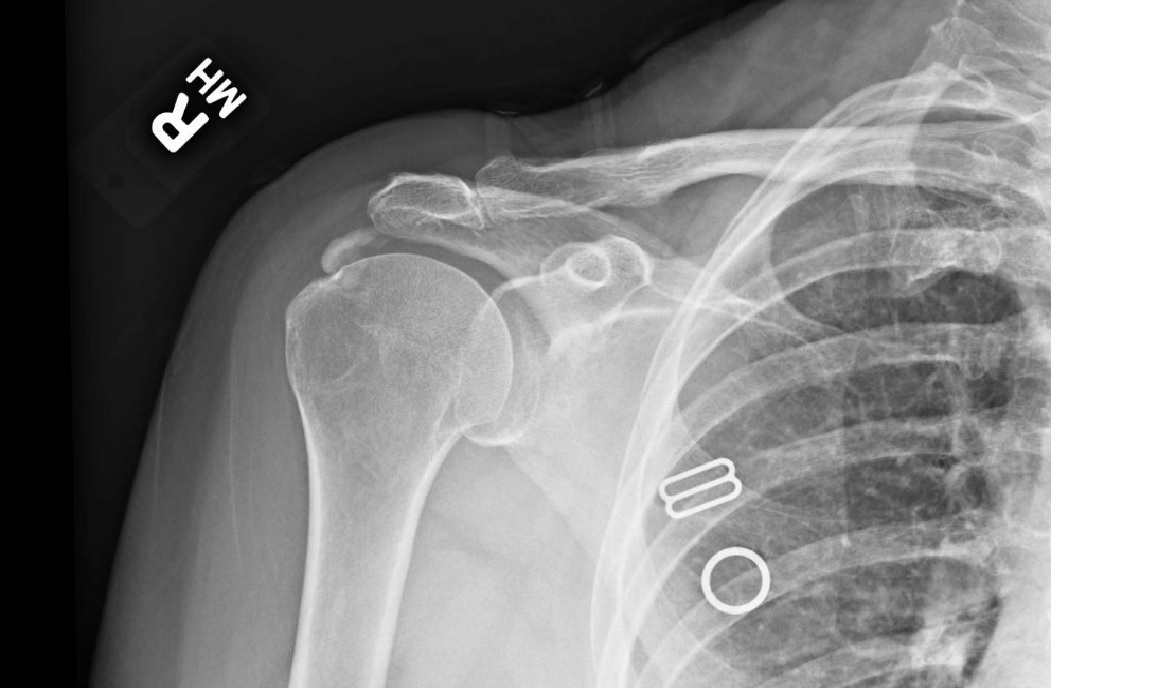
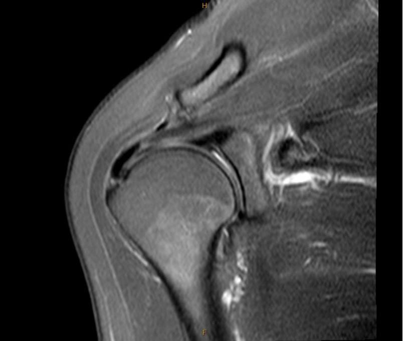
Each patient was treated conservatively for at least 3 months prior to the procedure. Conservative treatment methods performed included physical therapy, NSAIDs, intra-articular corticosteroid injections, and activity modification. UTD was performed on eighteen patients who failed conservative therapies for symptomatic calcific tendinopathy of the shoulder. The level of sedation was performed at the request of the patient with the supervision of an anesthesiologist. Thus, some patients underwent general anesthesia, and some underwent local anesthesia. Patients were diagnosed by clinical assessment and preoperative imaging studies, and were excluded with any rotator cuff tears, labral tears, or degenerative changes (radiographic imaging (Figure 1), magnetic resonance imaging (MRI) (Figure 2)). All patients were managed in a single outpatient orthopedic clinic between 8/20/2020 and 03/02/2022 by the same sports medicine trained orthopedic surgeon. Retrospective patient chart review was performed to determine age, sex, past medical history, natural history of calcific tendinopathy, treatment history, and imaging study results.


UTD
After induction of anesthesia, a patient is positioned supine. With gentle shoulder extension off the table, the calcific deposit clears the acromion and can be well-visualized by sonogram. Once the calcific deposit is identified, three milliliters of 0.25% Marcaine and three milliliters of 1% lidocaine are injected via a 25-gauge needle to anesthetize the skin, subcutaneous layer, and bursae overlying the calcific deposit within the rotator cuff tendon. An 11-blade scalpel is used to make a small incision and the two-inch Tenex needle (TX2) is guided with ultrasound into the calcific deposit in the tendon (Figure 3). The Tenex machine (Tenex Health, Lake Forest, CA) is activated to irrigate, debride with high frequency ultrasound, and aspirate the deposit. The needle is manipulated through the calcific deposit until resolution, as visualized with ultrasound– generally requiring three to five minutes of Tenex machine time. Once the Tenex needle is removed, mastisol, Steristrips, and a sterile dressing are applied, and the arm is placed in a sling.

Post-procedural evaluation
Clinical evaluation was performed at two weeks, six weeks, and three months post-procedure. A 10-question shoulder score survey was sent electronically via email more than six months post-procedure to all eighteen patients who underwent UTD. Patients were asked the following questions regarding symptoms both prior to and six months after UTD: timing and intensity of pain, level of functional limitation, and overall satisfaction with the procedure. Objective active shoulder range of motion and strength of forward flexion scores were determined by the attending physician during clinical evaluations. Survey questions were drafted based on the UCLA Shoulder Score (Moorthy et al. 2021). An optimum score of 35 indicates no pain with full function, range of motion, strength, and patient satisfaction. Range of motion scores were obtained by the attending physician during physical examinations at routine follow-up appointments. Per the UCLA Shoulder Score, scores <27 are classified as fair/poor and >27 are classified as great/excellent.
Results
Of the 18 patients treated with UTD for calcific tendinopathy of the shoulder, nine responded to the electronic survey. Respondents’ ages ranged from 39 to 72 with an average age of 58.1 years old. Most respondents were female, 88.9% (8/9). 44.4% (4/9) reported a history of comorbid endocrine disease including three patients with hypothyroidism and one patient with type 2 diabetes mellitus (T2DM). Two patients reported onset of shoulder symptoms related to an injury. None of the patients experienced post-procedural complications.
Table 1 demonstrates pain scores before and after UTD for each patient. Survey question analysis revealed that 77.8% (7/9) patients reported improved pain, 44.4% (4/9) reported improved function, and 5/9 (55.6%) experienced both improved strength and range of motion, with 66.7% (6/9) reporting complete patient satisfaction. 44.4% (4/9) of patients improved to a score > 27, classified as great/excellent per the UCLA Shoulder Score. Statistical analysis performed via t-test revealed significant improvement in shoulder scores when comparing pre-procedural and post-procedural data (p <.01).
Discussion
Although many patients with calcific tendinopathy of the shoulder are often adequately managed with conservative modalities, a subset of patients exist for which traditional therapy fails and quality of life is altered through persistent pain, limited range of motion, and decreased strength and functionality (De Carli et al. 2014).
This case series supplements a previous study reporting improved subjective pain (rated 1-10) after UTD for shoulder calcific tendinopathy (Erickson and Jagim 2020). In addition to reducing pain, our findings suggest that UTD is effective in improving patients’ shoulder function, strength, and range of motion using modified UCLA Shoulder Scores to measure comprehensive clinical outcomes.
Our data support previous literature suggesting UTD as an effective treatment option for pain in patients with rotator cuff calcific tendinopathy. Of the nine survey respondents in this study, seven reported pain reduction at least six months after the procedure. The previously mentioned pilot case report found rapid and significant pain reduction in all eight of their study patients, but their results did not extend past three months post-procedure (Erickson and Jagim 2020). A previous study on patients who underwent UTD for chronic elbow tendinosis found greater pain reduction 12-months post-procedure compared to six weeks, which could indicate that more time is needed for patients to experience maximum pain reduction (Barnes, Beckley, and Smith 2015). However, their study did not specifically examine shoulder calcific tendinopathy. Additionally, the duration of symptom attenuation resulting from UTD for calcific tendinopathy of the shoulder is not yet well-established in the literature. Duration of symptomatic resolution is likely to vary between patients due to factors such as extent of tendon involvement, but current research shows UTD is effective at reducing pain in most patients for at least several months (De Carli et al. 2014). Our findings indicate that pain reduction at six months is likely but may not be guaranteed in all patients as 22.2% (2/9) of respondents denied reduction of pain.
Our study builds on the current understanding of UTD for shoulder calcific tendinopathy by providing evidence of its ability to improve shoulder function, strength, and motion. Notably, we found improvement in UCLA Shoulder Score in the majority (66.7%, 6/9) of patients. The remaining study participants did not receive a significant benefit from the procedure (33.3%, 3/9). Previous research has established that pain is the most important parameter disturbing the daily living of patients with shoulder disease (Kim et al. 2020). Although most of the patients in our study reported improvement, not every patient who reported pain reduction necessarily reported increased function, strength, and motion. Only 44.4% (4/9) reported improved function, with 55.6% (5/9) demonstrated improved strength and greater range of motion during physical examination. Perhaps some patients did not benefit due to additional pathology, arthritis, and referred pain. Possible alternative explanations for these results could be that these patients did not have sufficient pain reduction to meet their preoperative expectations or perhaps their recovery took longer than they initially expected. Regardless, most of our patients experienced clinical improvement from the procedure.
For patients who did not reap improvement in function, strength, and range of motion, it is important to consider how comorbid conditions might impact the results of this study. Calcific tendinitis has previously been classified into two subtypes: type I (idiopathic) and type II (secondary/endocrine related) (Harvie, Pollard, and Carr 2007). Thyroid disorders, T2DM, and disorders of estrogen metabolism are endocrine diseases known to be associated with calcific tendinitis, with affected patients exhibiting significantly longer natural history of calcific tendinopathy (Harvie, Pollard, and Carr 2007). Our study found that 40% (2/5) of patients who denied improved function were affected by comorbid endocrine disease (i.e. T2DM, hypothyroidism). Incomplete management of comorbid conditions may have hindered efficacy of UTD in study patients who reported no change in function and/or strength and range of motion. Therefore, physicians must consider a patient’s past medical history for possible confounding endocrine and musculoskeletal pathology when evaluating UTD as a treatment option for calcific tendinopathy of the shoulder.
This study possesses some limitations. The cohort is small (n=9) and is limited to a single outpatient clinic. Recall bias is possible through patient reporting of pre-procedural symptoms, due to administration of the shoulder score survey more than six months after UTD. The information available by retrospective chart review was limited. This study did not consider sizes of calcific deposits or duration of symptoms prior to UTD, factors that could have influenced interpretation of the reported shoulder scores. Future studies should include a control group to avoid the natural diminution of results.
This study is unique because it provides evidence for UTD as a lasting treatment alternative for resistant shoulder calcific tendinopathy through evaluation of symptoms ≥6 months post-procedure. It also provides commentary on possible reasons for suboptimal responses to treatment in some patients. Additional case reports are warranted to further corroborate the efficacy of the procedure and the relief provided to patients long term. Further investigation into the efficacy of UTD in patients with and without comorbid endocrine disease is also warranted to enhance clinical understanding and recommendations in this group. Though standard conservative therapies should be prioritized for most patients, UTD should generally be considered as an effective, safe, and possibly definitive treatment alternative to failed conservative measures in patients without coexisting shoulder pathology.