Loading [Contrib]/a11y/accessibility-menu.js
Skip to main content
null
J Orthopaedic Experience & Innovation
  • Menu
  • Articles
    • Brief Report
    • Case Report
    • Data Paper
    • Editorial
    • Hand
    • Meeting Reports/Abstracts
    • Methods Article
    • Product Review
    • Research Article
    • Review Article
    • Review Articles
    • Systematic Review
    • All
  • For Authors
  • Editorial Board
  • About
  • Issues
  • Blog
  • "Open Mic" Topic Sessions
  • Advertisers
  • Recorded Content
  • CME
  • JOEI KOL Connect
  • search

RSS Feed

Enter the URL below into your favorite RSS reader.

https://journaloei.scholasticahq.com/feed
Methods Article
November 14, 2024 EDT

Removal of Well Fixed, Fluted Cementless Femoral Stem. Revisiting The Antegrade Drilling Technique for Easier Stem Extraction.

Timour El-Husseini, MD, Ahmed N Mahmoud, MD, Daniel S Horwitz, MD,
Revision Hip ArthroplastyCementless StemFluted StemTapered Stem
Copyright Logoccby-nc-nd-4.0 • https://doi.org/10.60118/001c.121293
J Orthopaedic Experience & Innovation
El-Husseini, Timour, Ahmed N Mahmoud, and Daniel S Horwitz. 2024. “Removal of Well Fixed, Fluted Cementless Femoral Stem. Revisiting The Antegrade Drilling Technique for Easier Stem Extraction.” Journal of Orthopaedic Experience & Innovation, November. https:/​/​doi.org/​10.60118/​001c.121293.
Save article as...▾
Download all (6)
  • Click here : https://joeipub.com/learning
    Download
  • Figure 1. A) A cementless, tapered fluted femoral stem. B) Note the longitudinal splines along the stem. C) Formulated cross-section of a conical, fluted stem showing the stem splines.
    Download
  • Figure 2. A-C) Explanted fluted stem. Note the bone ongrowth throughout the stem length. D, E) Bony ongrowth is more noticed on the distal portion of the stem.
    Download
  • Figure 3. A) Conceptualized cross-section of a tapered, fluted stem with bony integration between the 8 stem splines ribs. At the points of contact between the rib tip and the femoral cortex, the bone may be very thin. B) When the femoral cortex is directly osteotomized without preparation, the created stress risers may lead to potential comminution (C) in the femoral cortex.
    Download
  • Figure 4. A-G) Case 1: A) Plain pelvic radiographs of a 42-year-old female who presented with recurrent left prosthetic hip dislocation and left lower limb shortening. Upon revision, the stem was found to be markedly anteverted. B, C) Intraoperative C-arm radiographs showing well fixed fluted stem. D, E) In this case, Wagner Osteotomy was performed without adequate femoral preparation to dis-engage the well-fixed stem, leading to femoral cortical comminution. Note the bone ongrowth over the stem and between the stem splines. F, G) Intraoperative C-arm radiograph showing femoral cortical comminution after removal of the femoral stem. The iatrogenic fractures were fixed with cerclage wires and an external fixator. H-L) Case 2: H) Plain radiograph of a case of a 48-year-old male who presented with infected THA. I) After exposure of the proximal portion of the stem, multiple longitudinal drills were performed through flutes, guided by the C arm, to complete the holes between splines. J) Conceptualized cross-section of the proximal end of the stem as viewed from upwards, illustrates the placement of a drill bit or K-wire between splines before longitudinal drilling, with the possibility of tilting the drill bit during the process (blue arrows). K) Trochanteric osteotomy with intact bone after extraction of the stem. L) Plain radiograph after removal of implants and insertion of antibiotic beads. Note the integrity of trochanteric osteotomy, compared to the previous case.
    Download
  • Video 1. Antegrade Drilling
    Download

Sorry, something went wrong. Please try again.

If this problem reoccurs, please contact Scholastica Support

Error message:

undefined

View more stats

Abstract

In this study, we aimed to revisit and highlight the technique of antegrade drilling of the femur-stem interface during revision hip arthroplasty for extraction of the fluted cementless tapered stems, and to highlight the risk for femoral comminution with unplanned stem extraction. The unique design of fluted stems creates several stress risers in the femoral cortex, and care should be taken while performing a trochanteric osteotomy for stem extraction in such cases in order to avoid unplanned bone fracture, which could compromise the stability of the new implant.

Click here : https://joeipub.com/learning

Introduction

Whenever indicated, extraction of a well-fixed femoral stem is a challenging procedure that may lead to iatrogenic femoral cortical fractures with an incidence ranging from 12-82.8%. (Masri, Mitchell, and Duncan 2005; Matthys, Van Meirhaeghe, and Pattyn 2021). To reduce the risk of iatrogenic fracture, it is essential to carefully identify the reason behind the revision and plan for the utilized surgical technique. Accordingly, the particular stem type selected as well as the tools and implants used will be taken into account. (Barrack and Burnett 2005; Haynes et al. 2016).

Cementless fluted, tapered femoral stems were introduced to allow for rigid primary stability, provided by the roughened surface and the longitudinal splines along the stem (Figure 1) and secondary stability through osseointegration (bone ongrowth) along the stem, in revision hip arthroplasty (Abdel et al. 2017). Since its introduction, several studies reported excellent survival results (Masri, Mitchell, and Duncan 2005; Zang et al. 2019; Otero et al. 2020; Park et al. 2021) despite their various designs. The reported adequate osseointegration with these stems (Figure 2), providing long-term implant stability (Park et al. 2021), has rendered them the implants of choice in revision hip arthroplasty among many surgeons (Wagner and Wagner 2000; Otero et al. 2020; Park et al. 2021).

Figure 1
Figure 1.A) A cementless, tapered fluted femoral stem. B) Note the longitudinal splines along the stem. C) Formulated cross-section of a conical, fluted stem showing the stem splines.
Figure 2
Figure 2.A-C) Explanted fluted stem. Note the bone ongrowth throughout the stem length. D, E) Bony ongrowth is more noticed on the distal portion of the stem.

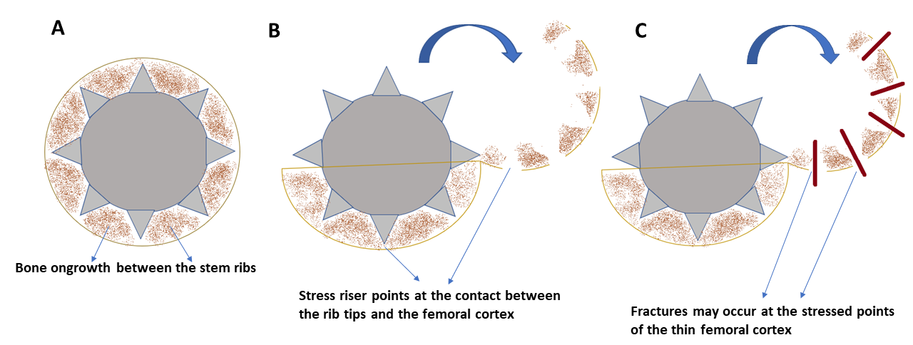
Given these stem characteristics and the remarkable osseointegration, extraction of fluted, tapered cementless stems during revision surgery can be technically demanding. The stem splines may have already created several stress risers at their contact with the femoral cortex. Unplanned insertion of osteotomes/ chisels in the stem-bone interface, and unplanned extended femoral osteotomy (King et al. 2008) in these settings may lead to iatrogenic perforations and fractures of the femoral cortex (Figure 3, 4A-G), which can compromise the stability of the new revision femoral stem.

Figure 3
Figure 3.A) Conceptualized cross-section of a tapered, fluted stem with bony integration between the 8 stem splines ribs. At the points of contact between the rib tip and the femoral cortex, the bone may be very thin. B) When the femoral cortex is directly osteotomized without preparation, the created stress risers may lead to potential comminution (C) in the femoral cortex.

Despite the presence of several reports that discuss how to revise femoral stems (Burstein et al. 2004; Masri, Mitchell, and Duncan 2005; Laffosse 2016; Khan et al. 2015), there is only one study that discussed in detail the K-wire drilling technique for extraction of cementless femoral stems in general (Hafez 2017). The purpose of this study is to revisit the K wire extraction technique, particularly for the tapered fluted cementless stems, giving their unique design. We believe this technique is simple and reproducible and does not require specific advanced surgical instruments.

TECHNIQUE

The patient is positioned, and surgical exposure is performed through the approach of the surgeon’s discretion. After exposure of the proximal portion of the stem, a trial of stem extraction using the stem applicator and back hammering may be attempted if the stem is felt to be loose. In cases where it is well fixed, further exposure of the proximal portion of the stem is performed using burrs, fine bone rongeurs and/or chisels until clearly viewing the proximal portion of the flutes or splines of the stem. This may necessitate performing bone trimming around the proximal, non-ridged portion of the stem in order to visualize the splines (ridges) and flutes.

At this stage, after proper exposure of the proximal portion of the stem, until viewing the proximal end of the splines, multiple forward and backward longitudinal drills were passed through the flutes between the splines downwards, guided by the C arm. Repeated drill passages are performed from proximal to distal until completing all the holes between splines (see Video 1 in the Supplementary Materials). Either a 1.8-2.2mm K-wire or 2-2.5mm drill bit is used and longitudinal drilling is performed along the 8 holes (Figure 4 I,J). The drill bit or K wire may be tilted between the splines during drilling. Additionally, a fine chisel may be then used in the same manner between the splines from proximal to distal to further free the stem. At this stage, the stem is almost loose and can usually be extracted utilizing the applicator and back hammering, or through an extended trochanteric osteotomy that should leave a single, intact femoral cortex (Figure 4 K). This is followed by fixation of the osteotomy with cerclage wire (Figure 4 L) and insertion of a spacer or revision THA stem as planned. A longer and larger, diaphyseal fitting stem is usually utilized when revising a fluted stem, and utilization of a stem with distal transfixing screws is a viable option.

Figure 4
Figure 4.A-G) Case 1: A) Plain pelvic radiographs of a 42-year-old female who presented with recurrent left prosthetic hip dislocation and left lower limb shortening. Upon revision, the stem was found to be markedly anteverted. B, C) Intraoperative C-arm radiographs showing well fixed fluted stem. D, E) In this case, Wagner Osteotomy was performed without adequate femoral preparation to dis-engage the well-fixed stem, leading to femoral cortical comminution. Note the bone ongrowth over the stem and between the stem splines. F, G) Intraoperative C-arm radiograph showing femoral cortical comminution after removal of the femoral stem. The iatrogenic fractures were fixed with cerclage wires and an external fixator. H-L) Case 2: H) Plain radiograph of a case of a 48-year-old male who presented with infected THA. I) After exposure of the proximal portion of the stem, multiple longitudinal drills were performed through flutes, guided by the C arm, to complete the holes between splines. J) Conceptualized cross-section of the proximal end of the stem as viewed from upwards, illustrates the placement of a drill bit or K-wire between splines before longitudinal drilling, with the possibility of tilting the drill bit during the process (blue arrows). K) Trochanteric osteotomy with intact bone after extraction of the stem. L) Plain radiograph after removal of implants and insertion of antibiotic beads. Note the integrity of trochanteric osteotomy, compared to the previous case.

Expected Outcomes

The longitudinal drilling technique can assist the surgeon in extracting a well-fixed, fluted cementless femoral stem while decreasing the risk for iatrogenic femoral fracture.

Complications

Potential complications include iatrogenic femoral metaphyseal and femoral shaft perforation with the wire, drill bit or wire breakage inside the femoral canal. Careful placement of the wires or drill bits inside the flutes through proper proximal exposure may decrease the chance of iatrogenic perforation or hardware breakage.

Discussion

This study describes a reproducible method for extraction of well fixed, uncemented, fluted conical stems. Although this technique is utilized by many surgeons it has only been described once in detail for uncemented femoral stems in general (Hafez 2017). Given the unique design of fluted stems, we believe this technique is particularly useful in such stems and we aimed to add to the existing literature for more popularization of the technique. The presence of flutes and splines in such stems can guide the drilling technique while decreasing the chance of femoral cortical perforation and K wire bending or breakage.

Extraction of well-fixed cementless stems in revision THA is a technically challenging procedure. Preserving a well-fixed original stem during revision surgery, when acetabular-side revision alone is adequate, has the potential to reduce time, blood loss, and minimize the risk of additional tissue injury and bone damage (Hafez 2017). However, stem removal is performed for multiple indications during the revision surgery, including infection, recurrent instability due to stem version and offset defects, stem breakage, taper damage, limb length discrepancy due to stem subsidence or mal insertion causing lengthening, femoral osteolysis, taper mismatch with the revision system, bony ankylosis, or during the revision of excessively medialized or mispositioned cup (Laffosse 2016; Randhawa et al. 2009; De Thomasson et al. 2001; Wahl et al. 2021; Reina et al. 2013).

Given the unique stem design and bone ongrowth, unplanned stem removal may result in unexpected bony damage. The proposed technique of axial drilling allows for a more predictable stem extraction, minimizing bone damage. Since it does not require specific instruments and could be tried in a timely fashion, we believe this technique is reproducible and helpful to be performed whenever extraction of a fluted, tapered stem is required.

Conclusion

The longitudinal drilling technique along the stem flutes is a reproducible technique that allows for partial loosening of well-integrated, fluted cementless tapered stems. This technique allows for easier stem extraction minimizing the risk of significant bony comminution or damage.

Conflicts of Interest

None to declare.

Submitted: April 27, 2024 EDT

Accepted: July 14, 2024 EDT

References

Abdel, M. P., U. Cottino, D. R. Larson, A. D. Hanssen, D. G. Lewallen, and D. J. Berry. 2017. “Modular Fluted Tapered Stems in Aseptic Revision Total Hip Arthroplasty.” The Journal of Bone and Joint Surgery. American Volume 99 (10): 873–81. https:/​/​doi.org/​10.2106/​JBJS.16.00423.
Google Scholar
Barrack, R. L., and S. J. Burnett. 2005. “Preoperative Planning for Revision Total Hip Arthroplasty.” The Journal of Bone and Joint Surgery. American Volume 87 (12): 2800–2811. https:/​/​doi.org/​10.2106/​00004623-200512000-00028.
Google Scholar
De Thomasson, E., O. Guingand, R. Terracher, and C. Mazel. 2001. “Perioperative Complications after Total Hip Revision Surgery and Their Predictive Factors. A Series of 181 Consecutive Procedures.” Revue de Chirurgie Orthopedique et Reparatrice de L’appareil Moteur 87 (5): 477–88.
Google Scholar
Hafez, M. A. 2017. “K-Wire Technique for the Removal of Uncemented Femoral Stem in Revision THA.” Techniques in Orthopaedics 32 (2): 105–7. https:/​/​doi.org/​10.1097/​BTO.0000000000000202.
Google Scholar
Haynes, J. A., J. B. Stambough, A. A. Sassoon, S. R. Johnson, J. C. Clohisy, and R. M. Nunley. 2016. “Contemporary Surgical Indications and Referral Trends in Revision Total Hip Arthroplasty: A 10-Year Review.” The Journal of Arthroplasty 31 (3): 622–25. https:/​/​doi.org/​10.1016/​j.arth.2015.09.026.
Google Scholar
Khan, R. J., L. McGonagle, A. R. Wallis, A. S. Sidhu, D. P. Fick, and B. Nivbrant. 2015. “A New Technique in Revision Hip Arthroplasty for Vancouver B Periprosthetic Fractures.” Techniques in Orthopaedics 30 (1): 49–53. https:/​/​doi.org/​10.1097/​BTO.0000000000000096.
Google Scholar
King, S., M. E. Berend, M. A. Ritter, E. M. Keating, P. M. Faris, and J. B. Meding. 2008. “Extended Femoral Osteotomy and Proximally Coated Prosthesis for Hip Revision.” Orthopedics (Online) 31 (1): 67. https:/​/​doi.org/​10.3928/​01477447-20080101-21.
Google Scholar
Laffosse, J. M. 2016. “Removal of Well-Fixed Fixed Femoral Stems.” Orthopaedics & Traumatology: Surgery & Research 102 (1): S177–87. https:/​/​doi.org/​10.1016/​j.otsr.2015.06.029.
Google Scholar
Masri, B. A., P. A. Mitchell, and C. P. Duncan. 2005. “Removal of Solidly Fixed Implants during Revision Hip and Knee Arthroplasty.” JAAOS-Journal of the American Academy of Orthopaedic Surgeons 13 (1): 18–27. https:/​/​doi.org/​10.5435/​00124635-200501000-00004.
Google Scholar
Matthys, F., J. Van Meirhaeghe, and C. Pattyn. 2021. “Fracture Risk during Extraction of Well-Fixed Extended Cementless Stems: Porous versus Hydroxyapatite Coated.” Acta Orthopaedica Belgica 87 (1): 41–45. https:/​/​doi.org/​10.52628/​87.1.06.
Google Scholar
Otero, J. E., J. R. Martin, T. M. Rowe, S. M. Odum, and J. B. Mason. 2020. “Radiographic and Clinical Outcomes of Modular Tapered Fluted Stems for Femoral Revision for Paprosky III and IV Femoral Defects or Vancouver B2 and B3 Femoral Fractures.” The Journal of Arthroplasty 35 (4): 1069–73. https:/​/​doi.org/​10.1016/​j.arth.2019.11.039.
Google Scholar
Park, K. S., S. Y. Jin, J. H. Lim, and T. R. Yoon. 2021. “Long-Term Outcomes of Cementless Femoral Stem Revision with the Wagner Cone Prosthesis.” Journal of Orthopaedic Surgery and Research 16 (June):1–8. https:/​/​doi.org/​10.1186/​s13018-021-02457-8.
Google Scholar
Randhawa, K., F. S. Hossain, B. Smith, C. Mauffrey, and T. Lawrence. 2009. “A Prospective Study of Hip Revision Surgery Using the Exeter Long-Stem Prosthesis: Function, Subsidence, and Complications for 57 Patients.” Journal of Orthopaedics and Traumatology 10:159–65. https:/​/​doi.org/​10.1007/​s10195-009-0068-0.
Google Scholar
Reina, N., C. Delaunay, P. Chiron, N. Ramdane, and M. Hamadouche. 2013. “Infection as a Cause of Primary Total Hip Arthroplasty Revision and Its Predictive Factors.” Orthop Traumatol Surg Res 99 (5): 555–61. https:/​/​doi.org/​10.1016/​j.otsr.2013.07.001.
Google Scholar
Wagner, H., and M. Wagner. 2000. “Cone Prosthesis for the Hip Joint.” Archives of Orthopaedic and Trauma Surgery 120 (1–2): 88–95. https:/​/​doi.org/​10.1007/​pl00021223.
Google Scholar
Wahl, P., T. Solinger, M. Schläppi, and E. Gautier. 2021. “Removal of an Osteointegrated Broken Uncemented Femoral Stem after Hip Arthroplasty.” Journal of Orthopaedic Surgery and Research 16:1–4. https:/​/​doi.org/​10.1186/​s13018-021-02365-x.
Google Scholar
Zang, J., K. Uchiyama, M. Moriya, K. Fukushima, N. Takahira, and M. Takaso. 2019. “Long-Term Outcomes of Wagner Self-Locking Stem with Bone Allograft for Paprosky Type II and III Bone Defects in Revision Total Hip Arthroplasty: A Mean 15.7-Year Follow-Up.” Journal of Orthopaedic Surgery 27 (2). https:/​/​doi.org/​10.1177/​2309499019854156.
Google Scholar

This website uses cookies

We use cookies to enhance your experience and support COUNTER Metrics for transparent reporting of readership statistics. Cookie data is not sold to third parties or used for marketing purposes.

Powered by Scholastica, the modern academic journal management system